



Phthalaldehyde

Based on 1 Customer Validation
Phthalaldehyde (Phthaldialdehyde) reacts with proteins containing primary amines and blocked amino-terminal peptides of amino acids. Phthalaldehyde stabilizes bacterial outer membranes and cell walls, increases the optical density of bacterial cell suspensions, and inhibits bacterial lysis induced by ethylenediaminetetraacetic acid and sodium dodecyl sulfate. Phthalaldehyde exhibits bactericidal activity against Gram-negative vegetative bacteria.
商品は「研究用試薬」です。人や動物の医療用・臨床診断用・食品用の製品ではありません。
研究用途以外に使用した場合、当社は一切の責任を負いかねます。
- 純度 : 99.80%
- CAS 番号: 643-79-8
- 分子式: C8H6O2
- 分子量:134.13
-
保管条件:
4°C, stored under nitrogen
* In solvent : -80°C, 6 months; -20°C, 1 month (stored under nitrogen)


To place orders, for customer services and technical support, please contact: MedChemExpress USA
Tel: 609-228-6898 E-mail: [email protected] [email protected]


-
 生物活性
生物活性
-
化学情報
-
溶剤 & 溶解度
- プロトコル
- 純度とドキュメンテーション
- 参考文献
-
 Help & FAQs


-
HY-B2176
ATPEndogenous Metabolite -

HY-W012669R
Phthalaldehyde (Standard)Biochemical Assay Reagent -
HY-66019
FITCFluorochrome -
HY-D0017
Dansyl chlorideFluorescent Dye -
HY-D0715
FluorescamineFluorescent Dye -
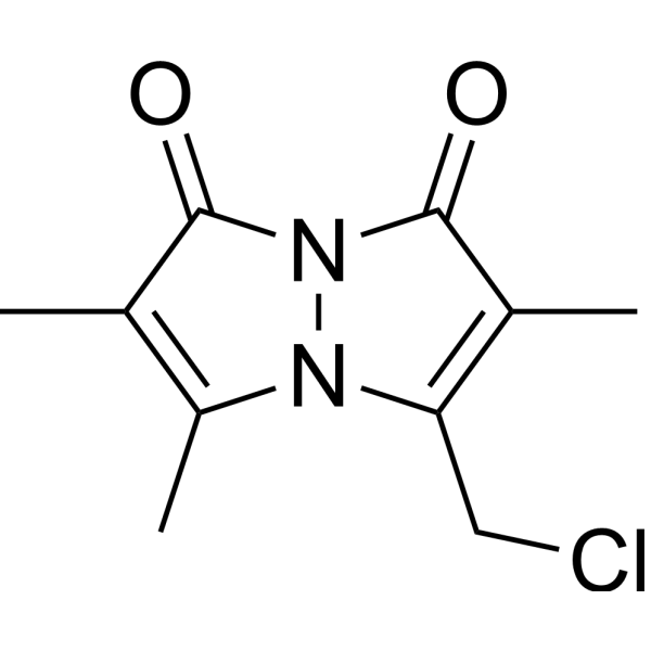
HY-101899
MonochlorobimaneGSH Fluorescent Dye -

HY-W089645
2,2,2-Trifluoro-N-methyl-N-(trimethylsilyl)acetamideEster Product -
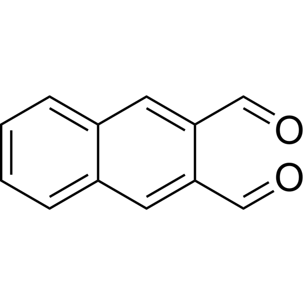
HY-W016288
Naphthalene-2,3-dicarboxaldehydeAntifungal Agents -
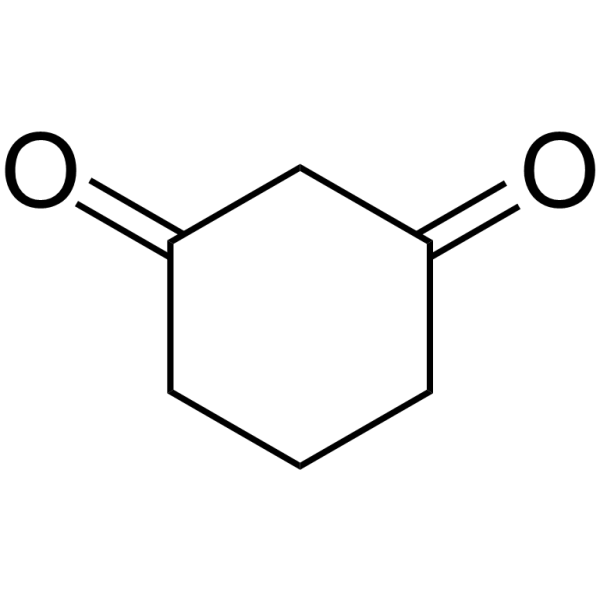
HY-W007466
Cyclohexane-1,3-dioneDrug Intermediate -
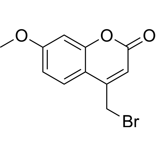
HY-D0036
Br-MmcFluorescent Label -
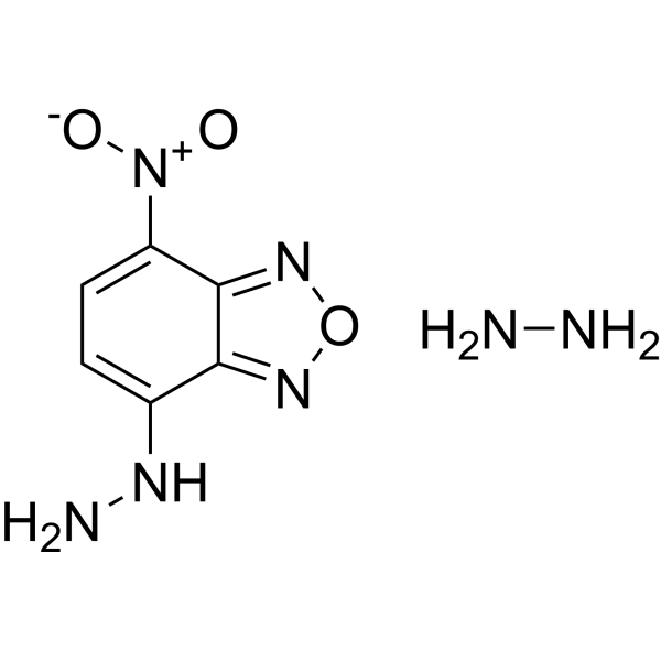
HY-W273413
4-Hydrazino-7-nitro-benzofurazan hydrazine adductFluorescent Reagent -

HY-DY1007
FITC (solution)Fluorochrome


生物活性
製品説明
体外実験
Phthalaldehyde (0.0025% (w/v); 1-180 min) reacts with lysine, glycine, and histidine to produce distinct UV absorption peaks at both acidic and alkaline pH, with reaction rates accelerating at higher pH; specific peak patterns and development times vary by amino acid[1].
Phthalaldehyde (0.05-0.2% (w/v); 2 h at 20 °C) induces dark color changes in pellets of Gram-negative (E. coli, P. aeruginosa) and Gram-positive (S. aureus) bacteria, with darker colors observed at alkaline pH and higher OPA concentration[1].
Phthalaldehyde (0.1% (w/v); 30 min at 20 °C) significantly inhibits EDTA-Tris induced lysis of Pseudomonas aeruginosa PA01[1].
Phthalaldehyde (0.1% (w/v); 30 min at 20 °C) significantly inhibits lysostaphin-induced lysis of Staphylococcus aureus NCTC 6571, with efficacy comparable to glutaraldehyde[1].
Phthalaldehyde (0.25-0.5% (w/v); 60 min) significantly increases the absorbance of Escherichia coli ATCC 9481, Staphylococcus aureus NCTC 6571, and Pseudomonas aeruginosa PA01 suspensions, with a greater increase in E. coli absorbance observed at the 0.5% (w/v) concentration[1].
Phthalaldehyde detects the blocked amino-terminal tryptic peptide of rat liver acyl-CoA oxidase by forming a fluorescent Schiff base with the peptide's free α-amino group[2].
MedChemExpress (MCE) has not independently confirmed the accuracy of these methods. They are for reference only.


化学情報
-
CAS 番号 643-79-8
-
性状 Solid
-
分子量 134.13
-
分子式 C8H6O2
-
Color Off-white to yellow
-
SMILES
O=CC1=CC=CC=C1C=O
-
別名
Phthaldialdehyde
-
輸送条件
Room temperature in continental US; may vary elsewhere.
-
保管条件
4°C, stored under nitrogen
* In solvent : -80°C, 6 months; -20°C, 1 month (stored under nitrogen)


溶剤 & 溶解度
体外:
DMSO : 200 mg/mL (1491.09 mM; Need ultrasonic; Hygroscopic DMSO has a significant impact on the solubility of product, please use newly opened DMSO)
Please refer to the solubility information to select the appropriate solvent. Once prepared, please aliquot and store the solution to prevent product inactivation from repeated freeze-thaw cycles.
Storage method and period of stock solution: -80°C, 6 months; -20°C, 1 month (stored under nitrogen). When stored at -80°C, please use it within 6 months. When stored at -20°C, please use it within 1 month.
Please refer to the solubility information to select the appropriate solvent. Once prepared, please aliquot and store the solution to prevent product inactivation from repeated freeze-thaw cycles.
Storage method and period of stock solution: -80°C, 6 months; -20°C, 1 month (stored under nitrogen). When stored at -80°C, please use it within 6 months. When stored at -20°C, please use it within 1 month.
濃度 (開始) × 体積 (開始) = 濃度 (終了) × 体積 (終了)


純度とドキュメンテーション
-
データシート (279 KB)
-
SDS (789 KB)
- English - EN (789 KB)
- Français - FR (789 KB)
- Deutsch - DE (789 KB)
- Norwegian - NO (789 KB)
- Español - ES (789 KB)
- Swedish - SV (789 KB)
- Italian - IT (789 KB)
- Korean - KR (789 KB)
- Portuguese - PT (789 KB)
-
取扱説明書 (2659 KB)
参考文献
[1]. Walsh SE, et al. Studies on the mechanisms of the antibacterial action of ortho-phthalaldehyde. J Appl Microbiol. 1999;87(5):702-710. [Content Brief]
[2]. Miyazawa S, et al. Complete nucleotide sequence of cDNA and predicted amino acid sequence of rat acyl-CoA oxidase. J Biol Chem. 1987;262(17):8131-8137. [Content Brief]


Complete Stock Solution Preparation Table
Please refer to the solubility information to select the appropriate solvent. Once prepared, please aliquot and store the solution to prevent product inactivation from repeated freeze-thaw cycles.
Storage method and period of stock solution: -80°C, 6 months; -20°C, 1 month (stored under nitrogen). When stored at -80°C, please use it within 6 months. When stored at -20°C, please use it within 1 month.
| Optional Solvent | Concentration Solvent Mass | 1 mg | 5 mg | 10 mg | 25 mg |
|---|---|---|---|---|---|
| DMSO | 1 mM | 7.4555 mL | 37.2773 mL | 74.5545 mL | 186.3863 mL |
| 5 mM | 1.4911 mL | 7.4555 mL | 14.9109 mL | 37.2773 mL | |
| 10 mM | 0.7455 mL | 3.7277 mL | 7.4555 mL | 18.6386 mL | |
| 15 mM | 0.4970 mL | 2.4852 mL | 4.9703 mL | 12.4258 mL | |
| 20 mM | 0.3728 mL | 1.8639 mL | 3.7277 mL | 9.3193 mL | |
| 25 mM | 0.2982 mL | 1.4911 mL | 2.9822 mL | 7.4555 mL | |
| 30 mM | 0.2485 mL | 1.2426 mL | 2.4852 mL | 6.2129 mL | |
| 40 mM | 0.1864 mL | 0.9319 mL | 1.8639 mL | 4.6597 mL | |
| 50 mM | 0.1491 mL | 0.7455 mL | 1.4911 mL | 3.7277 mL | |
| 60 mM | 0.1243 mL | 0.6213 mL | 1.2426 mL | 3.1064 mL | |
| 80 mM | 0.0932 mL | 0.4660 mL | 0.9319 mL | 2.3298 mL | |
| 100 mM | 0.0746 mL | 0.3728 mL | 0.7455 mL | 1.8639 mL |


Phthalaldehyde 関連分類
HY-W012669 関連分類
 Powered by Bioz
Keywords
- Phthalaldehyde
- 643-79-8
- Phthaldialdehyde
- Environmental Pollutants
- Biochemical Assay Reagents
- primary amine-containing proteins
- Escherichia coli ATCC 9481
- Gram-negative vegetative bacteria
- bacterial outer membranes
- Pseudomonas aeruginosa PA01
- amino acids
- rat liver acyl-CoA oxidase
- rat acyl-CoA oxidase
- bacterial cell walls
- Staphylococcus aureus NCTC 6571
- Inhibitor
- inhibitor
- inhibit