



PRO-F
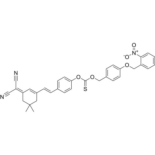
PRO-F is a photoactivable H2S donor with ROS scavenging ability. PRO-F can be activated by light to produce fluorescent signal, for real-time tracking of released H2S. PRO-F activation doesn’t consume endogenous substances. deliver H2S in an intracellular environment to protect cells from excessive reactive oxygen species (ROS) induced damage. PRO-F shows enhancement on chronic wound healing, researched in diabetic models as well.
For research use only. We do not sell to patients.
- Formula: C34H29N3O5S
- Molecular Weight:591.68
-
Storage:
Please store the product under the recommended conditions in the Certificate of Analysis.

To place orders, for customer services and technical support, please contact: MedChemExpress USA
Tel: 609-228-6898 E-mail: [email protected] [email protected]


-
 Biological Activity
-
Chemical Information
- Protocols
- Purity & Documentation
- References
-
 Help & FAQs




Biological Activity
Description
In Vitro
PRO-F (20 μM; 0-500s) can be activated by light activation (365 nm) time-dependently, λex=530 nm, λem=676 nm[1].
PRO-F (20 μM; 5 min, 10 min, 15 min) produces red fluorescent signal in NDF and HaCaT cells[1].
MedChemExpress (MCE) has not independently confirmed the accuracy of these methods. They are for reference only. Further protocols information, click here.


Chemical Information
-
Molecular Weight 591.68
-
Formula C34H29N3O5S
-
SMILES
CC1(C)CC(/C=C/C2=CC=C(OC(OCC3=CC=C(OCC4=CC=CC=C4[N+]([O-])=O)C=C3)=S)C=C2)=C/C(C1)=C(C#N)/C#N
-
Shipping
Room temperature in continental US; may vary elsewhere.
-
Storage
Please store the product under the recommended conditions in the Certificate of Analysis.

Protocols
-
ROS/oxidative-stress fluorescent staining
ROS/oxidative-stress fluorescent staining uses cell-permeant fluorogenic probes that become fluorescent after oxidation inside cells or tissues; commonly used examples include DCFH-DA/DCFDA for broad cellular oxidant detection, DHE for superoxide-related signal detection, MitoSOX for mitochondrial superoxide-related signal detection, and CellROX probes for oxidative-stress-associated fluorescence readouts. The assay detects probe oxidation rather than a single ROS species unless the probe and analysis method have been chemically validated for that species. DCFH-DA enters cells, is deacetylated by intracellular esterases to DCFH, and produces fluorescent DCF after oxidation, so the readout is used as an operational measure of total cellular oxidative stress rather than a species-specific ROS measurement. DHE and MitoSOX can report superoxide-related oxidation, but red fluorescence alone can include non-specific ethidium-like oxidation products; HPLC or optimized spectral approaches are
-
Cell migration
Cell migration is a method that plays an important role in wound healing, cell differentiation, embryonic development, etc.
-
Cell-Exclusion Zone Migration Assay
The Cell-Exclusion Zone (CEZ) migration assay is an in vitro 2D cell migration method in which a defined cell-free area is created using removable physical barriers such as silicone stoppers, allowing cells to be seeded around the barrier and subsequently migrate into the cleared zone after barrier removal. This approach enables quantification of collective cell migration by monitoring repopulation of the initially cell-free region over time using microscopy-based imaging. Compared with scratch-based wound healing assays, barrier-based exclusion methods are designed to avoid mechanical damage to the extracellular matrix and reduce injury-induced effects on boundary cells, thereby improving interpretability of migration behavior in vitro. The assay readout is typically the progressive reduction in the cell-free area or the number of cells invading the exclusion zone, reflecting coordinated cell motility relevant to physiological processes such as wound healing, epithelial repair, and ca


Purity & Documentation
References

Calculators
Concentration (start) × Volume (start) = Concentration (final) × Volume (final)


PRO-F Related Classifications
HY-149837 Related Classifications
 Powered by Bioz