



Sapienic acid-d19

Based on 1 Customer Validation
Sapienic acid-d19 is a deuterated labeled Sapienic acid. Sapienic acid is a fatty acid commonly found on the skin and in mucosa. Sapienic acid has variable antimicrobial activities against Gram-positive and Gram-negative bacteria found on the skin and in the oral cavity. Sapienic acid is active against Streptococcus sanguinis, Streptococcus mitis and Fusobacterium nucleatum with MBC values of 31.3 μg/mL, 375.0 μg/mL and 93.8 μg/mL, respectively.
For research use only. We do not sell to patients.
- Purity : 99.0%
- CAS No.: 2692623-89-3
- Formula: C16H11D19O2
- Molecular Weight:273.53
-
Storage:
Solution, -20°C, 2 years


To place orders, for customer services and technical support, please contact: MedChemExpress USA
Tel: 609-228-6898 E-mail: [email protected] [email protected]


-
 Biological Activity
-
Chemical Information
- Protocols
- Purity & Documentation
- References
-
 Help & FAQs


-
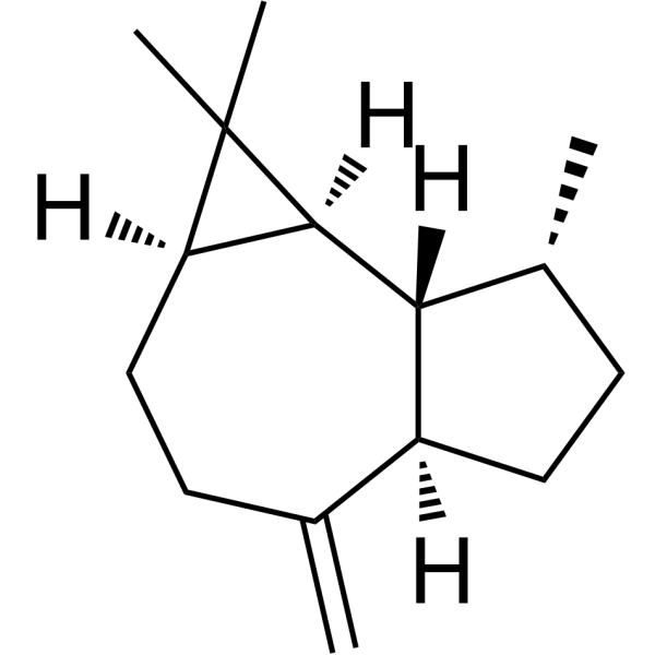
HY-N8411
AromadendreneAntibacterial Agent -
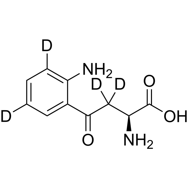
HY-104026S
L-Kynurenine-d4Stable Isotope -
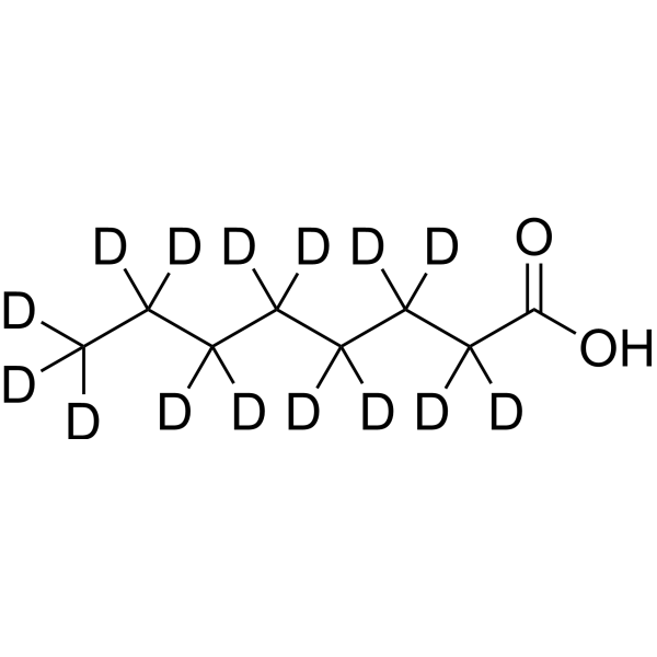
HY-41417S
Octanoic acid-d15Stable Isotope -

HY-N8411R
Aromadendrene (Standard)Reference Standard -
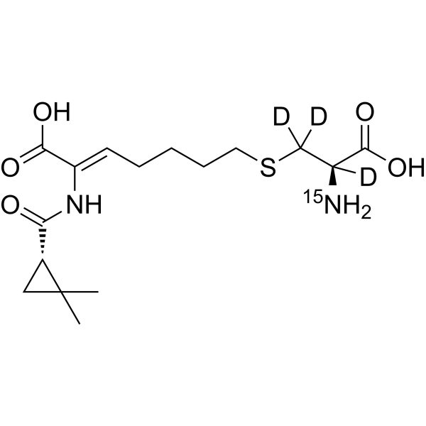
HY-A0166S
Cilastatin-15N,d3Stable Isotope -
HY-B0276S
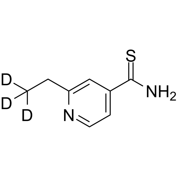
Ethionamide-d3Stable Isotope -
HY-B0117S
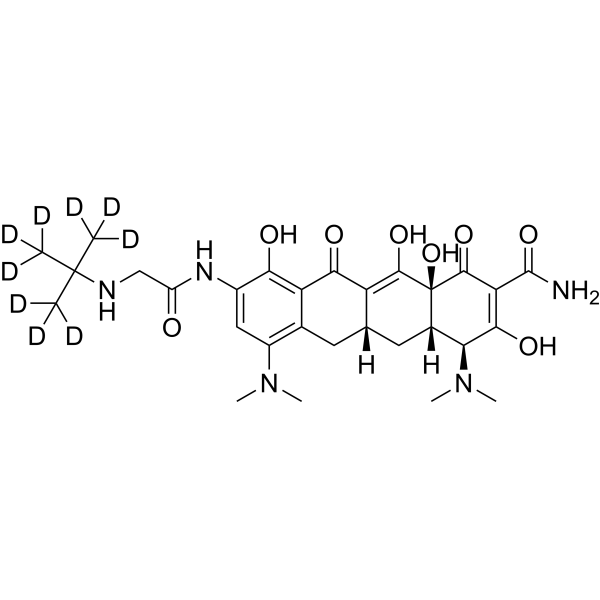
Tigecycline-d9Stable Isotope -
HY-41417S3
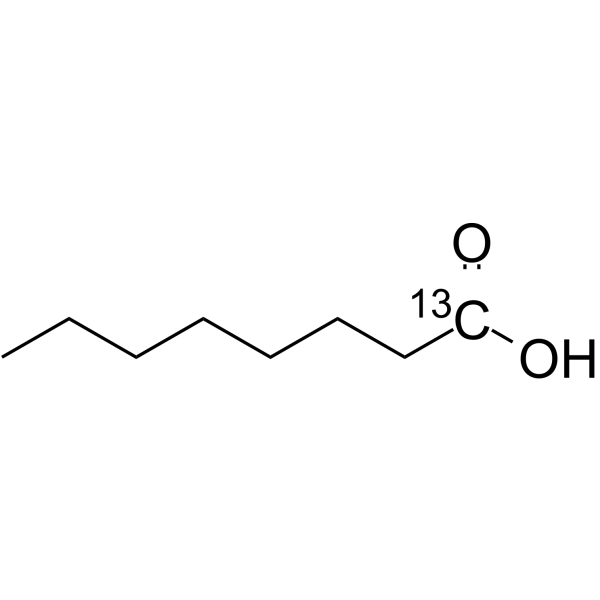
Octanoic acid-13CStable Isotope -

HY-169225
PDIC-NNSTING Activator -
HY-41417S1
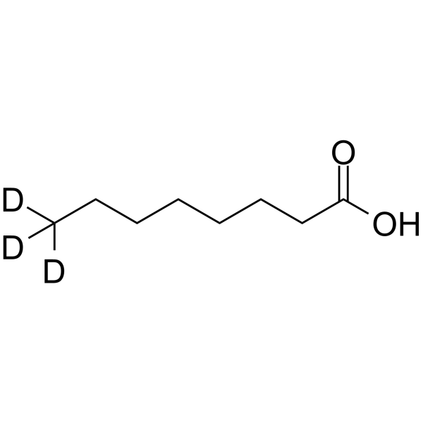
Octanoic acid-d3Stable Isotope -
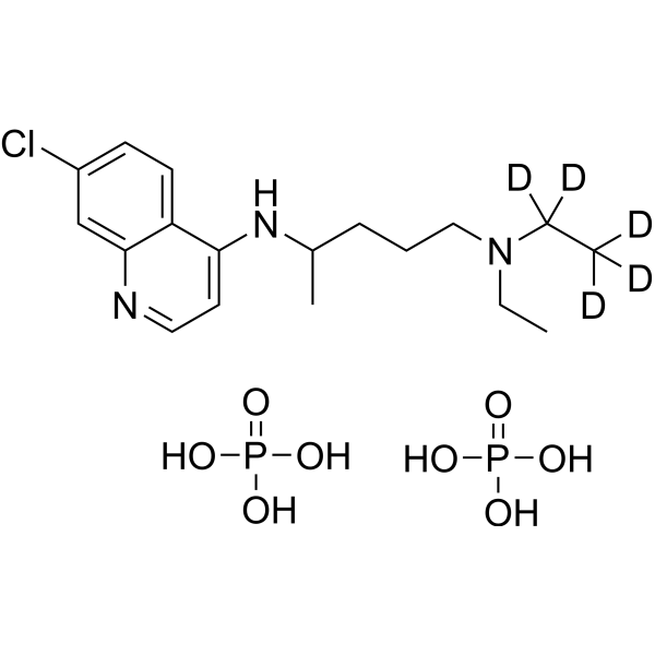
HY-17589S
Chloroquine-d5 diphosphateStable Isotope -
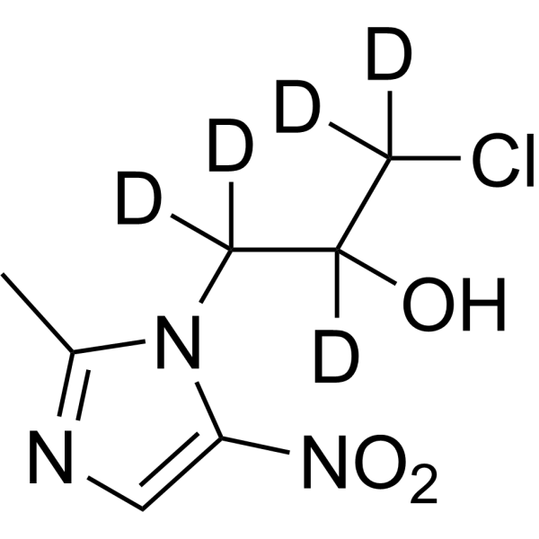
HY-B0508S
Ornidazole-d5Stable Isotope


Biological Activity
Description
In Vitro
Stable heavy isotopes of hydrogen, carbon, and other elements have been incorporated into drug molecules, largely as tracers for quantitation during the drug development process. Deuteration has gained attention because of its potential to affect the pharmacokinetic and metabolic profiles of drugs[1].
Sapienic acid (minimum bactericidal concentrations (MBCs) range, 31.3 to 375.0 μg/mL) is active against Streptococcus sanguinis, Streptococcus mitis, and Fusobacterium nucleatum but not active against Escherichia coli, Staphylococcus aureus, S. marcescens, P. aeruginosa, Corynebacterium bovis, Corynebacterium striatum, and Corynebacterium jeikeium (MBC > 500 μg/mL). Kinetic assays show that killing of S. sanguinis and S. mitis with sapienic acid is gradual and occurred within 24 h[2].
MedChemExpress (MCE) has not independently confirmed the accuracy of these methods. They are for reference only. Further protocols information, click here.
Application
1. This compound can be used as a tracer
2. This compound can be used as an internal standard for quantitative analysis by NMR, GC-MS, or LC-MS.


Chemical Information
-
CAS No. 2692623-89-3
-
Unlabeled CAS 17004-51-2
-
Appearance Liquid
-
Molecular Weight 273.53
-
Formula C16H11D19O2
-
Color Colorless to light yellow
-
SMILES
[2H]C([2H])([2H])C([2H])([2H])C([2H])([2H])C([2H])([2H])C([2H])([2H])C([2H])([2H])C([2H])([2H])C([2H])([2H])C([2H])([2H])/C=C\CCCCC(O)=O
-
Shipping
Room temperature in continental US; may vary elsewhere.
-
Storage
Solution, -20°C, 2 years

Protocols
-
Research Protocol for Infectious Diseases
Infectious-disease experiments test how pathogens interact with host barriers, innate immune receptors, inflammatory signaling, pathogen replication, and tissue injury; pattern-recognition receptors such as TLRs, RIG-I-like receptors, NOD-like receptors, and inflammasomes detect microbial molecules and activate NF-κB, interferon, and cytokine responses. The central hypothesis is that infection severity reflects the balance between pathogen burden and host response: protective inflammation restricts pathogen growth, whereas excessive or mislocalized inflammation contributes to tissue damage and disease phenotype. Unresolved questions include which host pathways are protective versus pathogenic, why some infection models fail to translate to human disease, and which combined readouts best predict clinically relevant infection outcomes.
-
Gram Staining of Tissue Sections
Gram staining of tissue sections is a histochemical technique used to differentiate Gram-positive and Gram-negative bacteria within histological specimens based on differences in bacterial cell wall structure and dye retention, adapted from classical bacteriological Gram staining into tissue-compatible “histological Gram stain” variants. In tissue applications, modifications of the Brown-Hopps and Brown-Brenn methods are commonly used to improve differentiation of microorganisms embedded within host connective tissue and to reduce overstaining or loss of Gram-negative signal, which are known limitations of earlier approaches. The principle relies on crystal violet-iodine complex retention in Gram-positive organisms and subsequent decolorization and counterstaining steps that allow contrast visualization of Gram-negative organisms against tissue background.


Purity & Documentation
-
Data Sheet (279 KB)
-
SDS (393 KB)
- English - EN (393 KB)
- Français - FR (393 KB)
- Deutsch - DE (393 KB)
- Norwegian - NO (393 KB)
- Español - ES (393 KB)
- Swedish - SV (393 KB)
- Italian - IT (393 KB)
- Korean - KR (393 KB)
- Portuguese - PT (393 KB)
-
Handling Instructions (2659 KB)
References
[1]. Russak EM, et al. Impact of Deuterium Substitution on the Pharmacokinetics of Pharmaceuticals. Ann Pharmacother. 2019 Feb;53(2):211-216. [Content Brief]
[2]. Fischer CL, et al. Antibacterial activity of sphingoid bases and fatty acids against Gram-positive and Gram-negative bacteria. Antimicrob Agents Chemother. 2012 Mar;56(3):1157-61. [Content Brief]

Calculators
Concentration (start) × Volume (start) = Concentration (final) × Volume (final)


Sapienic acid-d19 Related Classifications
HY-130187S Related Classifications
 Powered by Bioz