



SPP-86
Based on 2 publication(s) in Google Scholar
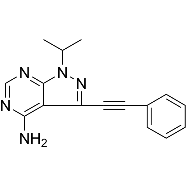
SPP-86 is a potent and selective cell permeable inhibitor of RET tyrosine kinase, with an IC50 of 8 nM. SPP-86 inhibits RET-induced phosphatidylinositide 3-kinases (PI3K)/Akt and MAPK signaling, also inhibits RET-induced estrogen receptorα (ERα) phosphorylation in MCF7 cells. SPP-86 is a click chemistry reagent, it contains an Alkyne group and can undergo copper-catalyzed azide-alkyne cycloaddition (CuAAc) with molecules containing Azide groups.
For research use only. We do not sell to patients.
- Purity : 99.71%
- CAS No.: 1357349-91-7
- Formula: C16H15N5
- Molecular Weight:277.32
-
Storage:Powder -20°C, 3 years , 4°C, 2 years ; In solvent -80°C, 6 months , -20°C, 1 month

To place orders, for customer services and technical support, please contact: MedChemExpress USA
Tel: 609-228-6898 E-mail: [email protected] [email protected]


-
 Biological Activity
-
Chemical Information
-
Solvent & Solubility
- Protocols
- Purity & Documentation
- References
-
 Help & FAQs


-
HY-107597
HalicinJNK Inhibitor -
HY-114370
SelpercatinibRET Kinase Inhibitor -
HY-112301
PralsetinibRET Inhibitor -
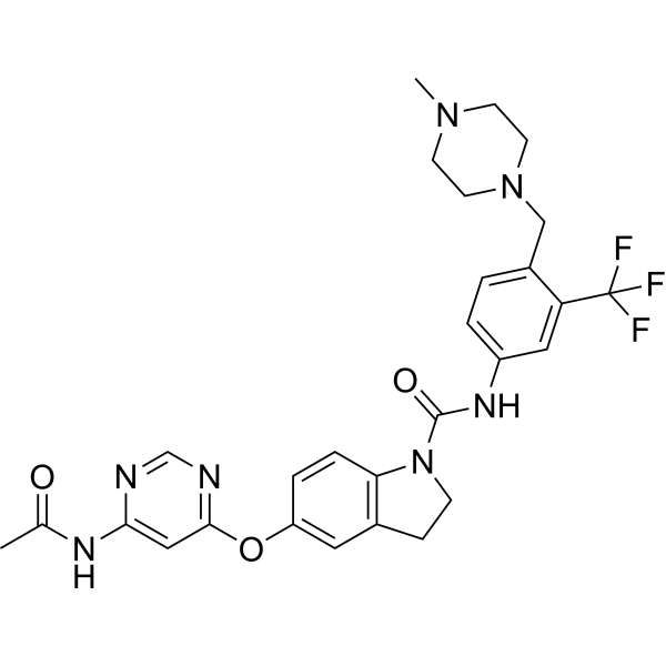
HY-18840
BBT594RET Inhibitor -
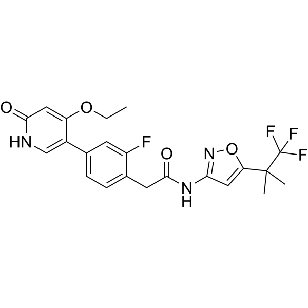
HY-100459
GSK3179106RET Kinase Inhibitor -
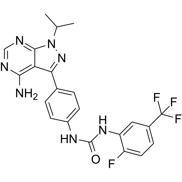
HY-101963
AD80Multikinase Inhibitor -
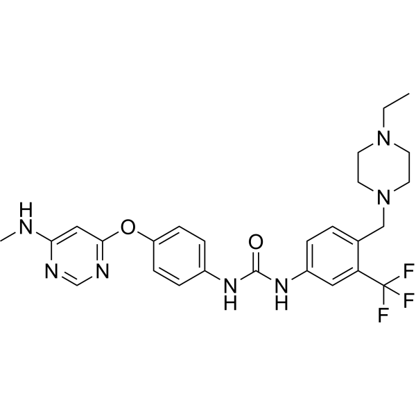
HY-15002
AST 487c-Kit Inhibitor -
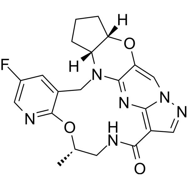
HY-145565
EnbezotinibRET Inhibitor -

HY-100459R
GSK3179106 (Standard)RET Kinase Inhibitor -
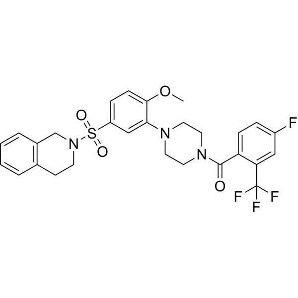
HY-153175
BT44RET Activator -
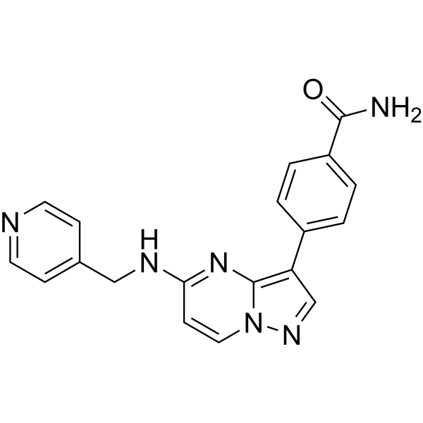
HY-148742
RET-IN-21RETV804M Inhibitor
Publications Citing Use of MedChemExpress (MCE) SPP-86
More

Biological Activity
Description
IC50 & Target
IC50: 8 nM (RET)[1].
Cellular Effect
|
Cell Line

|
Type | Value | Description | References |
|---|---|---|---|---|
| BaF3 | IC50 |
4500 nM
Compound: 2
|
Growth inhibition of mouse BA/F3 cells expressing Bcr-Abl T315I mutant after 48 hrs by MTT assay
Growth inhibition of mouse BA/F3 cells expressing Bcr-Abl T315I mutant after 48 hrs by MTT assay
|
[PMID: 26562217] |
| BaF3 | IC50 |
5020 nM
Compound: 2
|
Growth inhibition of mouse BA/F3 cells expressing wild-type Bcr-Abl after 48 hrs by MTT assay
Growth inhibition of mouse BA/F3 cells expressing wild-type Bcr-Abl after 48 hrs by MTT assay
|
[PMID: 26562217] |
| BaF3 | GI50 |
0.35 μM
Compound: 7a
|
Inhibition of TEL-fused Ret S891A mutant (unknown origin) expressed in mouse BaF3 cells assessed as cell growth inhibition after 48 hrs by MTT assay
Inhibition of TEL-fused Ret S891A mutant (unknown origin) expressed in mouse BaF3 cells assessed as cell growth inhibition after 48 hrs by MTT assay
|
[PMID: 26652860] |
| BaF3 | GI50 |
0.61 μM
Compound: 7a
|
Inhibition of wild type TEL-fused RET (unknown origin) expressed in mouse BA/F3 cells assessed as cell growth inhibition after 48 hrs by MTT assay
Inhibition of wild type TEL-fused RET (unknown origin) expressed in mouse BA/F3 cells assessed as cell growth inhibition after 48 hrs by MTT assay
|
[PMID: 26652860] |
| BaF3 | GI50 |
16.1 μM
Compound: 7a
|
Cytotoxicity against mouse BAF3 cells after 48 hrs by MTT assay
Cytotoxicity against mouse BAF3 cells after 48 hrs by MTT assay
|
[PMID: 26652860] |
| BaF3 | GI50 |
2.6 μM
Compound: 7a
|
Inhibition of TEL-fused Ret V804M mutant (unknown origin) expressed in mouse BA/F3 cells assessed as cell growth inhibition after 48 hrs by MTT assay
Inhibition of TEL-fused Ret V804M mutant (unknown origin) expressed in mouse BA/F3 cells assessed as cell growth inhibition after 48 hrs by MTT assay
|
[PMID: 26652860] |
| BaF3 | GI50 |
5.43 μM
Compound: 7a
|
Inhibition of TEL-fused Ret V804L mutant (unknown origin) expressed in mouse BaF3 cells assessed as cell growth inhibition after 48 hrs by MTT assay
Inhibition of TEL-fused Ret V804L mutant (unknown origin) expressed in mouse BaF3 cells assessed as cell growth inhibition after 48 hrs by MTT assay
|
[PMID: 26652860] |
| BaF3 | IC50 |
>1000 nM
Compound: 1
|
Antiproliferative activity against mouse BAF3 cells harboring CCDC6-RET fusion protein after 72 hrs by CCK8 assay
Antiproliferative activity against mouse BAF3 cells harboring CCDC6-RET fusion protein after 72 hrs by CCK8 assay
|
[PMID: 28404375] |
| HCT-116 | EC50 |
2.4 μM
Compound: 7a
|
Antiproliferative activity against human HCT116 cells
Antiproliferative activity against human HCT116 cells
|
[PMID: 22559926] |
| TPC1 | EC50 |
3.3 μM
Compound: 7a
|
Antiproliferative activity against human TPC1 cells
Antiproliferative activity against human TPC1 cells
|
[PMID: 22559926] |
| TT | GI50 |
2.54 μM
Compound: 7a
|
Inhibition of RET C634W mutant in human TT cells assessed as cell growth inhibition after 10 days by MTT assay
Inhibition of RET C634W mutant in human TT cells assessed as cell growth inhibition after 10 days by MTT assay
|
[PMID: 26652860] |
In Vitro
SPP86 (0-10 μM) inhibits MAPK signaling and proliferation in RET/PTC1 expressing TPC1 but not 8505C or C643 cells[1].
SPP86 (0-10 μM) inhibits RET- induced phosphatidylinositide 3-kinases (PI3K)/Akt and MAPK signaling and estrogen receptorα (ERα) phosphorylation in MCF7 cells[1].
MedChemExpress (MCE) has not independently confirmed the accuracy of these methods. They are for reference only. Further protocols information, click here.
-
Cell Line:Thyroid cancer derived cell lines expressing the RET/PTC1 rearrangement (TPC1), BRAFV600E (8505C) or RASG13R (C643) mutations.
-
Concentration:0-10 μM.
-
Incubation Time:90 min.
-
Result:Inhibited RET- induced ERK1/2 phosphorylation in thyroid cancer cell lines.
-
Cell Line:MCF7 cells (human breast cancer).
-
Concentration:0-10 μM.
-
Incubation Time:30 min.
-
Result:Inhibited RET- induced ERα phosphorylation and proliferation in MCF7 cells.


Chemical Information
-
CAS No. 1357349-91-7
-
Appearance Solid
-
Molecular Weight 277.32
-
Formula C16H15N5
-
Color Off-white to light yellow
-
SMILES
CC(C)N1C2=NC=NC(N)=C2C(C#CC3=CC=CC=C3)=N1
-
Shipping
Room temperature in continental US; may vary elsewhere.
-
Storage
Powder -20°C 3 years 4°C 2 years In solvent -80°C 6 months -20°C 1 month


Publications (2)
-
Journal Impact Factor
-
Most Recent
-
Drug Des Devel Ther
RET Inhibitor SPP86 Triggers Apoptosis and Activates the DNA Damage Response Through the Suppression of Autophagy and the PI3K/AKT Signaling Pathway in Melanoma Cells. [Abstract]2025 Jan 7:19:67-82. PMID: 39803607 -
Biomolecules
GDF-15 Inhibits ADP-Induced Human Platelet Aggregation through the GFRAL/RET Signaling Complex. [Abstract]2023 Dec 27;14(1):38. PMID: 38254638


Solvent & Solubility
In Vitro:
DMSO : 100 mg/mL (360.59 mM; Need ultrasonic; Hygroscopic DMSO has a significant impact on the solubility of product, please use newly opened DMSO)
Please refer to the solubility information to select the appropriate solvent. Once prepared, please aliquot and store the solution to prevent product inactivation from repeated freeze-thaw cycles.
Storage method and period of stock solution: -80°C, 6 months; -20°C, 1 month. When stored at -80°C, please use it within 6 months. When stored at -20°C, please use it within 1 month.
Please refer to the solubility information to select the appropriate solvent. Once prepared, please aliquot and store the solution to prevent product inactivation from repeated freeze-thaw cycles.
Storage method and period of stock solution: -80°C, 6 months; -20°C, 1 month. When stored at -80°C, please use it within 6 months. When stored at -20°C, please use it within 1 month.
Concentration (start) × Volume (start) = Concentration (final) × Volume (final)
In Vivo:
Select the appropriate dissolution method based on your experimental animal and administration route.
- For the following dissolution methods, please ensure to first prepare a clear stock solution using an In Vitro approach and then sequentially add co-solvents:
- To ensure reliable experimental results, the clarified stock solution can be appropriately stored based on storage conditions. As for the working solution for In Vivo experiments, it is recommended to prepare freshly and use it on the same day.
- The percentages shown for the solvents indicate their volumetric ratio in the final prepared solution. If precipitation or phase separation occurs during preparation, heat and/or sonication can be used to aid dissolution.
Add each solvent one by one: 10% DMSO 40% PEG300 5% Tween-80 45% Saline
Solubility: ≥ 2.5 mg/mL (9.01 mM); Clear solution
This protocol yields a clear solution of ≥ 2.5 mg/mL (saturation unknown).
Taking 1 mL working solution as an example, add 100 μL DMSO stock solution (25.0 mg/mL) to 400 μL PEG300, and mix evenly; then add 50 μL Tween-80 and mix evenly; then add 450 μL Saline to adjust the volume to 1 mL.
Preparation of Saline: Dissolve 0.9 g sodium chloride in ddH₂O and dilute to 100 mL to obtain a clear Saline solution.
Add each solvent one by one: 10% DMSO 90% (20% SBE-β-CD in Saline)
Solubility: ≥ 2.5 mg/mL (9.01 mM); Clear solution
This protocol yields a clear solution of ≥ 2.5 mg/mL (saturation unknown).
Taking 1 mL working solution as an example, add 100 μL DMSO stock solution (25.0 mg/mL) to 900 μL 20% SBE-β-CD in Saline, and mix evenly.
Preparation of 20% SBE-β-CD in Saline (4°C, storage for one week): 2 g SBE-β-CD powder is dissolved in 10 mL Saline, completely dissolve until clear.
In Vivo Dissolution Calculator
Please enter the basic information of animal experiments:
-
-
-
-
Recommended: Prepare an additional quantity of animals to account for potential losses during experiments.
Please enter your animal formula composition:
-
%DMSO +
Recommended: Keep the proportion of DMSO in working solution below 2% if your animal is weak.
-
%+
-
+%Tween-80 + +
-
%Saline +
The co-solvents required include: DMSO, . All of co-solvents are available by MedChemExpress (MCE). , Tween 80. All of co-solvents are available by MedChemExpress (MCE).
Working solution concentration: 0.22 mg/mL
Method for preparing stock solution: mg drug dissolved in μL DMSO. Stock solution concentration: mg/mL.
1. Take μL DMSO stock solution;
2. Add μL .
μL , mix evenly;
3. Then add μL Tween 80, mix evenly;
4. Then add μL
Please ensure that the stock solution in the first step is dissolved to a clear state, and add co-solvents in sequence. You can use ultrasonic heating (ultrasonic cleaner, recommended frequency 20-40 kHz), vortexing, etc. to assist dissolution.

Protocols
-
Kinase activity and phosphorylation assays
Kinase activity assays measure the ability of kinases to transfer phosphate groups from ATP to specific substrates, while phosphorylation assays detect the presence and levels of phosphorylated proteins. Common methods include radiolabeled ATP incorporation (e. g. ,), ADP release detection via bioluminescence (e. g. ,[3]), enzyme-linked immunosorbent assays (ELISA) for phospho-specific epitopes (e. g. ,[6]), and microtiter-based formats for high-throughput screening (e. g. ,[8]). The ADP-Glo assay quantifies kinase activity by measuring ADP produced during phosphorylation using a luciferase-based system. Radiometric assays involve autoradiography or scintillation counting after incorporation of 32P-labeled ATP into substrate proteins. ELISA-based approaches rely on phospho-specific antibodies to detect activated kinases in cell lysates or purified samples.
-
Western Blot
Western blotting (WB) is a commonly used experimental method in molecular biology, biochemistry, and immunogenetics for identifying and quantifying target proteins. It combines gel electrophoresis with immunoassay, enabling researchers to analyze protein expression, post-translational modifications, and molecular weight.
-
EdU Incorporation Assay (Click Chemistry-Based DNA Synthesis Measurement)
The EdU incorporation assay measures DNA synthesis by adding the thymidine analog 5-ethynyl-2′-deoxyuridine to cells or tissues, where it is incorporated into newly synthesized DNA during S phase. Incorporated EdU is detected by copper-catalyzed azide-alkyne cycloaddition, in which a fluorescent azide covalently reacts with the ethynyl group on EdU, allowing S-phase cells to be detected by fluorescence microscopy, flow cytometry, or high-content imaging. EdU detection does not require DNA denaturation or anti-BrdU antibody access, which preserves sample structure and improves compatibility with immunostaining and multiparameter cytometry compared with BrdU-based detection. EdU can be cytotoxic in a cell-type- and exposure-dependent manner, so pulse duration, concentration, and continuous-labeling designs should be validated for each cell type.
-
Cell Cytotoxicity Assay
Cytotoxicity assays are usually based on the assessment of cell membrane damage, which can also be indirectly detected by measuring cell viability. Detection methods include MTT assay, CKK-8 assay, LDH assay and ATP assay, etc.
-
Research Protocol for Endocrine Diseases
Endocrine diseases often arise from disrupted hormone production, hormone signaling, or target-tissue responsiveness; for diabetes-focused endocrine disease models, insulin signaling regulates glucose uptake, hepatic glucose output, lipid metabolism, and β-cell compensation. Type 2 diabetes develops through interacting defects in insulin resistance, β-cell dysfunction, adipose inflammation, hepatic glucose overproduction, altered incretin signaling, and ectopic lipid metabolism. A major unresolved question is whether endocrine dysfunction is driven primarily by target-tissue insulin resistance, intrinsic β-cell failure, immune/inflammatory stress, or combined multi-organ failure that differs by disease stage.
-
Protocol for Kinase activity and phosphorylation assays
Kinase activity assays measure transfer of phosphate from ATP to a protein or peptide substrate, generating phosphorylated substrate, ADP, or incorporated radiolabeled phosphate as the readout; phosphorylation assays measure site-specific phosphorylation in cells or tissues as a proxy for kinase-pathway activation, inhibition, or substrate regulation. Phosphorylation can be detected by phospho-specific Western blot, immunoprecipitation kinase assay, phospho-immunofluorescence, phospho-flow cytometry, luminescent ADP detection, radiolabeled ATP incorporation, or reporter-based pathway assays, and these readouts can be applied to cancer cells, primary neurons, mouse tumors, organoids, inflammatory macrophages, ferroptosis studies, and mitophagy studies when the kinase target is biologically relevant.


Purity & Documentation
-
Data Sheet (273 KB)
-
SDS (393 KB)
- English - EN (393 KB)
- Français - FR (393 KB)
- Deutsch - DE (393 KB)
- Norwegian - NO (393 KB)
- Español - ES (393 KB)
- Swedish - SV (393 KB)
- Italian - IT (393 KB)
- Korean - KR (393 KB)
- Portuguese - PT (393 KB)
-
Handling Instructions (2659 KB)
References


Complete Stock Solution Preparation Table
Please refer to the solubility information to select the appropriate solvent. Once prepared, please aliquot and store the solution to prevent product inactivation from repeated freeze-thaw cycles.
Storage method and period of stock solution: -80°C, 6 months; -20°C, 1 month. When stored at -80°C, please use it within 6 months. When stored at -20°C, please use it within 1 month.
| Optional Solvent | Concentration Solvent Mass | 1 mg | 5 mg | 10 mg | 25 mg |
|---|---|---|---|---|---|
| DMSO | 1 mM | 3.6059 mL | 18.0297 mL | 36.0594 mL | 90.1486 mL |
| 5 mM | 0.7212 mL | 3.6059 mL | 7.2119 mL | 18.0297 mL | |
| 10 mM | 0.3606 mL | 1.8030 mL | 3.6059 mL | 9.0149 mL | |
| 15 mM | 0.2404 mL | 1.2020 mL | 2.4040 mL | 6.0099 mL | |
| 20 mM | 0.1803 mL | 0.9015 mL | 1.8030 mL | 4.5074 mL | |
| 25 mM | 0.1442 mL | 0.7212 mL | 1.4424 mL | 3.6059 mL | |
| 30 mM | 0.1202 mL | 0.6010 mL | 1.2020 mL | 3.0050 mL | |
| 40 mM | 0.0901 mL | 0.4507 mL | 0.9015 mL | 2.2537 mL | |
| 50 mM | 0.0721 mL | 0.3606 mL | 0.7212 mL | 1.8030 mL | |
| 60 mM | 0.0601 mL | 0.3005 mL | 0.6010 mL | 1.5025 mL | |
| 80 mM | 0.0451 mL | 0.2254 mL | 0.4507 mL | 1.1269 mL | |
| 100 mM | 0.0361 mL | 0.1803 mL | 0.3606 mL | 0.9015 mL |


SPP-86 Related Classifications
HY-110193 Related Classifications
 Powered by Bioz