



Tetrazolium violet

Based on 1 Customer Validation
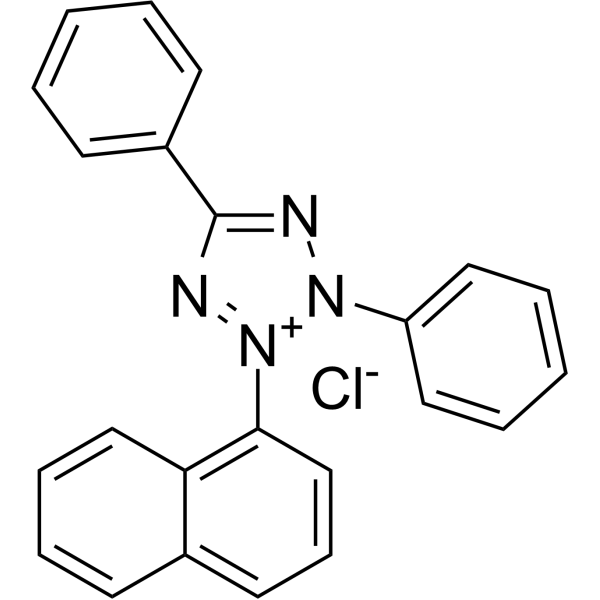
Tetrazolium violet is a redox indicator commonly used in various biochemical assays to measure cell viability and metabolic activity. Tetrazolium Violet has unique chemical properties that allow it to be reduced by cellular enzymes such as dehydrogenases to form a purple formazan product that can be detected spectrophotometrically. This makes it a useful tool for assessing cell health and growth in culture or tissue samples.
For research use only. We do not sell to patients.
- Purity : 99.68%
- CAS No.: 1719-71-7
- Formula: C23H17ClN4
- Molecular Weight:384.86
-
Storage:
RT, sealed storage, away from moisture and light.
In solvent -80°C, 1 year , -20°C, 6 months

To place orders, for customer services and technical support, please contact: MedChemExpress USA
Tel: 609-228-6898 E-mail: [email protected] [email protected]


-
 Biological Activity
-
Chemical Information
-
Solvent & Solubility
- Protocols
- Purity & Documentation
-
 Help & FAQs


-
HY-122131
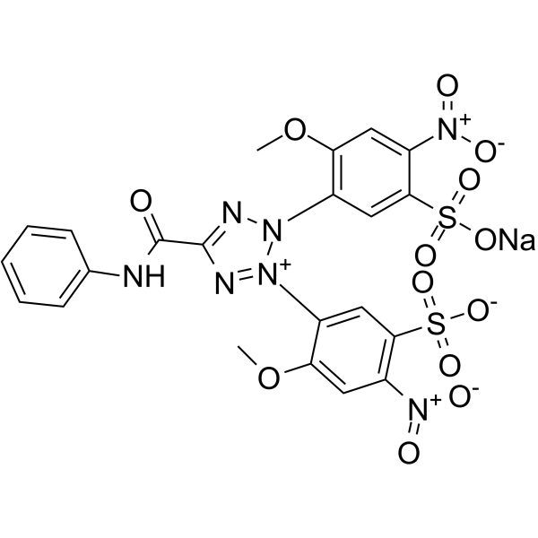
XTT sodiumTetrazolium Dye -
HY-D1181
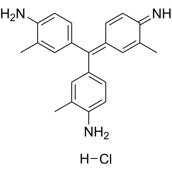
New FuchsinBiochemical Assay Reagent -
HY-15925
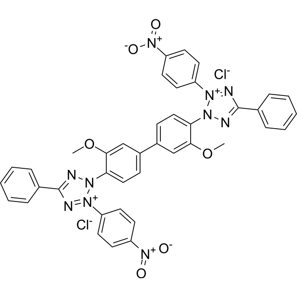
Nitro blue tetrazolium chlorideDehydrogenases Substrate -
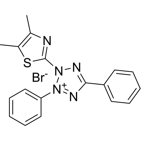
HY-15924
Thiazolyl BlueCytotoxicity Detection Reagent -
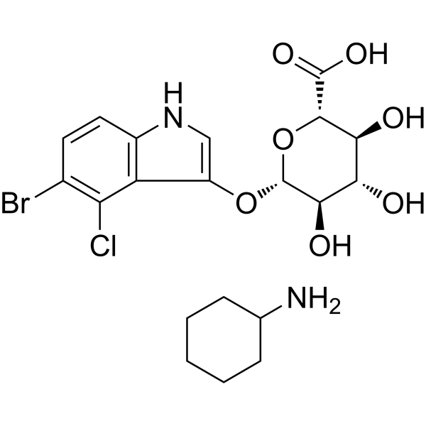
HY-15935B
X-Gluc cyclohexanamineβ-glucuronidase Assay Dye -
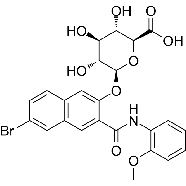
HY-137833
Naphthol AS-BI glucuronideBiochemical Assay Reagent -
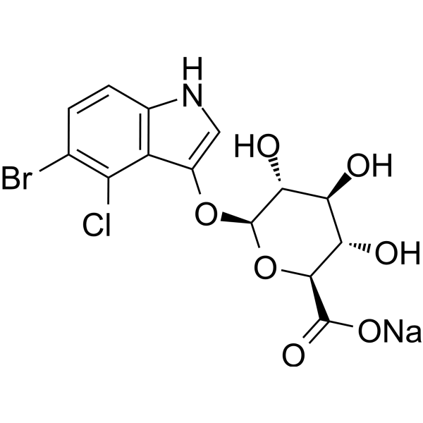
HY-15935C
X-Gluc sodiumβ-glucuronidase Assay Dye -
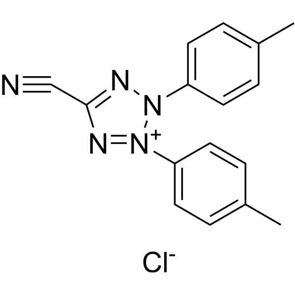
HY-D0016
5-Cyano-2,3-di-(p-tolyl)tetrazolium chlorideFluorescent Dye -
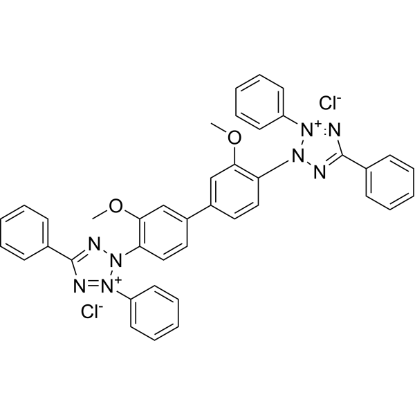
HY-D0007
Blue TetrazoliumChromogenic Dye -
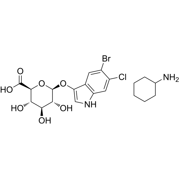
HY-137869
Magenta-β-D-GlcABiochemical Assay Reagent -
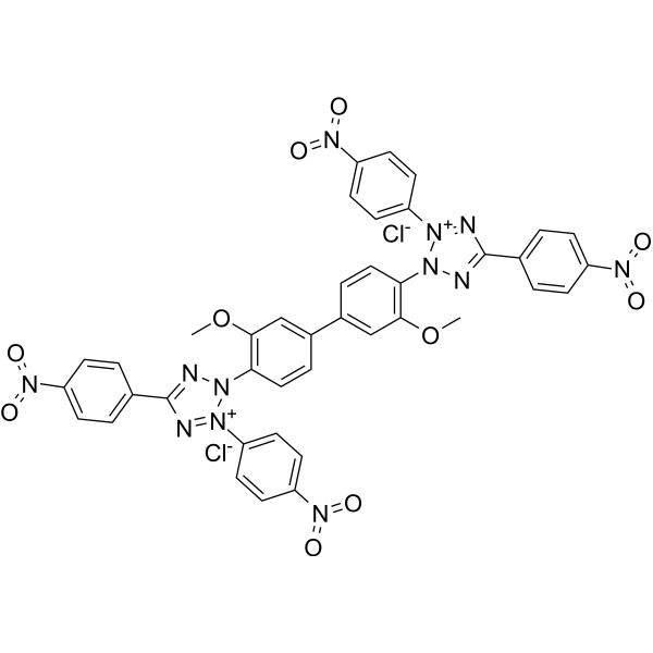
HY-D1193
Tetranitroblue tetrazolium chlorideBiochemical Assay Reagent -

HY-DY1086
Thiazolyl Blue (solution)Cytotoxicity Detection Reagent


Biological Activity
Description
Cellular Effect
|
Cell Line

|
Type | Value | Description | References |
|---|---|---|---|---|
| CHO | IC50 |
15 μM
Compound: tetrazolium violet
|
Toxicity in CHO cells
Toxicity in CHO cells
|
[PMID: 18078758] |
In Vitro
Tetrazolium Violet is a biochemical reagent that can be used as a biological material or organic compound for life science related research.
MedChemExpress (MCE) has not independently confirmed the accuracy of these methods. They are for reference only. Further protocols information, click here.


Chemical Information
-
CAS No. 1719-71-7
-
Appearance Solid
-
Molecular Weight 384.86
-
Formula C23H17ClN4
-
Color Off-white to light yellow
-
SMILES
C1(C2=CC=CC=C2)=NN(C3=CC=CC=C3)[N+](C4=C5C=CC=CC5=CC=C4)=N1.[Cl-]
-
Synonyms
Violet tetrazolium
-
Shipping
Room temperature in continental US; may vary elsewhere.
-
Storage
RT, sealed storage, away from moisture and light
In solvent -80°C 1 year -20°C 6 months


Solvent & Solubility
In Vitro:
DMSO : 50 mg/mL (129.92 mM; ultrasonic and warming and heat to 60°C; Hygroscopic DMSO has a significant impact on the solubility of product, please use newly opened DMSO)
Please refer to the solubility information to select the appropriate solvent. Once prepared, please aliquot and store the solution to prevent product inactivation from repeated freeze-thaw cycles.
Storage method and period of stock solution: -80°C, 1 year; -20°C, 6 months. When stored at -80°C, please use it within 1 year. When stored at -20°C, please use it within 6 months.
Please refer to the solubility information to select the appropriate solvent. Once prepared, please aliquot and store the solution to prevent product inactivation from repeated freeze-thaw cycles.
Storage method and period of stock solution: -80°C, 1 year; -20°C, 6 months. When stored at -80°C, please use it within 1 year. When stored at -20°C, please use it within 6 months.
Concentration (start) × Volume (start) = Concentration (final) × Volume (final)

Protocols
-
Cell Cytotoxicity Assay
Cytotoxicity assays are usually based on the assessment of cell membrane damage, which can also be indirectly detected by measuring cell viability. Detection methods include MTT assay, CKK-8 assay, LDH assay and ATP assay, etc.
-
Mammalian live/dead viability and cytotoxicity staining
Live/dead viability and cytotoxicity staining assays are based on the simultaneous detection of intracellular esterase activity in metabolically active (viable) cells and membrane integrity loss in non-viable cells. In commonly used dual-staining approaches, membrane-permeant fluorogenic substrates are converted by intracellular esterases into fluorescent products in live cells, while impermeant DNA-binding dyes selectively enter cells with compromised plasma membranes and label nucleic acids in dead or dying cells, enabling discrimination between viable and non-viable populations by fluorescence microscopy or flow cytometry.
-
ROS/oxidative-stress fluorescent staining
ROS/oxidative-stress fluorescent staining uses cell-permeant fluorogenic probes that become fluorescent after oxidation inside cells or tissues; commonly used examples include DCFH-DA/DCFDA for broad cellular oxidant detection, DHE for superoxide-related signal detection, MitoSOX for mitochondrial superoxide-related signal detection, and CellROX probes for oxidative-stress-associated fluorescence readouts. The assay detects probe oxidation rather than a single ROS species unless the probe and analysis method have been chemically validated for that species. DCFH-DA enters cells, is deacetylated by intracellular esterases to DCFH, and produces fluorescent DCF after oxidation, so the readout is used as an operational measure of total cellular oxidative stress rather than a species-specific ROS measurement. DHE and MitoSOX can report superoxide-related oxidation, but red fluorescence alone can include non-specific ethidium-like oxidation products; HPLC or optimized spectral approaches are
-
CCK-8/WST-8 Cell Proliferation Assay
The CCK-8/WST-8 assay is based on the reduction of the water-soluble tetrazolium salt WST-8 to a water-soluble formazan product by cellular dehydrogenases in metabolically active cells, where the generated formazan amount is proportional to the number of living cells and is quantified by measuring absorbance in the visible range, providing a colorimetric readout for cell viability and proliferation assessment. This class of tetrazolium-based assays improves upon earlier MTT-based systems by producing a water-soluble formazan, eliminating the need for organic solubilization steps and enabling direct spectrophotometric measurement in culture medium.
-
MTT Cell Proliferation Assay
The MTT assay is a colorimetric endpoint assay for estimating viable cell number, cell growth, cytotoxicity, or cell activation in cultured mammalian cells. Living cells reduce the yellow tetrazolium salt MTT into purple/blue formazan, while dead cells do not generate the same signal; the resulting color can be quantified with a multiwell spectrophotometer. MTT reduction is commonly interpreted as a readout of metabolic activity that often correlates with viable cell number, but it should not be treated as a direct cell-counting method unless the assay is optimized for the cell type and experimental condition. Studies show that MTT reduction can involve mitochondrial and non-mitochondrial reducing systems, and formazan may accumulate in intracellular lipid droplets rather than simply marking mitochondria.
-
Cell Viability Determination by MTT Colorimetric Assay
The following protocol uses the MTT colorimetric assay as a classic literature-established method for assessing cell viability/metabolic activity in cultured mammalian cells. MTT[3-(4,5-dimethylthiazol-2-yl)-2,5-diphenyltetrazolium bromide] is reduced by metabolically active cells to a colored formazan product; the amount of formazan is quantified spectrophotometrically and provides an indirect measure of metabolically active viable cells. Importantly, MTT reduction reflects cellular oxidoreductase/metabolic activity rather than an absolute direct count of living cells, so changes in cellular metabolism can alter the signal independently of cell number.


Purity & Documentation
-
Data Sheet (252 KB)
-
SDS (393 KB)
- English - EN (393 KB)
- Français - FR (393 KB)
- Deutsch - DE (393 KB)
- Norwegian - NO (393 KB)
- Español - ES (393 KB)
- Swedish - SV (393 KB)
- Italian - IT (393 KB)
- Korean - KR (393 KB)
- Portuguese - PT (393 KB)
-
Handling Instructions (2659 KB)


Complete Stock Solution Preparation Table
Please refer to the solubility information to select the appropriate solvent. Once prepared, please aliquot and store the solution to prevent product inactivation from repeated freeze-thaw cycles.
Storage method and period of stock solution: -80°C, 1 year; -20°C, 6 months. When stored at -80°C, please use it within 1 year. When stored at -20°C, please use it within 6 months.
| Optional Solvent | Concentration Solvent Mass | 1 mg | 5 mg | 10 mg | 25 mg |
|---|---|---|---|---|---|
| DMSO | 1 mM | 2.5983 mL | 12.9917 mL | 25.9835 mL | 64.9587 mL |
| 5 mM | 0.5197 mL | 2.5983 mL | 5.1967 mL | 12.9917 mL | |
| 10 mM | 0.2598 mL | 1.2992 mL | 2.5983 mL | 6.4959 mL | |
| 15 mM | 0.1732 mL | 0.8661 mL | 1.7322 mL | 4.3306 mL | |
| 20 mM | 0.1299 mL | 0.6496 mL | 1.2992 mL | 3.2479 mL | |
| 25 mM | 0.1039 mL | 0.5197 mL | 1.0393 mL | 2.5983 mL | |
| 30 mM | 0.0866 mL | 0.4331 mL | 0.8661 mL | 2.1653 mL | |
| 40 mM | 0.0650 mL | 0.3248 mL | 0.6496 mL | 1.6240 mL | |
| 50 mM | 0.0520 mL | 0.2598 mL | 0.5197 mL | 1.2992 mL | |
| 60 mM | 0.0433 mL | 0.2165 mL | 0.4331 mL | 1.0826 mL | |
| 80 mM | 0.0325 mL | 0.1624 mL | 0.3248 mL | 0.8120 mL | |
| 100 mM | 0.0260 mL | 0.1299 mL | 0.2598 mL | 0.6496 mL |


Tetrazolium violet Related Classifications
HY-131490 Related Classifications
 Powered by Bioz