



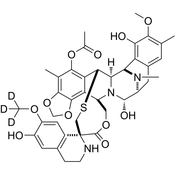
Trabectedin-d3

Based on 1 Customer Validation
Trabectedin D3 (Ecteinascidin 743 D3) is deuterium labeled Trabectedin. Trabectedin is a tetrahydroisoquinoline alkaloid with potent antitumor activity. Trabectedin binds to the minor groove of DNA, blocks transcription of stress-induced proteins, induces DNA backbone cleavage and cancer cells apoptosis, and increases the generation of ROS in MCF-7 and MDA-MB-453 cells. Trabectedin has tje potential for soft tissue sarcoma and ovarian cancer treatment.
Nos produits utilisent uniquement pour la recherche. Nous ne vendons pas aux patients.
- Pureté : 98.04%
- Formule: C39H40D3N3O11S
- Masse moléculaire:764.86
-
Stockage:
-20°C, sealed storage, away from moisture and light
* In solvent : -80°C, 6 months; -20°C, 1 month (sealed storage, away from moisture and light)

To place orders, for customer services and technical support, please contact: MedChemExpress USA
Tel: 609-228-6898 E-mail: [email protected] [email protected]


-
 Activité biologique
-
Chemical Information
-
Solvant et solubilité
- Protocole
- Pureté et documentation
- Références
-
 Help & FAQs




Activité biologique
Description
In Vitro
Stable heavy isotopes of hydrogen, carbon, and other elements have been incorporated into drug molecules, largely as tracers for quantitation during the drug development process. Deuteration has gained attention because of its potential to affect the pharmacokinetic and metabolic profiles of drugs.
MedChemExpress (MCE) has not independently confirmed the accuracy of these methods. They are for reference only.
Application
1. This compound can be used as a tracer
2. This compound can be used as an internal standard for quantitative analysis by NMR, GC-MS, or LC-MS.


Chemical Information
-
Unlabeled CAS 114899-77-3
-
Appearance Solid
-
Masse moléculaire 764.86
-
Formule C39H40D3N3O11S
-
Color Off-white to light yellow
-
SMILES
CC1=C(OCO2)C2=C([C@H](COC3=O)N4[C@]([C@@H]5SC[C@]63C(C=C(OC([2H])([2H])[2H])C(O)=C7)=C7CCN6)([H])[C@@H]8N(C)[C@@H](CC9=CC(C)=C(OC)C(O)=C98)[C@@H]4O)C5=C1OC(C)=O
-
Synonyms
Ecteinascidin 743-d3; ET-743-d3
-
Livraison
Room temperature in continental US; may vary elsewhere.
-
Stockage
-20°C, sealed storage, away from moisture and light
* In solvent : -80°C, 6 months; -20°C, 1 month (sealed storage, away from moisture and light)


Solvant et solubilité
In Vitro:
DMSO : 33.33 mg/mL (43.58 mM; Need ultrasonic; Hygroscopic DMSO has a significant impact on the solubility of product, please use newly opened DMSO)
Please refer to the solubility information to select the appropriate solvent. Once prepared, please aliquot and store the solution to prevent product inactivation from repeated freeze-thaw cycles.
Storage method and period of stock solution: -80°C, 6 months; -20°C, 1 month (sealed storage, away from moisture and light). When stored at -80°C, please use it within 6 months. When stored at -20°C, please use it within 1 month.
Please refer to the solubility information to select the appropriate solvent. Once prepared, please aliquot and store the solution to prevent product inactivation from repeated freeze-thaw cycles.
Storage method and period of stock solution: -80°C, 6 months; -20°C, 1 month (sealed storage, away from moisture and light). When stored at -80°C, please use it within 6 months. When stored at -20°C, please use it within 1 month.
Concentration (start) × Volume (start) = Concentration (final) × Volume (final)


Pureté et documentation
-
Fiche technique (283 KB)
-
SDS (252 KB)
- English - EN (252 KB)
- Français - FR (252 KB)
- Deutsch - DE (252 KB)
- Norwegian - NO (252 KB)
- Español - ES (252 KB)
- Swedish - SV (252 KB)
- Italian - IT (252 KB)
- Korean - KR (252 KB)
- Portuguese - PT (252 KB)
-
Instruction de manipulation (2659 KB)
Références
[1]. Takahashi N, et al. Sequence-dependent synergistic cytotoxicity of ecteinascidin-743 and NSC 125973 in human breast cancer cell linesin vitro and in vivo. Cancer Res. 2002 Dec 1;62(23):6909-15. [Content Brief]
[2]. Atmaca H, et al. A diverse induction of apoptosis by trabectedin in MCF-7 (HER2-/ER+) and MDA-MB-453 (HER2+/ER-) breast cancer cells. Toxicol Lett. 2013 Jun 20;221(2):128-136. [Content Brief]
[3]. Germano G, et al. Antitumor and anti-inflammatory effects of trabectedin on human myxoid liposarcoma cells. Cancer Res. 2010 Mar 15;70(6):2235-44. [Content Brief]


Complete Stock Solution Preparation Table
Please refer to the solubility information to select the appropriate solvent. Once prepared, please aliquot and store the solution to prevent product inactivation from repeated freeze-thaw cycles.
Storage method and period of stock solution: -80°C, 6 months; -20°C, 1 month (sealed storage, away from moisture and light). When stored at -80°C, please use it within 6 months. When stored at -20°C, please use it within 1 month.
| Optional Solvent | Concentration Solvent Mass | 1 mg | 5 mg | 10 mg | 25 mg |
|---|---|---|---|---|---|
| DMSO | 1 mM | 1.3074 mL | 6.5371 mL | 13.0743 mL | 32.6857 mL |
| 5 mM | 0.2615 mL | 1.3074 mL | 2.6149 mL | 6.5371 mL | |
| 10 mM | 0.1307 mL | 0.6537 mL | 1.3074 mL | 3.2686 mL | |
| 15 mM | 0.0872 mL | 0.4358 mL | 0.8716 mL | 2.1790 mL | |
| 20 mM | 0.0654 mL | 0.3269 mL | 0.6537 mL | 1.6343 mL | |
| 25 mM | 0.0523 mL | 0.2615 mL | 0.5230 mL | 1.3074 mL | |
| 30 mM | 0.0436 mL | 0.2179 mL | 0.4358 mL | 1.0895 mL | |
| 40 mM | 0.0327 mL | 0.1634 mL | 0.3269 mL | 0.8171 mL |


Trabectedin-d3 Related Classifications
HY-50936S Related Classifications
 Powered by Bioz