



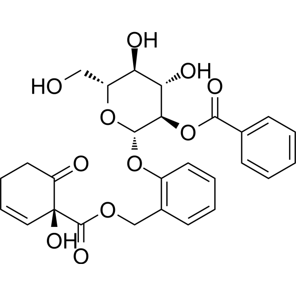
Tremulacin

Based on 1 Customer Validation
Tremulacin is a 5-LOX inhibitor. Tremulacin reduces the biosynthesis of LTB4 and slow-reacting substances of anaphylaxis. Tremulacin alleviates carrageenan-induced paw edema in rats, croton oil-induced ear edema in mice, and acetic acid-induced writhing response in mice. Tremulacin inhibits TNF-α-stimulated ROS production and MMP-1 expression, and promotes collagen secretion in human dermal fibroblasts. Tremulacin is investigated for studies on inflammation-related diseases.
For research use only. We do not sell to patients.
- Purity : 96.41%
- CAS No.: 29836-40-6
- Formula: C27H28O11
- Molecular Weight:528.50
-
Storage:Powder -20°C, 3 years ; In solvent -80°C, 6 months , -20°C, 1 month

To place orders, for customer services and technical support, please contact: MedChemExpress USA
Tel: 609-228-6898 E-mail: [email protected] [email protected]


-
 Biological Activity
-
Chemical Information
-
Solvent & Solubility
- Protocols
- Purity & Documentation
- References
-
 Help & FAQs


All Leukotriene Receptor Isoforms
More

Biological Activity
Description
|
5-LOX |
LTB4 |
MMP-1 |
In Vitro
Tremulacin (10 μM; 30 min) inhibits LTB4 biosynthesis by 30% in leukocytes from rat pleural cavities, but exerts no effect on HHT biosynthesis in platelets, and reduces SRS-A biosynthesis by 41% in isolated longitudinal muscle strips from the ileum of sensitized guinea pigs[1].
Tremulacin (1-100 μM; 24 h) reduces cell viability at the concentration of 100 μM[2].
Tremulacin (1-30 μM; pre-incubated for 1 h prior to TNF-α treatment) inhibits TNF-α-induced ROS production in normal human skin fibroblasts, suppresses TNF-α-induced MMP-1 secretion in normal human dermal fibroblasts, and partially restores COLIA1 secretion[2].
Tremulacin exhibits weak antiviral activity against HSV-1 (EC50 = 87 μM), HSV-2 (EC50 = 86 μM) and HIV-1 (EC50 = 52 μM)[3].
MedChemExpress (MCE) has not independently confirmed the accuracy of these methods. They are for reference only.
-
Cell Line:normal human dermal fibroblasts (HDFs)
-
Concentration:1, 3, 10, 30, 100 μM
-
Incubation Time:24 h
-
Result:Showed no significant changes in cell viability at concentrations of 1, 3, 10, and 30 μM.
Caused a significant decrease in cell viability at 100 μM.
-
Cell Line:TNF-α-stimulated normal human dermal fibroblasts (HDFs)
-
Concentration:1, 3, 10, 30 μM
-
Incubation Time:1 h (pre-incubation before 24 h TNF-α treatment)
-
Result:Reduced TNF-α-induced MMP-1 secretion to 1.03 ng/mL at 1 μM.
Reduced TNF-α-induced MMP-1 secretion to 1.66 ng/mL at 3 μM.
Reduced TNF-α-induced MMP-1 secretion to 1.28 ng/mL at 10 μM.
Reduced TNF-α-induced MMP-1 secretion to 1.21 ng/mL at 30 μM.
Increased TNF-α-reduced COLIA1 secretion to 0.82 ng/mL at 3 μM.
In Vivo
Tremulacin (100 mg/kg; subcutaneous injection; single administration) inhibits croton oil-induced ear swelling in Kunming mice by 43%[1].
Tremulacin (100 mg/kg; subcutaneous injection; single administration) prolongs the latency and reduces the frequency of acetic acid-induced writhing responses in Kunming mice[1].
MedChemExpress (MCE) has not independently confirmed the accuracy of these methods. They are for reference only.
-
Animal Model:Wistar (male, 180-220 g, carrageenan-induced paw edema model/ sodium carboxymethylcellulose-induced peritoneal leukocyte migration model)[1]
-
Dosage:100 mg/kg
-
Administration:s.c.; single dose
-
Result:Significantly reduced paw swelling for at least 6 hours, with a maximum inhibition rate of approximately 55%.
Reduced peritoneal leukocyte counts from 1.2×10⁴ cells/mL in controls to 6×10³ cells/mL in treated rats, resulting in a 50% inhibition rate.
-
Animal Model:Kun Ming (male, 18-22 g, croton oil-induced ear edema model)[1]
-
Dosage:100 mg/kg
-
Administration:s.c.; single dose
-
Result:Decreased ear edema by 43%, reducing the mean ear weight increase from 18.1 mg in controls to 10.3 mg in treated mice.
-
Animal Model:Kun Ming (male, 18-22 g, acetic acid-induced writhing model)[1]
-
Dosage:100 mg/kg
-
Administration:s.c.; single dose
-
Result:Increased the mean time to onset of writhing from 2.43 minutes in controls to 3.75 min.
Reduced the mean number of writhing responses over 15 minutes from 34.86 in controls to 19.75.
- RNA extraction experimental
- Mitochondrial membrane-potential fluorescent assay
- ROS/oxidative-stress fluorescent staining
- Carrageenan-Induced Paw Edema
- TPA/Croton Oil Ear Edema and Dermatitis
- Mitochondrial membrane-potential and mitochondrial mass staining
- Research Protocol for Inflammation-related Diseases


Chemical Information
-
CAS No. 29836-40-6
-
Appearance Solid
-
Molecular Weight 528.50
-
Formula C27H28O11
-
Color White to off-white
-
SMILES
O=C(C1=CC=CC=C1)O[C@H]([C@H]([C@@H]([C@H](O2)CO)O)O)[C@@H]2OC3=C(C=CC=C3)COC([C@]4(C(CCC=C4)=O)O)=O
-
Structure Classification
-
Shipping
Room temperature in continental US; may vary elsewhere.
-
Storage
Powder -20°C 3 years In solvent -80°C 6 months -20°C 1 month


Solvent & Solubility
In Vitro:
DMSO : 100 mg/mL (189.21 mM; Need ultrasonic; Hygroscopic DMSO has a significant impact on the solubility of product, please use newly opened DMSO)
Please refer to the solubility information to select the appropriate solvent. Once prepared, please aliquot and store the solution to prevent product inactivation from repeated freeze-thaw cycles.
Storage method and period of stock solution: -80°C, 6 months; -20°C, 1 month. When stored at -80°C, please use it within 6 months. When stored at -20°C, please use it within 1 month.
Please refer to the solubility information to select the appropriate solvent. Once prepared, please aliquot and store the solution to prevent product inactivation from repeated freeze-thaw cycles.
Storage method and period of stock solution: -80°C, 6 months; -20°C, 1 month. When stored at -80°C, please use it within 6 months. When stored at -20°C, please use it within 1 month.
Concentration (start) × Volume (start) = Concentration (final) × Volume (final)
In Vivo:
Select the appropriate dissolution method based on your experimental animal and administration route.
- For the following dissolution methods, please ensure to first prepare a clear stock solution using an In Vitro approach and then sequentially add co-solvents:
- To ensure reliable experimental results, the clarified stock solution can be appropriately stored based on storage conditions. As for the working solution for In Vivo experiments, it is recommended to prepare freshly and use it on the same day.
- The percentages shown for the solvents indicate their volumetric ratio in the final prepared solution. If precipitation or phase separation occurs during preparation, heat and/or sonication can be used to aid dissolution.
Add each solvent one by one: 10% DMSO 90% (20% SBE-β-CD in Saline)
Solubility: ≥ 2.5 mg/mL (4.73 mM); Clear solution
This protocol yields a clear solution of ≥ 2.5 mg/mL (saturation unknown).
Taking 1 mL working solution as an example, add 100 μL DMSO stock solution (25.0 mg/mL) to 900 μL 20% SBE-β-CD in Saline, and mix evenly.
Preparation of 20% SBE-β-CD in Saline (4°C, storage for one week): 2 g SBE-β-CD powder is dissolved in 10 mL Saline, completely dissolve until clear.
In Vivo Dissolution Calculator
Please enter the basic information of animal experiments:
-
-
-
-
Recommended: Prepare an additional quantity of animals to account for potential losses during experiments.
Please enter your animal formula composition:
-
%DMSO +
Recommended: Keep the proportion of DMSO in working solution below 2% if your animal is weak.
-
%+
-
+%Tween-80 + +
-
%Saline +
The co-solvents required include: DMSO, . All of co-solvents are available by MedChemExpress (MCE). , Tween 80. All of co-solvents are available by MedChemExpress (MCE).
Working solution concentration: 0.22 mg/mL
Method for preparing stock solution: mg drug dissolved in μL DMSO. Stock solution concentration: mg/mL.
1. Take μL DMSO stock solution;
2. Add μL .
μL , mix evenly;
3. Then add μL Tween 80, mix evenly;
4. Then add μL
Please ensure that the stock solution in the first step is dissolved to a clear state, and add co-solvents in sequence. You can use ultrasonic heating (ultrasonic cleaner, recommended frequency 20-40 kHz), vortexing, etc. to assist dissolution.


Purity & Documentation
-
Data Sheet (287 KB)
-
SDS (394 KB)
- English - EN (394 KB)
- Français - FR (394 KB)
- Deutsch - DE (394 KB)
- Norwegian - NO (394 KB)
- Español - ES (394 KB)
- Swedish - SV (394 KB)
- Italian - IT (394 KB)
- Korean - KR (394 KB)
- Portuguese - PT (394 KB)
-
Handling Instructions (2659 KB)
References
[1]. Cheng GF, et al. Antiinflammatory effects of Tremulacin, a Salicin-related substance isolated from Populus tomentosa Carr. leaves. Phytomedicine. 1994;1(3):209-211. [Content Brief]
[2]. Ahn SY, et al. Potential skin anti-aging effects of main phenolic compounds, tremulacin and tremuloidin from Salix chaenomeloides leaves on TNF-α-stimulated human dermal fibroblasts. Chem Biol Interact. 2024;402:111192. [Content Brief]
[3]. Ishikawa T, et al. Isolation of salicin derivatives from Homalium cochinchinensis and their antiviral activities. J Nat Prod. 2004 Apr;67(4):659-63. [Content Brief]


Complete Stock Solution Preparation Table
Please refer to the solubility information to select the appropriate solvent. Once prepared, please aliquot and store the solution to prevent product inactivation from repeated freeze-thaw cycles.
Storage method and period of stock solution: -80°C, 6 months; -20°C, 1 month. When stored at -80°C, please use it within 6 months. When stored at -20°C, please use it within 1 month.
| Optional Solvent | Concentration Solvent Mass | 1 mg | 5 mg | 10 mg | 25 mg |
|---|---|---|---|---|---|
| DMSO | 1 mM | 1.8921 mL | 9.4607 mL | 18.9215 mL | 47.3037 mL |
| 5 mM | 0.3784 mL | 1.8921 mL | 3.7843 mL | 9.4607 mL | |
| 10 mM | 0.1892 mL | 0.9461 mL | 1.8921 mL | 4.7304 mL | |
| 15 mM | 0.1261 mL | 0.6307 mL | 1.2614 mL | 3.1536 mL | |
| 20 mM | 0.0946 mL | 0.4730 mL | 0.9461 mL | 2.3652 mL | |
| 25 mM | 0.0757 mL | 0.3784 mL | 0.7569 mL | 1.8921 mL | |
| 30 mM | 0.0631 mL | 0.3154 mL | 0.6307 mL | 1.5768 mL | |
| 40 mM | 0.0473 mL | 0.2365 mL | 0.4730 mL | 1.1826 mL | |
| 50 mM | 0.0378 mL | 0.1892 mL | 0.3784 mL | 0.9461 mL | |
| 60 mM | 0.0315 mL | 0.1577 mL | 0.3154 mL | 0.7884 mL | |
| 80 mM | 0.0237 mL | 0.1183 mL | 0.2365 mL | 0.5913 mL | |
| 100 mM | 0.0189 mL | 0.0946 mL | 0.1892 mL | 0.4730 mL |


Tremulacin Related Classifications
HY-N11072 Related Classifications
 Powered by Bioz
Keywords
- Tremulacin
- 29836-40-6
- Lipoxygenase
- Leukotriene Receptor
- Reactive Oxygen Species (ROS)
- MMP
- cyclooxygenase
- carrageenan-induced paw edema
- croton oil-induced ear edema
- 5-lipoxygenase
- HIV-1
- acetic acid-induced writhing responses
- Slow Reaction Substance of Anaphylaxis
- human dermal fibroblasts
- leukotriene B4
- HSV-1
- Inhibitor
- inhibitor
- inhibit