



UniPR129

Based on 1 Customer Validation
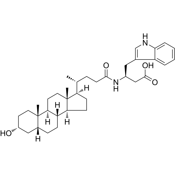
UniPR129 is a potent and orally active Eph/ephrin antagonist. UniPR129 can inhibit EphA2-ephrin-A1 interaction with an IC50 of 945 nM and a Ki of 370 nM. UniPR129 can inhibit angiogenesis and show antitumor and neuroprotective effect. UniPR129 can be used for the researches of cancer and neurological disease, such as colorectal cancer and optic neuropathy.
For research use only. We do not sell to patients.
- Purity : 99.09%
- CAS No.: 1639159-47-9
- Formula: C36H52N2O4
- Molecular Weight:576.81
-
Storage:Powder -20°C, 3 years , 4°C, 2 years ; In solvent -80°C, 6 months , -20°C, 1 month

To place orders, for customer services and technical support, please contact: MedChemExpress USA
Tel: 609-228-6898 E-mail: [email protected] [email protected]


-
 Biological Activity
-
Chemical Information
-
Solvent & Solubility
- Protocols
- Purity & Documentation
- References
-
 Help & FAQs


-
HY-18833
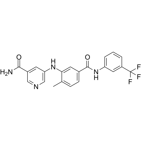
ALW-II-49-7EphB2 Inhibitor -
HY-13258
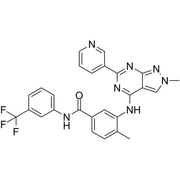
NVP-BHG712 isomerEphrin Receptor -
HY-18007
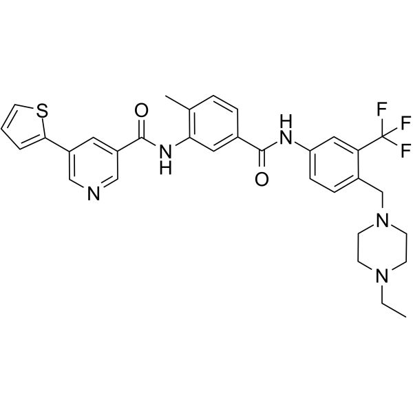
ALW-II-41-27Eph2 Inhibitor -
HY-114199
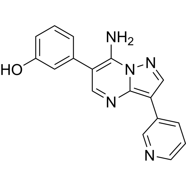
Eph inhibitor 1Eph Inhibitor -
HY-P2264
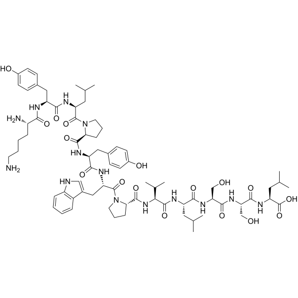
KYL peptideEphA4 Antagonist -
HY-133178
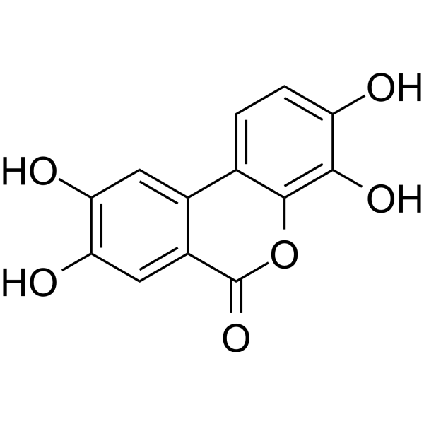
Urolithin DEphA Receptor Antagonist -
HY-16265
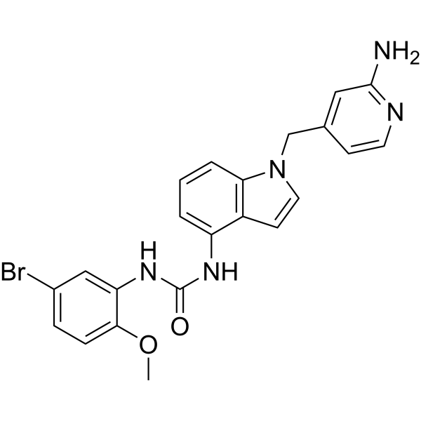
JI-101EphB4, VEGFR-2 and PDGFR-β Inhibitor -
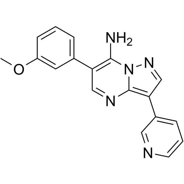
HY-131005
Eph inhibitor 2Eph Inhibitor -
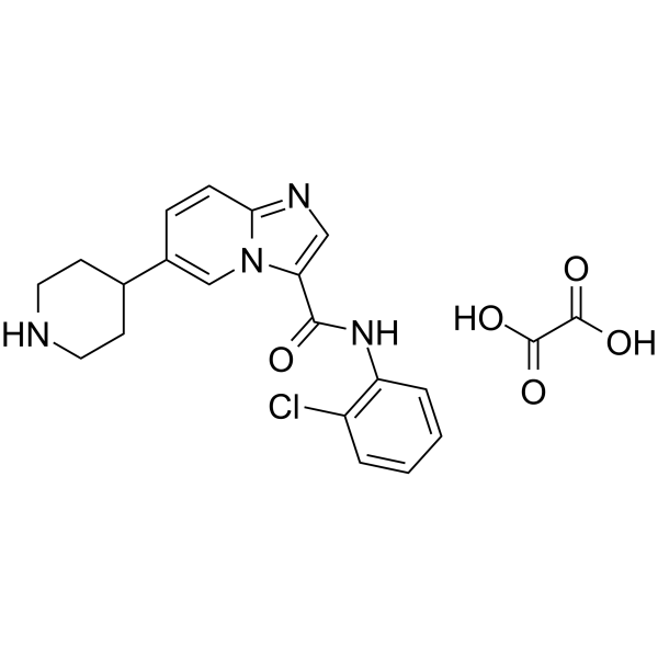
HY-107460
LDN-211904 oxalateEphB3 Inhibitor -
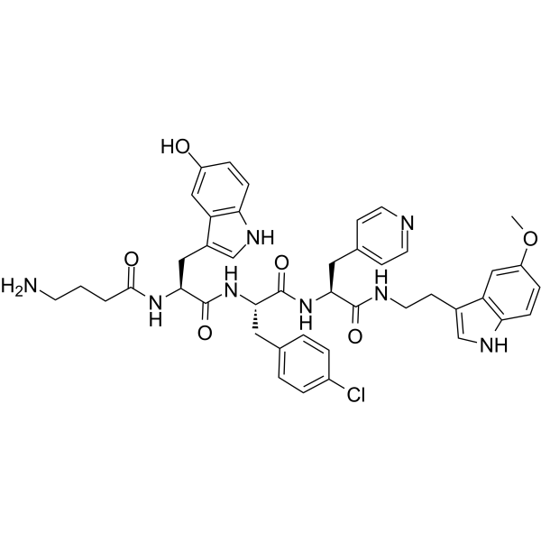
HY-P0177
123C4Ephrin Receptor Agonist -
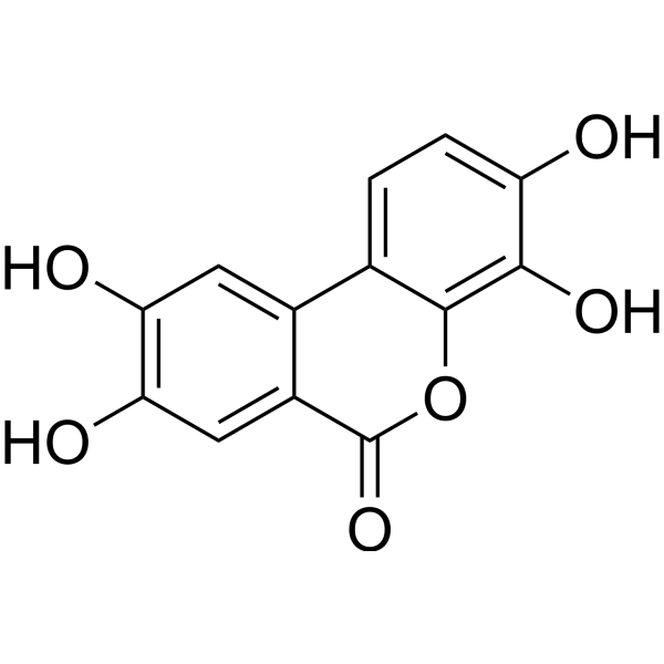
HY-133178R
Urolithin D (Standard)Standard of Urolithin D


Biological Activity
Description
IC50 & Target
[4]|
EphA8 0.84 μM (IC50) |
EphA2 1.12 μM (IC50) |
EphA7 1.24 μM (IC50) |
EphA1 1.39 μM (IC50) |
EphA3 1.43 μM (IC50) |
EphA6 1.53 μM (IC50) |
EphA5 1.57 μM (IC50) |
EphA4 1.58 μM (IC50) |
EphB4 2.60 μM (IC50) |
EphB1 2.75 μM (IC50) |
EphB2 2.96 μM (IC50) |
EphB5 3.01 μM (IC50) |
EphB3 3.74 μM (IC50) |
In Vitro
UniPR129 (1.5-25 μM, 20 mins) inhibits EphA2 activation in PC3 cells[1].
UniPR129 (6-50 μM, 2 h) does not show any toxic effect in PC3 cells[1].
UniPR129 (1.56-12.5, 20 mins) inhibits PC3 cells retraction[1].
UniPR129 (1.5-25, 15 h) inhibits angiogenesis in HUVECs[1].
UniPR129 (50 μM) shows neuroprotection in retinas isolated from optic nerve crush (ONC) injury mices model[3].
MedChemExpress (MCE) has not independently confirmed the accuracy of these methods. They are for reference only. Further protocols information, click here.
-
Cell Line:PC3 cells
-
Concentration:1.56, 3.12, 6.25 and 12.5 μM
-
Incubation Time:20 mins
-
Result:Inhibited the rounding effect of ephrin-A1-Fc
In Vivo
MedChemExpress (MCE) has not independently confirmed the accuracy of these methods. They are for reference only.
-
Animal Model:APC min/J mice[2]
-
Dosage:30 mg/kg
-
Administration:Orally administration, every other day for 8 weeks
-
Result:Reduced the formation of adenomas in the ileum of 1.6 times.
Reduced tumor volume.


Chemical Information
-
CAS No. 1639159-47-9
-
Appearance Solid
-
Molecular Weight 576.81
-
Formula C36H52N2O4
-
Color White to off-white
-
SMILES
C[C@@]12[C@](CC[C@]2([H])[C@H](C)CCC(N[C@H](CC(O)=O)CC3=CNC4=CC=CC=C34)=O)([H])[C@@]5([H])[C@]([C@@]6([C@](C[C@H](O)CC6)([H])CC5)C)([H])CC1
-
Shipping
Room temperature in continental US; may vary elsewhere.
-
Storage
Powder -20°C 3 years 4°C 2 years In solvent -80°C 6 months -20°C 1 month


Solvent & Solubility
In Vitro:
DMSO : 33.33 mg/mL (57.78 mM; ultrasonic and warming and heat to 60°C; Hygroscopic DMSO has a significant impact on the solubility of product, please use newly opened DMSO)
Please refer to the solubility information to select the appropriate solvent. Once prepared, please aliquot and store the solution to prevent product inactivation from repeated freeze-thaw cycles.
Storage method and period of stock solution: -80°C, 6 months; -20°C, 1 month. When stored at -80°C, please use it within 6 months. When stored at -20°C, please use it within 1 month.
Please refer to the solubility information to select the appropriate solvent. Once prepared, please aliquot and store the solution to prevent product inactivation from repeated freeze-thaw cycles.
Storage method and period of stock solution: -80°C, 6 months; -20°C, 1 month. When stored at -80°C, please use it within 6 months. When stored at -20°C, please use it within 1 month.
Concentration (start) × Volume (start) = Concentration (final) × Volume (final)

Protocols
-
Human pluripotent stem cell endothelial-cell differentiation
Human pluripotent stem cell endothelial differentiation is based on stepwise developmental patterning: early activation of WNT/GSK3β inhibition promotes mesodermal or vascular progenitor entry, followed by endothelial specification using VEGF-related signaling, BMP4, FGF2, Notch modulation, or cAMP depending on the published protocol. Endothelial differentiation is read out by acquisition of CD31, CD34, VE-cadherin/CD144, KDR/VEGFR2, vWF, Tie2, NOS3, acetylated LDL uptake, tube/network formation, barrier function, and in vivo vessel-forming capacity where tested.
-
Cell Cytotoxicity Assay
Cytotoxicity assays are usually based on the assessment of cell membrane damage, which can also be indirectly detected by measuring cell viability. Detection methods include MTT assay, CKK-8 assay, LDH assay and ATP assay, etc.
-
Vascular/Branching Fractal Analysis
Vascular/branching fractal analysis quantifies the geometric complexity of vessel trees or vascular networks from segmented 2D images, commonly by converting vessels into binary and/or skeletonized maps and estimating fractal dimension using box-counting or related approaches. Fractal dimension is interpreted as an image-derived readout of vascular branching complexity, space filling, or density, and has been applied to retinal photographs, fluorescein angiography, OCT angiography, capillary perfusion maps, and in vitro Matrigel angiogenesis networks. The assay readout is generated from vessel-positive pixels after image preprocessing, vessel segmentation, binarization, and optional skeletonization; reported outputs include fractal dimension, vessel density, branchpoint density, endpoint density, vessel length density, tortuosity, and generation-based branching metrics when VESGEN-style analysis is used. The biological interpretation is limited to quantitative vascular patterning and s
-
Endothelial Tube Formation Assay
Endothelial tube formation assay evaluates the ability of endothelial cells to attach, migrate, align, and organize into capillary-like networks when cultured on gelled basement membrane extract or Matrigel; the readout is the morphology and quantity of tube-like networks, which reflects an in vitro endothelial morphogenesis step related to angiogenesis. Basement membrane extract/Matrigel provides laminin-rich extracellular matrix cues that support endothelial differentiation into capillary-like structures, but it can contain biologically active growth factors, so growth-factor-reduced matrix is preferred when testing defined angiogenic stimulators or inhibitors.
-
Research Protocol for Neurological Diseases
PINK1/Parkin-mediated mitophagy pathway is a mitochondrial quality-control signaling axis in which mitochondrial depolarization stabilizes PINK1 on damaged mitochondria, activates Parkin recruitment and E3 ubiquitin ligase activity, promotes ubiquitination of outer mitochondrial membrane proteins, recruits selective autophagy adaptors, and drives lysosomal degradation of damaged mitochondria. In neurological disease research, this pathway is experimentally important because neurons, especially dopaminergic neurons, are highly dependent on mitochondrial integrity, and defective mitochondrial turnover can lead to mitochondrial dysfunction, oxidative stress, impaired neuronal survival, α-synuclein accumulation, and neuroinflammatory damage-associated signals. The genetic disease link is strongest in Parkinson’s disease because mutations in PRKN/parkin cause autosomal recessive juvenile parkinsonism, mutations in PINK1 cause hereditary early-onset Parkinson’s disease, and Drosophila studie
-
Endothelial Cell Migration/Angiogenic Sprouting Assay
Endothelial cell migration and angiogenic sprouting assays are in vitro (and partially ex vivo-adapted) functional models that quantify the ability of endothelial cells to undergo coordinated migration, extracellular matrix invasion, and multicellular organization into capillary-like sprouts in response to pro-angiogenic stimuli such as VEGF, bFGF, or conditioned microenvironments. These assays are used to model early angiogenic events including tip-cell formation, directional migration, and lumen-like sprout extension, which collectively reflect angiogenic activation and vascular morphogenesis processes observed in vivo.


Purity & Documentation
-
Data Sheet (274 KB)
-
SDS (251 KB)
- English - EN (251 KB)
- Français - FR (251 KB)
- Deutsch - DE (251 KB)
- Norwegian - NO (251 KB)
- Español - ES (251 KB)
- Swedish - SV (251 KB)
- Italian - IT (251 KB)
- Korean - KR (251 KB)
- Portuguese - PT (251 KB)
-
Handling Instructions (2659 KB)
References
[1]. Hassan-Mohamed I, et al. UniPR129 is a competitive small molecule Eph-ephrin antagonist blocking in vitro angiogenesis at low micromolar concentrations. Br J Pharmacol. 2014 Dec;171(23):5195-208. [Content Brief]
[2]. Corrado M, et al. Evaluation of the Anti-Tumor Activity of Small Molecules Targeting Eph/Ephrins in APC min/J Mice. Pharmaceuticals (Basel). 2020 Apr 16;13(4):69. [Content Brief]
[3]. Strong TA, et al. Activation of multiple Eph receptors on neuronal membranes correlates with the onset of optic neuropathy. Eye Vis (Lond). 2023 Oct 2;10(1):42. [Content Brief]
[4]. Giorgio C, et al. Pharmacological evaluation of new bioavailable small molecules targeting Eph/ephrin interaction. Biochem Pharmacol. 2018 Jan;147:21-29. [Content Brief]


Complete Stock Solution Preparation Table
Please refer to the solubility information to select the appropriate solvent. Once prepared, please aliquot and store the solution to prevent product inactivation from repeated freeze-thaw cycles.
Storage method and period of stock solution: -80°C, 6 months; -20°C, 1 month. When stored at -80°C, please use it within 6 months. When stored at -20°C, please use it within 1 month.
| Optional Solvent | Concentration Solvent Mass | 1 mg | 5 mg | 10 mg | 25 mg |
|---|---|---|---|---|---|
| DMSO | 1 mM | 1.7337 mL | 8.6684 mL | 17.3367 mL | 43.3418 mL |
| 5 mM | 0.3467 mL | 1.7337 mL | 3.4673 mL | 8.6684 mL | |
| 10 mM | 0.1734 mL | 0.8668 mL | 1.7337 mL | 4.3342 mL | |
| 15 mM | 0.1156 mL | 0.5779 mL | 1.1558 mL | 2.8895 mL | |
| 20 mM | 0.0867 mL | 0.4334 mL | 0.8668 mL | 2.1671 mL | |
| 25 mM | 0.0693 mL | 0.3467 mL | 0.6935 mL | 1.7337 mL | |
| 30 mM | 0.0578 mL | 0.2889 mL | 0.5779 mL | 1.4447 mL | |
| 40 mM | 0.0433 mL | 0.2167 mL | 0.4334 mL | 1.0835 mL | |
| 50 mM | 0.0347 mL | 0.1734 mL | 0.3467 mL | 0.8668 mL |


UniPR129 Related Classifications
HY-123607 Related Classifications
 Powered by Bioz