
DMUP
Based on 1 Customer Validation
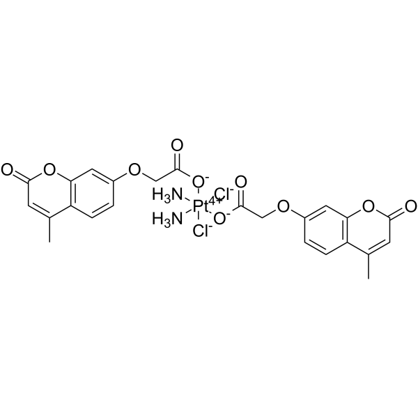
DMUP is a potent CD47-SIRPα axis inhibitor. DMUP induces apoptosis and increases the macrophage phagocytosis in A549 cells. DMUP decreases the expression of CD47 and SIRPα protein. DMUP shows antitumor activity.
For research use only. We do not sell to patients.
- Purity : 98.0%
- CAS No.: 2364350-07-0
- Formula: C24H24Cl2N2O10Pt
- Molecular Weight:766.44
-
Storage:Powder -20°C, 3 years , 4°C, 2 years ; In solvent -80°C, 6 months , -20°C, 1 month
Biological Activity
Description
In Vitro
DMUP (72 h) shows antiproliferative activity with IC50s of 0.92, 3.58, 6.29, 1.54 μM for A549, A549/DDP, PANC-1, HepG2 cells, respectively[1].
DMUP (5 μM, 24 h) arrests the cell cycle in S phase in A549 cells[1].
DMUP (5 μM, 48 h) induces apoptosis in A549 cells[1].
DMUP (5 μM, 24 h) decreases the expression of CD47 and SIRPα protein[1].
DMUP (5 μM, 4 h) increases the macrophage phagocytosis in A549-GFP cells[1].
MedChemExpress (MCE) has not independently confirmed the accuracy of these methods. They are for reference only. Further protocols information, click here.
-
Cell Line:A549, A549/DDP, PANC-1, HepG2 cells
-
Concentration:0-64 µM
-
Incubation Time:72 h
-
Result:Showed antiproliferative activity with IC50s of 0.92, 3.58, 6.29, 1.54 µM for A549, A549/DDP, PANC-1, HepG2 cells, respectively.
-
Cell Line:A549 cells
-
Concentration:5 µM
-
Incubation Time:24 h
-
Result:Arrested the cell cycle in the S phase.
-
Cell Line:A549 cells
-
Concentration:5 µM
-
Incubation Time:48 h
-
Result:Induced cell apoptosis.
-
Cell Line:A549, THP-1 cells
-
Concentration:5 µM
-
Incubation Time:24 h
-
Result:Decreased the expression of CD47 and SIRPα protein.
In Vivo
DMUP (5, 10, 20, 40 mg/kg, i.v.,every two days for 18 days) shows no toxicity in mice[1].
MedChemExpress (MCE) has not independently confirmed the accuracy of these methods. They are for reference only.
-
Animal Model:ICR mice[1]
-
Dosage:5, 10, 20, 40 mg/kg
-
Administration:i.v., every two days, 18 days
-
Result:Showed low toxicity with LD50 of greater than 20 mg/kg.
-
Animal Model:A549 xenograft BALB/c nude mice[1]
-
Dosage:10 mg/kg
-
Administration:i.v., every other day, 17 day
-
Result:Showed antitumor activity.
Chemical Information
-
CAS No. 2364350-07-0
-
Appearance Solid
-
Molecular Weight 766.44
-
Formula C24H24Cl2N2O10Pt
-
Color White to light yellow
-
SMILES
O=C1OC2=C(C(C)=C1)C=CC(OCC([O-][Pt+4]([Cl-])([O-]C(COC3=CC(OC(C=C4C)=O)=C4C=C3)=O)([NH3])([NH3])[Cl-])=O)=C2
-
Shipping
Room temperature in continental US; may vary elsewhere.
-
Storage
Powder -20°C 3 years 4°C 2 years In solvent -80°C 6 months -20°C 1 month
Protocols
-
Phagocytosis Functional Assay
A phagocytosis functional assay measures the ability of phagocytic cells, such as neutrophils, macrophages, monocytes, or microglia/macrophages, to bind and internalize particulate targets including bacteria, yeast particles, beads, or myelin particles. Fluorescent flow-cytometry assays detect target uptake as fluorescence associated with gated phagocytes, while pH-sensitive dyes such as pHrodo increase signal in acidic phagosomal compartments and therefore preferentially report internalized particles rather than particles remaining outside the cell. Microscopy or high-content imaging can be used to confirm intracellular localization and, in some protocols, to follow uptake kinetics.
-
RNA extraction experimental
By lysing cells, releasing RNA, and removing impurities such as proteins and DNA, high-purity RNA products are finally obtained. The commonly used traditional method is the guanidine isothiocyanate/phenol/chloroform method (Trizol), which is suitable for a variety of animal materials including animal tissues, microorganisms, cultured cells, etc., and most plant materials.
-
Apoptosis
Apoptosis, also called programmed cell death, is generally characterized by distinct morphological characteristics.
-
TUNEL staining for apoptotic DNA fragmentation
TUNEL staining detects DNA strand breaks by using terminal deoxynucleotidyl transferase to add labeled nucleotides to exposed 3′-OH DNA termini, generating either microscopic staining in fixed cells or tissue sections, or fluorescence/cytometric signal in cell suspensions. TUNEL positivity reflects DNA fragmentation but should not be interpreted alone as definitive apoptosis, because TUNEL can also label necrotic, autolytic, mechanically damaged, or DNA-repair-associated DNA breaks.
-
Annexin V plus membrane-impermeant dye apoptosis staining
Annexin V-based apoptosis assays rely on the detection of phosphatidylserine (PS) externalization from the inner leaflet of the plasma membrane to the outer leaflet, an early biochemical hallmark of apoptosis. Fluorescently labeled Annexin V binds PS in a calcium-dependent manner, enabling identification of early apoptotic cells by flow cytometry or fluorescence microscopy. When combined with a membrane-impermeant DNA-binding dye (e. g. , propidium iodide), this approach allows discrimination between viable (Annexin V−/dye−), early apoptotic (Annexin V+/dye−), and late apoptotic or necrotic (Annexin V+/dye+) cell populations by assessing membrane integrity and PS exposure.
-
Apoptosis Solutions
Apoptosis is a regulated, generally non-lytic cell-death pathway that removes unwanted, damaged, infected, or abnormal cells through coordinated morphological changes, caspase activation, DNA fragmentation, and membrane remodeling. The intrinsic apoptosis pathway is controlled mainly by mitochondrial outer membrane permeabilization, BCL-2 family proteins, cytochrome c release, apoptosome formation, caspase-9 activation, and downstream executioner caspase-3/7 activation. The extrinsic apoptosis pathway is initiated by death receptors such as Fas, TNFR, and TRAIL receptors, which recruit adaptor proteins and activate caspase-8 before engaging executioner caspases or mitochondrial amplification through BID cleavage. Apoptosis is linked to many phenotypes, including cancer cell killing, tissue homeostasis, immune regulation, neurodegeneration, infection response, and treatment-induced cytotoxicity; unresolved questions include how apoptosis interacts with necroptosis, pyroptosis, ferroptos
-
Subcutaneous Cell-Line-Derived Xenograft
Subcutaneous cell-line-derived xenograft (CDX) models are established by implanting cultured human cancer cell lines into immunodeficient mice, where the injected cells form localized tumors that can be monitored in vivo as a measure of tumorigenic potential, growth kinetics, and treatment response. These models are widely used in oncology research because they allow reproducible tumor formation and enable comparative assessment of tumor growth between different cell lines or genetic manipulations in a controlled in vivo microenvironment. Subcutaneous implantation of cancer cells in immunodeficient mice is a standard approach for evaluating tumor growth behavior and therapeutic response across multiple cancer types, including prostate, esophageal, pancreatic, and colon cancer models.
Purity & Documentation
-
Data Sheet (271 KB)
-
SDS (251 KB)
- English - EN (251 KB)
- Français - FR (251 KB)
- Deutsch - DE (251 KB)
- Norwegian - NO (251 KB)
- Español - ES (251 KB)
- Swedish - SV (251 KB)
- Italian - IT (251 KB)
- Korean - KR (251 KB)
- Portuguese - PT (251 KB)
-
Handling Instructions (2659 KB)
References
Calculators
Concentration (start) × Volume (start) = Concentration (final) × Volume (final)

