



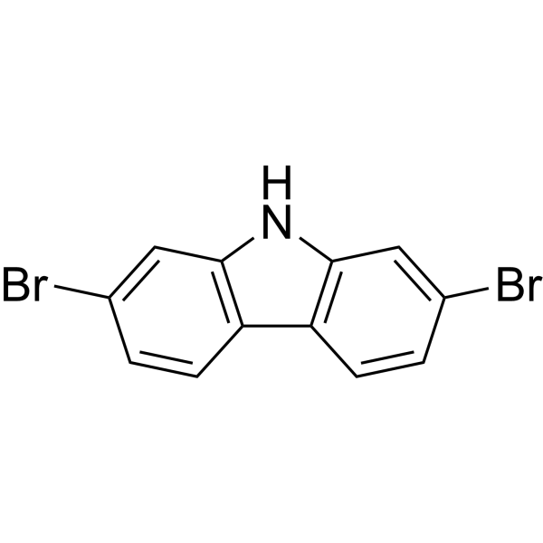
2,7-Dibromocarbazole

Based on 1 Customer Validation
2,7-Dibromocarbazole (2,7-DBCZ) is an orally active AhR agonist and MAOB inhibitor that can cross the blood-brain barrier. 2,7-Dibromocarbazole induces CYP1A/CYP1B1, developmental toxicity and oxidative toxicity, Akt phosphorylation, apoptosis, mitochondrial depolarization, and calcium elevation. 2,7-Dibromocarbazole disrupts dopamine homeostasis, promotes α-synuclein aggregation and transcriptomic dysregulation, leading to hepatic lipid accumulation, liver lesions, brain injury, and angiogenesis/energy metabolism disorders, while activating ERα and inhibiting GRα. 2,7-Dibromocarbazole can be used in research on cardiotoxicity and Parkinson's disease.
For research use only. We do not sell to patients.
- Purity : 99.96%
- CAS No.: 136630-39-2
- Formula: C12H7Br2N
- Molecular Weight:325.00
-
Storage:Powder -20°C, 3 years , 4°C, 2 years ; In solvent -80°C, 6 months , -20°C, 1 month

To place orders, for customer services and technical support, please contact: MedChemExpress USA
Tel: 609-228-6898 E-mail: [email protected] [email protected]


-
 Biological Activity
-
Chemical Information
-
Solvent & Solubility
- Protocols
- Purity & Documentation
- References
-
 Help & FAQs





Biological Activity
Description
|
MAO-B |
AhR |
CYP1A |
CYP1B1 |
Akt |
α-synuclein |
ERα |
GRα |
In Vitro
2,7-Dibromocarbazole (10 μM-1 nM; 48 h) showed no cytotoxicity against recombinant HepG2 cells[1].
2,7-Dibromocarbazole (10 μM-0.1 μM; 48 h) showed weak AhR agonist activity in a recombinant HepG2 luciferase reporter gene assay[1].
2,7-Dibromocarbazole (2,7-DBCZ) (10 nM-100 μM; 48 h) reduces H9c2 cell viability, with 100 μM being cytotoxic[2].
2,7-Dibromocarbazole (10 μM; 48 h) demethylates the Ang2 promoter in HUVECs, reducing region 2 by approximately 11%[7].
2,7-Dibromocarbazole (0.1-2 μM; 96 h) induced developmental toxicity and pericardial edema in wild-type zebrafish embryos, with a 96-h LC50 of 581.8 μg/L and an EC50 for pericardial edema of 201.5 μg/L[8].
2,7-Dibromocarbazole upregulates the expression of AhR1 and CYP1A in zebrafish embryos, consistent with AhR activation[8].
2,7-Dibromocarbazole (2,7-DBCZ) (10 μM) inhibits the viability of normal HepG2 cells[1].
2,7-Dibromocarbazole (1 μM; 48 h) does not induce CYP1A1 and inhibits the expression of AHR, PAI-2, and HSP90 in normal HepG2 cells[1].
2,7-Dibromocarbazole (10 μM; 48 h) induces early and late apoptosis in H9c2 cells and inhibits mitochondrial membrane potential[2].
2,7-Dibromocarbazole (10 μM; 48 h) dysregulated apoptosis-related genes in H9c2 cells, including upregulation of CD40LG and ANXA5[2].
2,7-Dibromocarbazole (10 μM; 48 h) co-modulates apoptosis, Akt phosphorylation, and Ca2+ elevation in H9c2 cells together with SC79/BAPTA-AM/Ppp2ca RNAi (10-100 μM; 1-3 h pre-exposure)[2].
2,7-Dibromocarbazole (10 μM; 48 h) increases the p-Akt/Akt ratio, Akt signaling gene expression, and intracellular Ca2+ in H9c2 cells[2].
2,7-Dibromocarbazole (100 nM-10 μM; 24 h) inhibits HUVEC tube formation, with 1 μM reducing tube size and length by 1.69-fold and 1.52-fold, respectively, and 10 μM eliminating tube structures[7].
2,7-Dibromocarbazole (100 nM-10 μM; 48 h) alters angiogenesis-related gene expression in HUVECs, most strongly upregulating Ang2 while decreasing VEGFB[7].
2,7-Dibromocarbazole (10 μM; 48 h post-transfection) inhibited HUVEC tube formation, whereas Ang2 silencing restored it[7].
MedChemExpress (MCE) has not independently confirmed the accuracy of these methods. They are for reference only. Further protocols information, click here.
-
Cell Line:H9c2 rat cardiomyocytes
-
Concentration:10 nM-100 μM; 10 μM
-
Incubation Time:48 h
-
Result:Reduced cell viability significantly versus control at 100 μM after 48 h.
Showed no significant cytotoxicity at other concentrations.
Selected 10 μM as the in vitro working concentration.
-
Cell Line:H9c2 rat cardiomyocytes
-
Concentration:10 μM
-
Incubation Time:48 h
-
Result:Increased early apoptotic cells by 44% and late-apoptotic cells by 57% relative to control after 48 h.
Showed 5.79% late-apoptotic, 2.68% early apoptotic, and 91.07% normal cells by flow cytometry.
Elevated J-monomers by 25%.
Reduced normalized MMP to 72% of control.
-
Cell Line:H9c2 rat cardiomyocytes
-
Concentration:10 μM
-
Incubation Time:48 h
-
Result:Activated CYP1A1, CYP1B1, CD40LG, and ANXA5 expression after 2,7-DBCZ exposure.
Suppressed this activation with CH-223191 HY-12684) co-exposure.
Decreased CYP1A1 expression by 27-48%.
Decreased CYP1B1 expression by 18.5-36.5%.
Decreased CD40LG expression by 56%.
Showed ANXA5 expression at 25% of the 2,7-DBCZ-exposed group.
In Vivo
2,7-Dibromocarbazole (2,7-DBCZ) (0.1-100 mg/kg/d; oral gavage; once daily; 14 days) induces cardiac tissue apoptosis and increases cardiac CYP1B1, CYP1A1, CD40LG, and ANXA5 expression in male Sprague-Dawley rats[2].
MedChemExpress (MCE) has not independently confirmed the accuracy of these methods. They are for reference only.
-
Animal Model:Tg(cyp1a-12DRE:EGFP) transgenic line (embryos, 0.5-1.0 h post fertilization)[1]
-
Dosage:100 nM, 1 μM, and 10 μM
-
Administration:waterborne exposure; renewal every 24 h; 96 h
-
Result:Induced EGFP fluorescence intensity of 4,310,000 at 10 μM.
Induced EGFP fluorescence intensity of 767,000 at 1 μM.
Induced EGFP fluorescence intensity of 530,000 at 100 nM.
Induced stronger EGFP fluorescence than TCDD by 4.50-fold at 10 μM.
Induced the utmost EGFP signal among tested PHCZs, equal to 79.9% of the TCDD group, at 1 μM.
Induced the highest EGFP signal among tested PHCZs, up to 55.3% of the TCDD group, at 100 nM.
Induced EGFP expression equal to 55.3% of that induced by TCDD at 100 nM (32.5 µg/L).
Ranked 1st among tested PHCZs for TCDD potency in the transgenic zebrafish assay.
-
Animal Model:Sprague-Dawley (male, six-week-old)[2]
-
Dosage:100 μg/kg/d; 100 mg/kg/d
-
Administration:i.g.; once daily; 14 days
-
Result:Reported cardiac tissue 2,7-DBCZ concentrations of 1.73 and 3.43 μg/kg after 14 days of intragastric infusion.
Increased apoptotic cardiac tissue cell proportions by 46.5% and 95% at 100 μg/kg/d and 100 mg/kg/d, respectively, versus corn oil controls.
Activated CYP1B1 expression by 17% in cardiac tissue at 100 μg/kg/d 2,7-DBCZ only.
Induced CYP1A1 and CYP1B1 expression by 409% and 65%, respectively, in cardiac tissue at 100 mg/kg/d 2,7-DBCZ.
Enhanced CD40LG and ANXA5 expression by 53% each in cardiac tissue at 100 mg/kg/d 2,7-DBCZ.


Chemical Information
-
CAS No. 136630-39-2
-
Appearance Solid
-
Molecular Weight 325.00
-
Formula C12H7Br2N
-
Color White to off-white
-
SMILES
BrC1=CC2=C(C=C1)C3=C(C=C(Br)C=C3)N2
-
Synonyms
2,7-DBCZ
-
Shipping
Room temperature in continental US; may vary elsewhere.
-
Storage
Powder -20°C 3 years 4°C 2 years In solvent -80°C 6 months -20°C 1 month


Solvent & Solubility
In Vitro:
DMSO : 100 mg/mL (307.69 mM; Need ultrasonic; Hygroscopic DMSO has a significant impact on the solubility of product, please use newly opened DMSO)
Please refer to the solubility information to select the appropriate solvent. Once prepared, please aliquot and store the solution to prevent product inactivation from repeated freeze-thaw cycles.
Storage method and period of stock solution: -80°C, 6 months; -20°C, 1 month. When stored at -80°C, please use it within 6 months. When stored at -20°C, please use it within 1 month.
Please refer to the solubility information to select the appropriate solvent. Once prepared, please aliquot and store the solution to prevent product inactivation from repeated freeze-thaw cycles.
Storage method and period of stock solution: -80°C, 6 months; -20°C, 1 month. When stored at -80°C, please use it within 6 months. When stored at -20°C, please use it within 1 month.
Concentration (start) × Volume (start) = Concentration (final) × Volume (final)

Protocols
-
Reproductive and Developmental Toxicity Study
Reproductive and developmental toxicity studies detect adverse effects of prenatal or peri/postnatal exposure on maternal condition, pregnancy maintenance, embryo-fetal survival, fetal growth, structural development, and offspring reproductive or developmental endpoints; classic rat protocols generate readouts by comparing treated groups with vehicle, pair-fed, or untreated controls for implantation, resorption, fetal weight, crown-rump length, external morphology, visceral morphology, skeletal ossification, anogenital distance, nipple/areola retention, and postnatal cohort outcomes.
-
Western Blot
Western blotting (WB) is a commonly used experimental method in molecular biology, biochemistry, and immunogenetics for identifying and quantifying target proteins. It combines gel electrophoresis with immunoassay, enabling researchers to analyze protein expression, post-translational modifications, and molecular weight.
-
Mitochondrial membrane-potential fluorescent assay
Mitochondrial membrane potential fluorescent assays estimate ΔΨm in living cells using lipophilic cationic dyes such as TMRM, TMRE, rhodamine 123, and JC-1, which accumulate in mitochondria according to membrane polarization; loss of signal after FCCP or CCCP treatment is interpreted as mitochondrial depolarization. TMRM/TMRE and rhodamine 123 are commonly used for semi-quantitative live-cell microscopy or flow cytometry, while JC-1 can report a shift from red aggregate fluorescence to green monomer fluorescence during depolarization; interpretation requires controls because dye concentration, quenching mode, cell type, dye efflux, and mitochondrial mass can affect fluorescence independently of ΔΨm.
-
Human pluripotent stem cell endothelial-cell differentiation
Human pluripotent stem cell endothelial differentiation is based on stepwise developmental patterning: early activation of WNT/GSK3β inhibition promotes mesodermal or vascular progenitor entry, followed by endothelial specification using VEGF-related signaling, BMP4, FGF2, Notch modulation, or cAMP depending on the published protocol. Endothelial differentiation is read out by acquisition of CD31, CD34, VE-cadherin/CD144, KDR/VEGFR2, vWF, Tie2, NOS3, acetylated LDL uptake, tube/network formation, barrier function, and in vivo vessel-forming capacity where tested.
-
Vascular/Branching Fractal Analysis
Vascular/branching fractal analysis quantifies the geometric complexity of vessel trees or vascular networks from segmented 2D images, commonly by converting vessels into binary and/or skeletonized maps and estimating fractal dimension using box-counting or related approaches. Fractal dimension is interpreted as an image-derived readout of vascular branching complexity, space filling, or density, and has been applied to retinal photographs, fluorescein angiography, OCT angiography, capillary perfusion maps, and in vitro Matrigel angiogenesis networks. The assay readout is generated from vessel-positive pixels after image preprocessing, vessel segmentation, binarization, and optional skeletonization; reported outputs include fractal dimension, vessel density, branchpoint density, endpoint density, vessel length density, tortuosity, and generation-based branching metrics when VESGEN-style analysis is used. The biological interpretation is limited to quantitative vascular patterning and s
-
Annexin V plus membrane-impermeant dye apoptosis staining
Annexin V-based apoptosis assays rely on the detection of phosphatidylserine (PS) externalization from the inner leaflet of the plasma membrane to the outer leaflet, an early biochemical hallmark of apoptosis. Fluorescently labeled Annexin V binds PS in a calcium-dependent manner, enabling identification of early apoptotic cells by flow cytometry or fluorescence microscopy. When combined with a membrane-impermeant DNA-binding dye (e. g. , propidium iodide), this approach allows discrimination between viable (Annexin V−/dye−), early apoptotic (Annexin V+/dye−), and late apoptotic or necrotic (Annexin V+/dye+) cell populations by assessing membrane integrity and PS exposure.
-
Kinase activity and phosphorylation assays
Kinase activity assays measure the ability of kinases to transfer phosphate groups from ATP to specific substrates, while phosphorylation assays detect the presence and levels of phosphorylated proteins. Common methods include radiolabeled ATP incorporation (e. g. ,), ADP release detection via bioluminescence (e. g. ,[3]), enzyme-linked immunosorbent assays (ELISA) for phospho-specific epitopes (e. g. ,[6]), and microtiter-based formats for high-throughput screening (e. g. ,[8]). The ADP-Glo assay quantifies kinase activity by measuring ADP produced during phosphorylation using a luciferase-based system. Radiometric assays involve autoradiography or scintillation counting after incorporation of 32P-labeled ATP into substrate proteins. ELISA-based approaches rely on phospho-specific antibodies to detect activated kinases in cell lysates or purified samples.
-
3T3-L1 preadipocyte-to-adipocyte differentiation
3T3-L1 preadipocytes are induced to differentiate after growth arrest using adipogenic media containing insulin, dexamethasone, and IBMX; differentiation is assessed by lipid-droplet accumulation, triglyceride increase, Oil Red O staining, and adipocyte-marker induction such as PPARγ and C/EBPα.
-
Apoptosis
Apoptosis, also called programmed cell death, is generally characterized by distinct morphological characteristics.
-
TUNEL staining for apoptotic DNA fragmentation
TUNEL staining detects DNA strand breaks by using terminal deoxynucleotidyl transferase to add labeled nucleotides to exposed 3′-OH DNA termini, generating either microscopic staining in fixed cells or tissue sections, or fluorescence/cytometric signal in cell suspensions. TUNEL positivity reflects DNA fragmentation but should not be interpreted alone as definitive apoptosis, because TUNEL can also label necrotic, autolytic, mechanically damaged, or DNA-repair-associated DNA breaks.
-
Human pluripotent stem cell midbrain dopaminergic neuron differentiation
Human pluripotent stem cells are directed toward midbrain dopaminergic neurons by first inducing a neural floor-plate-like progenitor state, then patterning cells with ventralizing SHH signaling and midbrain/WNT-FGF cues, and finally maturing progenitors into neurons expressing dopaminergic markers such as TH, NURR1/NR4A2, PITX3, DAT/SLC6A3, VMAT2/SLC18A2, GIRK2/KCNJ6, FOXA2, LMX1A, and EN1. The main readouts are loss of pluripotency, acquisition of FOXA2+/LMX1A+ midbrain floor-plate progenitors, emergence of βIII-tubulin+/MAP2+ neurons, and production of TH+ dopaminergic neurons with molecular, dopamine-release, and electrophysiological features of midbrain dopaminergic identity.
-
Endothelial Tube Formation Assay
Endothelial tube formation assay evaluates the ability of endothelial cells to attach, migrate, align, and organize into capillary-like networks when cultured on gelled basement membrane extract or Matrigel; the readout is the morphology and quantity of tube-like networks, which reflects an in vitro endothelial morphogenesis step related to angiogenesis. Basement membrane extract/Matrigel provides laminin-rich extracellular matrix cues that support endothelial differentiation into capillary-like structures, but it can contain biologically active growth factors, so growth-factor-reduced matrix is preferred when testing defined angiogenic stimulators or inhibitors.
-
Apoptosis Solutions
Apoptosis is a regulated, generally non-lytic cell-death pathway that removes unwanted, damaged, infected, or abnormal cells through coordinated morphological changes, caspase activation, DNA fragmentation, and membrane remodeling. The intrinsic apoptosis pathway is controlled mainly by mitochondrial outer membrane permeabilization, BCL-2 family proteins, cytochrome c release, apoptosome formation, caspase-9 activation, and downstream executioner caspase-3/7 activation. The extrinsic apoptosis pathway is initiated by death receptors such as Fas, TNFR, and TRAIL receptors, which recruit adaptor proteins and activate caspase-8 before engaging executioner caspases or mitochondrial amplification through BID cleavage. Apoptosis is linked to many phenotypes, including cancer cell killing, tissue homeostasis, immune regulation, neurodegeneration, infection response, and treatment-induced cytotoxicity; unresolved questions include how apoptosis interacts with necroptosis, pyroptosis, ferroptos
-
Protocol for Kinase activity and phosphorylation assays
Kinase activity assays measure transfer of phosphate from ATP to a protein or peptide substrate, generating phosphorylated substrate, ADP, or incorporated radiolabeled phosphate as the readout; phosphorylation assays measure site-specific phosphorylation in cells or tissues as a proxy for kinase-pathway activation, inhibition, or substrate regulation. Phosphorylation can be detected by phospho-specific Western blot, immunoprecipitation kinase assay, phospho-immunofluorescence, phospho-flow cytometry, luminescent ADP detection, radiolabeled ATP incorporation, or reporter-based pathway assays, and these readouts can be applied to cancer cells, primary neurons, mouse tumors, organoids, inflammatory macrophages, ferroptosis studies, and mitophagy studies when the kinase target is biologically relevant.
-
Endothelial Cell Migration/Angiogenic Sprouting Assay
Endothelial cell migration and angiogenic sprouting assays are in vitro (and partially ex vivo-adapted) functional models that quantify the ability of endothelial cells to undergo coordinated migration, extracellular matrix invasion, and multicellular organization into capillary-like sprouts in response to pro-angiogenic stimuli such as VEGF, bFGF, or conditioned microenvironments. These assays are used to model early angiogenic events including tip-cell formation, directional migration, and lumen-like sprout extension, which collectively reflect angiogenic activation and vascular morphogenesis processes observed in vivo.
-
Lipid Droplets: Oil Red O/Sudan Dye Lipid Staining
Lipid droplets are intracellular organelles with a neutral-lipid core that stores triacylglycerols and sterol esters, and Oil Red O or Sudan dyes detect these hydrophobic lipid deposits by partitioning into retained lipids in fresh or frozen specimens. Oil Red O stains neutral triglycerides and lipids in frozen tissue sections or air-dried cytologic preparations, while Sudan Black B has also been used as a histochemical fat stain for lipid-rich tissue structures.


Purity & Documentation
-
Data Sheet (295 KB)
-
SDS (481 KB)
- English - EN (481 KB)
- Français - FR (481 KB)
- Deutsch - DE (481 KB)
- Norwegian - NO (481 KB)
- Español - ES (481 KB)
- Swedish - SV (481 KB)
- Italian - IT (481 KB)
- Korean - KR (481 KB)
- Portuguese - PT (481 KB)
-
Handling Instructions (2659 KB)
References


Complete Stock Solution Preparation Table
Please refer to the solubility information to select the appropriate solvent. Once prepared, please aliquot and store the solution to prevent product inactivation from repeated freeze-thaw cycles.
Storage method and period of stock solution: -80°C, 6 months; -20°C, 1 month. When stored at -80°C, please use it within 6 months. When stored at -20°C, please use it within 1 month.
| Optional Solvent | Concentration Solvent Mass | 1 mg | 5 mg | 10 mg | 25 mg |
|---|---|---|---|---|---|
| DMSO | 1 mM | 3.0769 mL | 15.3846 mL | 30.7692 mL | 76.9231 mL |
| 5 mM | 0.6154 mL | 3.0769 mL | 6.1538 mL | 15.3846 mL | |
| 10 mM | 0.3077 mL | 1.5385 mL | 3.0769 mL | 7.6923 mL | |
| 15 mM | 0.2051 mL | 1.0256 mL | 2.0513 mL | 5.1282 mL | |
| 20 mM | 0.1538 mL | 0.7692 mL | 1.5385 mL | 3.8462 mL | |
| 25 mM | 0.1231 mL | 0.6154 mL | 1.2308 mL | 3.0769 mL | |
| 30 mM | 0.1026 mL | 0.5128 mL | 1.0256 mL | 2.5641 mL | |
| 40 mM | 0.0769 mL | 0.3846 mL | 0.7692 mL | 1.9231 mL | |
| 50 mM | 0.0615 mL | 0.3077 mL | 0.6154 mL | 1.5385 mL | |
| 60 mM | 0.0513 mL | 0.2564 mL | 0.5128 mL | 1.2821 mL | |
| 80 mM | 0.0385 mL | 0.1923 mL | 0.3846 mL | 0.9615 mL | |
| 100 mM | 0.0308 mL | 0.1538 mL | 0.3077 mL | 0.7692 mL |


2,7-Dibromocarbazole Related Classifications
HY-34464 Related Classifications
 Powered by Bioz