



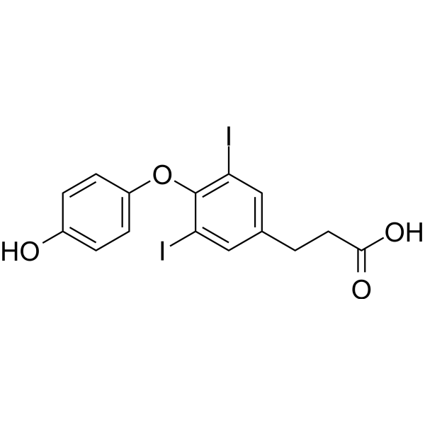
3,5-Diiodothyropropionic acid
Based on 1 publication(s) in Google Scholar
3,5-Diiodothyropropionic acid is a thyroid hormone analog, induces α-myosin heavy chain mRNA expression, binds to thyroid hormone receptor (TR), with Ka of 2.40 and 4.06 M-1 for TRα1 and TRβ1, respectively. 3,5-Diiodothyropropionic acid promotes angiogenesis in 3-D human dermal microvascular endothelial cell sprouting assay. 3,5-Diiodothyropropionic acid prevents myocardial arteriolar loss in thyroidectomized rats and enhances cardiac energy-generating capacity in postinfarction heart failure rats. 3,5-Diiodothyropropionic can be used in studies related to angiogenesis and heart failure.
For research use only. We do not sell to patients.
- Purity : 98.34%
- CAS No.: 1158-10-7
- Formula: C15H12I2O4
- Molecular Weight:510.06
-
Storage:Powder -20°C, 3 years ; In solvent -80°C, 6 months , -20°C, 1 month

To place orders, for customer services and technical support, please contact: MedChemExpress USA
Tel: 609-228-6898 E-mail: [email protected] [email protected]


-
 Biological Activity
-
Chemical Information
-
Solvent & Solubility
- Protocols
- Purity & Documentation
- References
-
 Help & FAQs


-
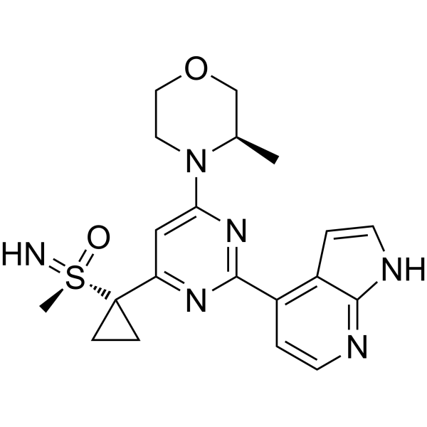
HY-19323
CeralasertibATR Inhibitor -

HY-P0059
TeriparatideParathyroid Hormone Agonist -
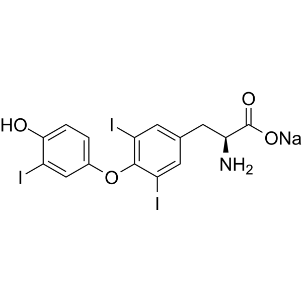
HY-A0070
Liothyronine sodiumTRα/TRβ Agonist -
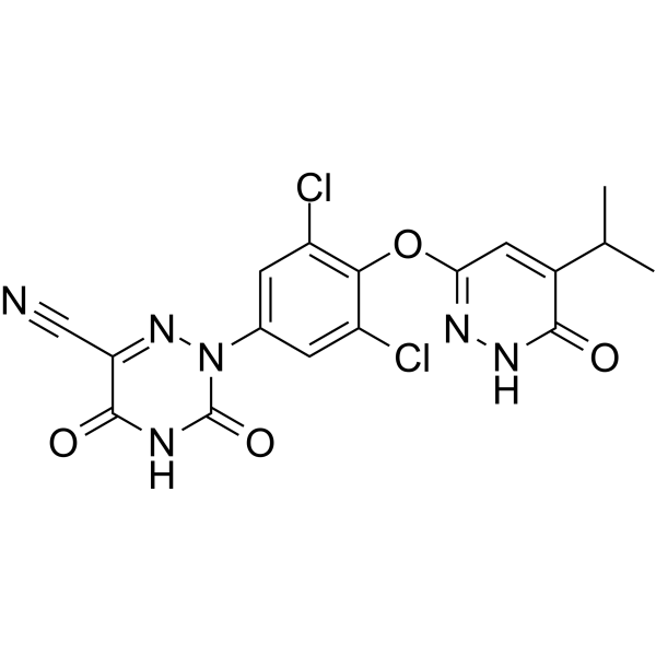
HY-12216
ResmetiromTHR-β Agonist -

HY-A0070R
Liothyronine sodium (Standard)TRα/TRβ Agonist -
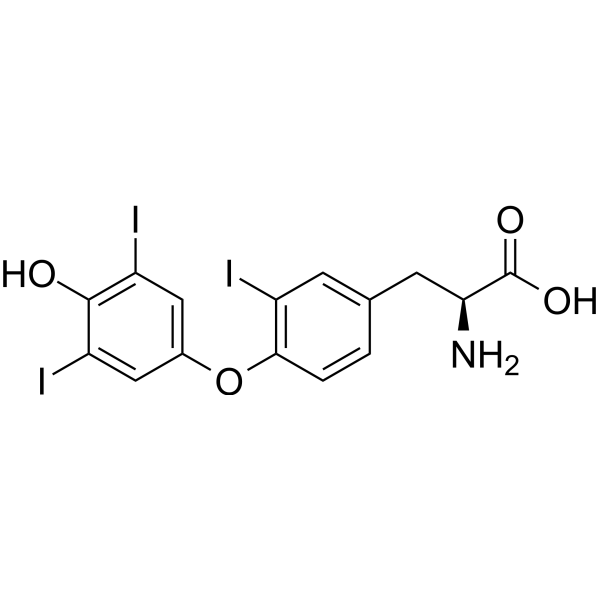
HY-W010696
Reverse T3Thyroid Hormone -

HY-P2426
(D-Trp12,Tyr34)-pTH (7-34) amide (bovine)PTH Antagonist -
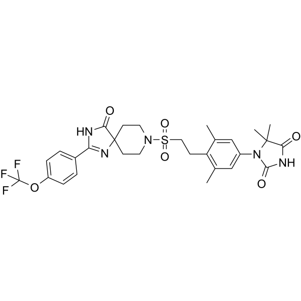
HY-100856
PCO371PTHR1 Agonist -

HY-W010696R
Reverse T3 (Standard)Thyroid Hormone -
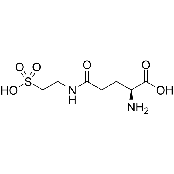
HY-106608
GlutaurineParathyroid Hormone -
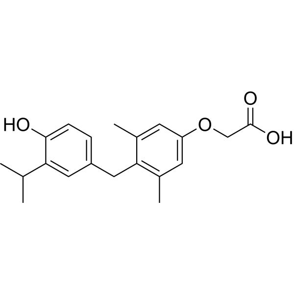
HY-14823
SobetiromeTRβ Agonist -
HY-10473
Eprotirome
Publications Citing Use of MedChemExpress (MCE) 3,5-Diiodothyropropionic acid
More

Biological Activity
Description
In Vitro
3,5-Diiodothyropropionic acid (0.1 μM) promotes angiogenesis in 3-D human dermal microvascular endothelial cell sprouting assay, with 2.1-fold increase in migrated cells and 2.0-fold increase in microvessel length [4].
3,5-Diiodothyropropionic acid (0.1 mM, 3 days) stimulates angiogenesis in 10-day-old chick embryo chorioallantoic membrane[4].
MedChemExpress (MCE) has not independently confirmed the accuracy of these methods. They are for reference only. Further protocols information, click here.
In Vivo
3,5-Diiodothyropropionic acid (375 μg/100 g, subcutaneous injection, once daily, 10 days) improves energy-generating capacity of the heart, and returns most downregulated metabolic genes toward normal in postinfarction heart failure rats[3].
3,5-Diiodothyropropionic acid (375 μg/100 g, subcutaneous injection once daily for 10 days, with Captopril (HY-B0368) 2 g/L in drinking water for 21 days) improves cardiac energy-generating capacity, and returns more genes toward normal compared to used alone in postinfarction heart failure rats[3].
MedChemExpress (MCE) has not independently confirmed the accuracy of these methods. They are for reference only.
-
Animal Model:Thyroidectomized male Sprague-Dawley rats (10.5 weeks old)[2]
-
Dosage:80 mg/60-day release pellets
-
Administration:Via subcutaneous slow-release pellets for 6 weeks
-
Result:Prevented a 31% reduction in myocardial arteriolar length density in thyroidectomized Sprague-Dawley rats, with the 5-15 μm arterioles well preserved.
Failed to prevent hypothyroidism-induced decreases in body temperature, body weight gain, heart weight and heart weight-to-body weight ratio.
Did not improve cardiac function parameters including posterior wall thickness, fractional shortening, +dP/dt, -dP/dt, and heart rate.
Increased serum triiodothyronine (T3) levels but kept serum thyroxine (T4) levels as low as the placebo group.
-
Animal Model:Adult male Spraque-Dawley rats (175-275 g) were anesthetized with methoxyflurane, subjected to left thoracotomy, and a ligature was placed around the proximal left coronary artery to induce myocardial infarction[3]
-
Dosage:375 μg/100 g
-
Administration:Subcutaneous injection once daily for 10 days
-
Result:Reversed the downregulation of most metabolic genes.
Restored the energy-generating capacity of the heart.
Improved cardiac function as evidenced by reduced left ventricular end-diastolic pressure.
-
Animal Model:Adult male Spraque-Dawley rats (175-275 g) were anesthetized with methoxyflurane, subjected to left thoracotomy, and a ligature was placed around the proximal left coronary artery to induce myocardial infarction [3]
-
Dosage:375 μg/100 g
-
Administration:Subcutaneous injection once daily for 10 days, with Captopril (HY-B0368) 2 g/L in drinking water for 21 days
-
Result:Reversed the downregulation of metabolic enzymes.
Restored the energy-generating capacity of the heart.
Improved cardiac function as evidenced by more genes returning toward normal compared to used alone.


Chemical Information
-
CAS No. 1158-10-7
-
Appearance Solid
-
Molecular Weight 510.06
-
Formula C15H12I2O4
-
Color White to off-white
-
SMILES
O=C(O)CCC1=CC(I)=C(OC2=CC=C(O)C=C2)C(I)=C1
-
Shipping
Room temperature in continental US; may vary elsewhere.
-
Storage
Powder -20°C 3 years In solvent -80°C 6 months -20°C 1 month


Publications (1)
-
Journal Impact Factor
-
Most Recent
-
Environ Sci Technol
IodoFinder: Machine Learning-Guided Recognition of Iodinated Chemicals in Nontargeted LC-MS/MS Analysis. [Abstract]2025 Mar 11;59(9):4530-4539. PMID: 40015982


Solvent & Solubility
In Vitro:
DMSO : 50 mg/mL (98.03 mM; Need ultrasonic; Hygroscopic DMSO has a significant impact on the solubility of product, please use newly opened DMSO)
Please refer to the solubility information to select the appropriate solvent. Once prepared, please aliquot and store the solution to prevent product inactivation from repeated freeze-thaw cycles.
Storage method and period of stock solution: -80°C, 6 months; -20°C, 1 month. When stored at -80°C, please use it within 6 months. When stored at -20°C, please use it within 1 month.
Please refer to the solubility information to select the appropriate solvent. Once prepared, please aliquot and store the solution to prevent product inactivation from repeated freeze-thaw cycles.
Storage method and period of stock solution: -80°C, 6 months; -20°C, 1 month. When stored at -80°C, please use it within 6 months. When stored at -20°C, please use it within 1 month.
Concentration (start) × Volume (start) = Concentration (final) × Volume (final)
In Vivo:
Select the appropriate dissolution method based on your experimental animal and administration route.
- For the following dissolution methods, please ensure to first prepare a clear stock solution using an In Vitro approach and then sequentially add co-solvents:
- To ensure reliable experimental results, the clarified stock solution can be appropriately stored based on storage conditions. As for the working solution for In Vivo experiments, it is recommended to prepare freshly and use it on the same day.
- The percentages shown for the solvents indicate their volumetric ratio in the final prepared solution. If precipitation or phase separation occurs during preparation, heat and/or sonication can be used to aid dissolution.
Add each solvent one by one: 10% DMSO 40% PEG300 5% Tween-80 45% Saline
Solubility: ≥ 2.5 mg/mL (4.90 mM); Clear solution
This protocol yields a clear solution of ≥ 2.5 mg/mL (saturation unknown).
Taking 1 mL working solution as an example, add 100 μL DMSO stock solution (25.0 mg/mL) to 400 μL PEG300, and mix evenly; then add 50 μL Tween-80 and mix evenly; then add 450 μL Saline to adjust the volume to 1 mL.
Preparation of Saline: Dissolve 0.9 g sodium chloride in ddH₂O and dilute to 100 mL to obtain a clear Saline solution.
Add each solvent one by one: 10% DMSO 90% (20% SBE-β-CD in Saline)
Solubility: ≥ 2.5 mg/mL (4.90 mM); Clear solution
This protocol yields a clear solution of ≥ 2.5 mg/mL (saturation unknown).
Taking 1 mL working solution as an example, add 100 μL DMSO stock solution (25.0 mg/mL) to 900 μL 20% SBE-β-CD in Saline, and mix evenly.
Preparation of 20% SBE-β-CD in Saline (4°C, storage for one week): 2 g SBE-β-CD powder is dissolved in 10 mL Saline, completely dissolve until clear.
In Vivo Dissolution Calculator
Please enter the basic information of animal experiments:
-
-
-
-
Recommended: Prepare an additional quantity of animals to account for potential losses during experiments.
Please enter your animal formula composition:
-
%DMSO +
Recommended: Keep the proportion of DMSO in working solution below 2% if your animal is weak.
-
%+
-
+%Tween-80 + +
-
%Saline +
The co-solvents required include: DMSO, . All of co-solvents are available by MedChemExpress (MCE). , Tween 80. All of co-solvents are available by MedChemExpress (MCE).
Working solution concentration: 0.22 mg/mL
Method for preparing stock solution: mg drug dissolved in μL DMSO. Stock solution concentration: mg/mL.
1. Take μL DMSO stock solution;
2. Add μL .
μL , mix evenly;
3. Then add μL Tween 80, mix evenly;
4. Then add μL
Please ensure that the stock solution in the first step is dissolved to a clear state, and add co-solvents in sequence. You can use ultrasonic heating (ultrasonic cleaner, recommended frequency 20-40 kHz), vortexing, etc. to assist dissolution.

Protocols
-
RT-PCR
Reverse transcription technology uses RNA as a template to synthesize DNA. RT-PCR is simple, specific and sensitive, and can be used to detect gene expression levels and expression differences in cells; detect RNA virus content; clone cDNA sequences of specific genes.
-
RNA extraction experimental
By lysing cells, releasing RNA, and removing impurities such as proteins and DNA, high-purity RNA products are finally obtained. The commonly used traditional method is the guanidine isothiocyanate/phenol/chloroform method (Trizol), which is suitable for a variety of animal materials including animal tissues, microorganisms, cultured cells, etc., and most plant materials.
-
Human pluripotent stem cell endothelial-cell differentiation
Human pluripotent stem cell endothelial differentiation is based on stepwise developmental patterning: early activation of WNT/GSK3β inhibition promotes mesodermal or vascular progenitor entry, followed by endothelial specification using VEGF-related signaling, BMP4, FGF2, Notch modulation, or cAMP depending on the published protocol. Endothelial differentiation is read out by acquisition of CD31, CD34, VE-cadherin/CD144, KDR/VEGFR2, vWF, Tie2, NOS3, acetylated LDL uptake, tube/network formation, barrier function, and in vivo vessel-forming capacity where tested.
-
Vascular/Branching Fractal Analysis
Vascular/branching fractal analysis quantifies the geometric complexity of vessel trees or vascular networks from segmented 2D images, commonly by converting vessels into binary and/or skeletonized maps and estimating fractal dimension using box-counting or related approaches. Fractal dimension is interpreted as an image-derived readout of vascular branching complexity, space filling, or density, and has been applied to retinal photographs, fluorescein angiography, OCT angiography, capillary perfusion maps, and in vitro Matrigel angiogenesis networks. The assay readout is generated from vessel-positive pixels after image preprocessing, vessel segmentation, binarization, and optional skeletonization; reported outputs include fractal dimension, vessel density, branchpoint density, endpoint density, vessel length density, tortuosity, and generation-based branching metrics when VESGEN-style analysis is used. The biological interpretation is limited to quantitative vascular patterning and s
-
Research Protocol for Endocrine Diseases
Endocrine diseases often arise from disrupted hormone production, hormone signaling, or target-tissue responsiveness; for diabetes-focused endocrine disease models, insulin signaling regulates glucose uptake, hepatic glucose output, lipid metabolism, and β-cell compensation. Type 2 diabetes develops through interacting defects in insulin resistance, β-cell dysfunction, adipose inflammation, hepatic glucose overproduction, altered incretin signaling, and ectopic lipid metabolism. A major unresolved question is whether endocrine dysfunction is driven primarily by target-tissue insulin resistance, intrinsic β-cell failure, immune/inflammatory stress, or combined multi-organ failure that differs by disease stage.
-
Endothelial Tube Formation Assay
Endothelial tube formation assay evaluates the ability of endothelial cells to attach, migrate, align, and organize into capillary-like networks when cultured on gelled basement membrane extract or Matrigel; the readout is the morphology and quantity of tube-like networks, which reflects an in vitro endothelial morphogenesis step related to angiogenesis. Basement membrane extract/Matrigel provides laminin-rich extracellular matrix cues that support endothelial differentiation into capillary-like structures, but it can contain biologically active growth factors, so growth-factor-reduced matrix is preferred when testing defined angiogenic stimulators or inhibitors.
-
Research Protocol for Cardiovascular Diseases
Cardiovascular disease can be modeled as maladaptive cardiac remodeling, where ischemic injury or pressure overload activates inflammatory signaling, fibroblast activation, extracellular-matrix deposition, cardiomyocyte hypertrophy, vascular remodeling, and progressive ventricular dysfunction. The TGF-β/SMAD axis is a central profibrotic pathway after myocardial injury and pressure overload, while innate immune and cytokine pathways regulate leukocyte recruitment, scar formation, and adverse remodeling. Key unresolved questions include which inflammatory signals are reparative versus harmful, when fibrosis is protective versus maladaptive, and whether pathway inhibition improves function without weakening necessary infarct healing or compensatory remodeling.
-
Real Time qPCR (Q-PCR)
Real-time quantitative PCR (qPCR) quantifies an amplifiable nucleic-acid target by monitoring fluorescence during PCR cycling rather than measuring product only after amplification. The increase in fluorescence tracks accumulation of PCR product, and the quantification cycle (Cq; historically also Ct/CP) is related to the initial amount of target: samples containing more starting target generally reach the defined fluorescence threshold in fewer cycles.
-
Endothelial Cell Migration/Angiogenic Sprouting Assay
Endothelial cell migration and angiogenic sprouting assays are in vitro (and partially ex vivo-adapted) functional models that quantify the ability of endothelial cells to undergo coordinated migration, extracellular matrix invasion, and multicellular organization into capillary-like sprouts in response to pro-angiogenic stimuli such as VEGF, bFGF, or conditioned microenvironments. These assays are used to model early angiogenic events including tip-cell formation, directional migration, and lumen-like sprout extension, which collectively reflect angiogenic activation and vascular morphogenesis processes observed in vivo.


Purity & Documentation
-
Data Sheet (277 KB)
-
SDS (393 KB)
- English - EN (393 KB)
- Français - FR (393 KB)
- Deutsch - DE (393 KB)
- Norwegian - NO (393 KB)
- Español - ES (393 KB)
- Swedish - SV (393 KB)
- Italian - IT (393 KB)
- Korean - KR (393 KB)
- Portuguese - PT (393 KB)
-
Handling Instructions (2659 KB)
References
[1]. Pennock GD, et al. Cardiac effects of 3,5-diiodothyropropionic acid, a thyroid hormone analog with inotropic selectivity. J Pharmacol Exp Ther. 1992 Oct;263(1):163-9. [Content Brief]
[2]. Liu Y, et al. Thyroid hormone analog 3,5-diiodothyropropionic acid promotes healthy vasculature in the adult myocardium independent of thyroid effects on cardiac function. Am J Physiol Heart Circ Physiol. 2009 May;296(5):H1551-7. [Content Brief]
[3]. Maitra N, et al. Regulation of gene expression in rats with heart failure treated with the thyroid hormone analog 3,5-diiodothyropropionic acid (DITPA) and the combination of DITPA and captopril. J Cardiovasc Pharmacol. 2007 Nov;50(5):526-34. [Content Brief]
[4]. Mousa SA, et al. Proangiogenesis action of the thyroid hormone analog 3,5-diiodothyropropionic acid (DITPA) is initiated at the cell surface and is integrin mediated. Endocrinology. 2006 Apr;147(4):1602-7. [Content Brief]


Complete Stock Solution Preparation Table
Please refer to the solubility information to select the appropriate solvent. Once prepared, please aliquot and store the solution to prevent product inactivation from repeated freeze-thaw cycles.
Storage method and period of stock solution: -80°C, 6 months; -20°C, 1 month. When stored at -80°C, please use it within 6 months. When stored at -20°C, please use it within 1 month.
| Optional Solvent | Concentration Solvent Mass | 1 mg | 5 mg | 10 mg | 25 mg |
|---|---|---|---|---|---|
| DMSO | 1 mM | 1.9606 mL | 9.8028 mL | 19.6055 mL | 49.0138 mL |
| 5 mM | 0.3921 mL | 1.9606 mL | 3.9211 mL | 9.8028 mL | |
| 10 mM | 0.1961 mL | 0.9803 mL | 1.9606 mL | 4.9014 mL | |
| 15 mM | 0.1307 mL | 0.6535 mL | 1.3070 mL | 3.2676 mL | |
| 20 mM | 0.0980 mL | 0.4901 mL | 0.9803 mL | 2.4507 mL | |
| 25 mM | 0.0784 mL | 0.3921 mL | 0.7842 mL | 1.9606 mL | |
| 30 mM | 0.0654 mL | 0.3268 mL | 0.6535 mL | 1.6338 mL | |
| 40 mM | 0.0490 mL | 0.2451 mL | 0.4901 mL | 1.2253 mL | |
| 50 mM | 0.0392 mL | 0.1961 mL | 0.3921 mL | 0.9803 mL | |
| 60 mM | 0.0327 mL | 0.1634 mL | 0.3268 mL | 0.8169 mL | |
| 80 mM | 0.0245 mL | 0.1225 mL | 0.2451 mL | 0.6127 mL |


3,5-Diiodothyropropionic acid Related Classifications
HY-126236 Related Classifications
 Powered by Bioz