



6,8-Diprenylgenistein

Based on 1 Customer Validation
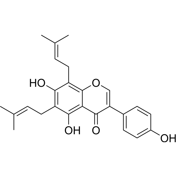
6,8-Diprenylgenistein is an isoflavone compound isolated from Cudrania tricuspidata. 6,8-Diprenylgenistein has antimicrobial and anti-obesity activity. 6,8-Diprenylgenistein inhibits the proliferation, migration and tubular formation of HLMEC induced by recombinant human vascular endothelial growth factor-A. 6,8-Diprenylgenistein can be used to study new therapeutic drugs for the prevention and treatment of oral cancer metastasis .
For research use only. We do not sell to patients.
- Purity : 99.55%
- CAS No.: 51225-28-6
- Formula: C25H26O5
- Molecular Weight:406.47
-
Storage:Powder -20°C, 3 years , 4°C, 2 years ; In solvent -80°C, 6 months , -20°C, 1 month

To place orders, for customer services and technical support, please contact: MedChemExpress USA
Tel: 609-228-6898 E-mail: [email protected] [email protected]


-
 Biological Activity
-
Chemical Information
- Protocols
- Purity & Documentation
- References
-
 Help & FAQs




Biological Activity
Description
Cellular Effect
|
Cell Line

|
Type | Value | Description | References |
|---|---|---|---|---|
| MCF7 | IC50 |
8.58 μM
Compound: 38
|
Cytotoxicity against human MCF7 cells assessed as reduction in cell viability after 24 hrs by MTS assay
Cytotoxicity against human MCF7 cells assessed as reduction in cell viability after 24 hrs by MTS assay
|
[PMID: 28522265] |
| RAW264.7 | IC50 |
19.2 μM
Compound: 10
|
Inhibition of LPS-induced nitric oxide production in mouse RAW264.7 cells by Griess assay
Inhibition of LPS-induced nitric oxide production in mouse RAW264.7 cells by Griess assay
|
[PMID: 19113968] |
| SW480 | IC50 |
8.08 μM
Compound: 38
|
Cytotoxicity against human SW480 cells assessed as reduction in cell viability after 24 hrs by MTS assay
Cytotoxicity against human SW480 cells assessed as reduction in cell viability after 24 hrs by MTS assay
|
[PMID: 28522265] |


Chemical Information
-
CAS No. 51225-28-6
-
Appearance Solid
-
Molecular Weight 406.47
-
Formula C25H26O5
-
Color Light yellow to yellow
-
SMILES
O=C1C(C2=CC=C(O)C=C2)=COC3=C(C/C=C(C)\C)C(O)=C(C/C=C(C)\C)C(O)=C13
-
Structure Classification
-
Shipping
Room temperature in continental US; may vary elsewhere.
-
Storage
Powder -20°C 3 years 4°C 2 years In solvent -80°C 6 months -20°C 1 month

Protocols
-
Research Protocol for Infectious Diseases
Infectious-disease experiments test how pathogens interact with host barriers, innate immune receptors, inflammatory signaling, pathogen replication, and tissue injury; pattern-recognition receptors such as TLRs, RIG-I-like receptors, NOD-like receptors, and inflammasomes detect microbial molecules and activate NF-κB, interferon, and cytokine responses. The central hypothesis is that infection severity reflects the balance between pathogen burden and host response: protective inflammation restricts pathogen growth, whereas excessive or mislocalized inflammation contributes to tissue damage and disease phenotype. Unresolved questions include which host pathways are protective versus pathogenic, why some infection models fail to translate to human disease, and which combined readouts best predict clinically relevant infection outcomes.
-
Cell migration
Cell migration is a method that plays an important role in wound healing, cell differentiation, embryonic development, etc.
-
Research Protocol for Metabolic Diseases
AMP-activated protein kinase, AMPK, is a conserved cellular energy sensor that responds to reduced cellular energy status and coordinates metabolism by increasing ATP-generating catabolic pathways while suppressing ATP-consuming anabolic processes. In metabolic disease research, the AMPK pathway is experimentally relevant because it regulates hepatic lipid synthesis, fatty acid oxidation, glucose production, skeletal-muscle glucose disposal, mTORC1-linked biosynthesis, autophagy, mitochondrial homeostasis, and whole-body energy balance. The central pathway logic is that energy stress, metformin, exercise-like stimulation, or direct AMPK activators increase AMPKα Thr172 phosphorylation and downstream substrate phosphorylation, including ACC and RAPTOR. Phosphorylation of ACC suppresses lipogenesis and supports fatty acid oxidation, whereas phosphorylation of RAPTOR suppresses mTORC1 signaling and links cellular energy status to growth and protein synthesis control. The pathway is linked
-
Orthotopic Cell-Line Xenograft
Orthotopic cell-line xenograft models involve implantation of human cancer cell lines into the anatomically corresponding organ of immunodeficient mice to reproduce tumor growth within a native microenvironment, enabling more clinically relevant tumor behavior compared with subcutaneous models. These models are widely used because orthotopic placement better recapitulates tumor progression, including invasion and metastatic spread, which are often underrepresented in heterotopic implantation systems. Compared with conventional xenografts, orthotopic implantation is described as more technically complex but provides improved simulation of tumor-microenvironment interactions and metastatic behavior, making it particularly valuable for translational oncology research. Surgical orthotopic implantation approaches have been emphasized as enabling faithful reproduction of clinical cancer features, including metastasis and disease progression patterns that align with the tumor’s organ of origi
-
Cell Viability Determination by MTT Colorimetric Assay
The following protocol uses the MTT colorimetric assay as a classic literature-established method for assessing cell viability/metabolic activity in cultured mammalian cells. MTT[3-(4,5-dimethylthiazol-2-yl)-2,5-diphenyltetrazolium bromide] is reduced by metabolically active cells to a colored formazan product; the amount of formazan is quantified spectrophotometrically and provides an indirect measure of metabolically active viable cells. Importantly, MTT reduction reflects cellular oxidoreductase/metabolic activity rather than an absolute direct count of living cells, so changes in cellular metabolism can alter the signal independently of cell number.


Purity & Documentation
-
Data Sheet (267 KB)
-
SDS (252 KB)
- English - EN (252 KB)
- Français - FR (252 KB)
- Deutsch - DE (252 KB)
- Norwegian - NO (252 KB)
- Español - ES (252 KB)
- Swedish - SV (252 KB)
- Italian - IT (252 KB)
- Korean - KR (252 KB)
- Portuguese - PT (252 KB)
-
Handling Instructions (2659 KB)
References

Calculators
Concentration (start) × Volume (start) = Concentration (final) × Volume (final)


6,8-Diprenylgenistein Related Classifications
HY-N2692 Related Classifications
 Powered by Bioz