



β-Amyloid (1-40) (rat)
Based on 2 publication(s) in Google Scholar
β-Amyloid (1-40) (rat) is a rat form of the amyloid β-peptide, which accumulates as an insoluble extracellular deposit around neurons, giving rise to the senile plaques associated with Alzheimer's disease (AD). β-Amyloid (1-40) (rat) increases 45Ca2+ influx, induces neurodegeneration in the rat hippocampal neurons of the CA1 subfield. β-Amyloid (1-40) (rat) induces apoptosis. β-Amyloid (1-40) (rat) can be used for the research of Alzheimer's disease.
For research use only. We do not sell to patients.
- CAS No.: 144409-98-3
- Formula: C190H291N51O57S
- Molecular Weight:4233.76
-
Storage:
Sealed storage, away from moisture and light.
Powder -80°C, 2 years , -20°C, 1 year* The compound is unstable in solutions, freshly prepared is recommended.


To place orders, for customer services and technical support, please contact: MedChemExpress USA
Tel: 609-228-6898 E-mail: [email protected] [email protected]


-
 Biological Activity
-
Chemical Information
-
Solvent & Solubility
- Protocols
- Purity & Documentation
- References
-
 Help & FAQs


-
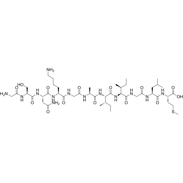
HY-P0128
β-Amyloid (25-35)Amyloid β Peptide -
HY-P1388
β-Amyloid (1-42), (rat/mouse)β-Amyloid (1-42) Peptide -

HY-P0128A
β-Amyloid (25-35), HFIP-treatedAmyloid β Peptide -

HY-P1363A
β-Amyloid (1-42), humanAmyloid β Peptide -

HY-P1363B
β-Amyloid (1-42), human, HFIP-treatedβ-Amyloid (1-42) Peptide -
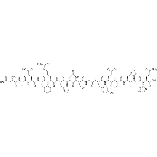
HY-P1046
β-Amyloid (1-15)Peptide Fragment -

HY-P1362
β-Amyloid (42-1), humanInactive Form of Aβ(1-42) -
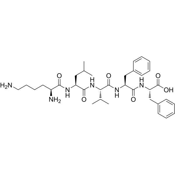
HY-P5124
β-Amyloid peptide(16-20)Abeta Inhibitor -
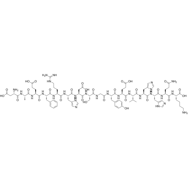
HY-P1466
β-Amyloid (1-16)β-Amyloid -

HY-P1378
β-Amyloid (1-43)(human)Peptide -
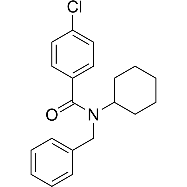
HY-19370
FPS-ZM1Amyloid-β Inhibitor -

HY-P1051
β-Amyloid (12-28)Amyloid Peptide
Publications Citing Use of MedChemExpress (MCE) β-Amyloid (1-40) (rat)
More

Biological Activity
Description
In Vitro
β-Amyloid (1-40) (rat) (1 μM; 1 h) increases 45Ca2+ influx and elevates Ca2+ in cortical synaptosomes[1].
β-Amyloid (1-40) (rat) (3 nM) induces neurodegeneration in the rat hippocampal neurons of the CA1 subfield and induces apoptosis[2].
β-Amyloid Aggregation Guidelines (Following is our recommended protocol. This protocol only provides a guideline, and should be modified according to your specific needs).
1. Solid Aβ peptide is dissolved in cold hexafluoro-2-propanol (HFIP). The peptide is incubated at room temperature for at least 1 h to establish monomerization and randomization of structure.
2. The HFIP is removed by evaporation, and the resulting peptide is stored as a film at -20 or -80 °C.
3. The resulting film is dissolved in anhydrous DMSO at 5 mM and then dilutes into the appropriate concentration and buffer (serum- and phenol red-free culture medium) with vortexing.
4. Next, the solution is age 48 h at 4-8 °C. The sample is then centrifuged at 14000g for 10 min at 4-8 °C; the soluble oligomers are in the supernatant. The supernatant is diluted 10-200-fold for experiments.
Methods vary depends on the downstream applications.
MedChemExpress (MCE) has not independently confirmed the accuracy of these methods. They are for reference only. Further protocols information, click here.
In Vivo
MedChemExpress (MCE) has not independently confirmed the accuracy of these methods. They are for reference only.
-
Animal Model:Swiss and C57BL/6 mice[3]
-
Dosage:1.7 mg
-
Administration:Intracerebroventrical injection; for 7 days
-
Result:Presented spatial learning and memory impairments.


Chemical Information
-
CAS No. 144409-98-3
-
Appearance Solid
-
Molecular Weight 4233.76
-
Formula C190H291N51O57S
-
Color White to off-white
-
Sequence
Asp-Ala-Glu-Phe-Gly-His-Asp-Ser-Gly-Phe-Glu-Val-Arg-His-Gln-Lys-Leu-Val-Phe-Phe-Ala-Glu-Asp-Val-Gly-Ser-Asn-Lys-Gly-Ala-Ile-Ile-Gly-Leu-Met-Val-Gly-Gly-Val-Val
-
Sequence Shortening
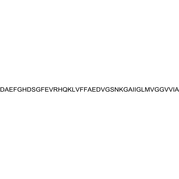
DAEFGHDSGFEVRHQKLVFFAEDVGSNKGAIIGLMVGGVV
-
Shipping
Room temperature in continental US; may vary elsewhere.
-
Storage
Sealed storage, away from moisture and light
Powder -80°C 2 years -20°C 1 year * The compound is unstable in solutions, freshly prepared is recommended.


Publications (2)
-
Journal Impact Factor
-
Most Recent
-
Sci Rep
Amyloid-β slows cilia movement along the ventricle, impairs fluid flow, and exacerbates its neurotoxicity in explant culture. [Abstract]2023 Aug 21;13(1):13586. PMID: 37605005 -
Med Phys
Cerebrovascular mechanisms between type 2 diabetes and Alzheimer's disease: Insights from ultrasound localization microscopy. [Abstract]2025 Nov;52(11):e70131. PMID: 41206345


Solvent & Solubility
In Vitro:
DMSO : 50 mg/mL (11.81 mM; Need ultrasonic; Hygroscopic DMSO has a significant impact on the solubility of product, please use newly opened DMSO)
Please refer to the solubility information to select the appropriate solvent. The compound is unstable in solutions, freshly prepared is recommended.
Please refer to the solubility information to select the appropriate solvent. The compound is unstable in solutions, freshly prepared is recommended.
Concentration (start) × Volume (start) = Concentration (final) × Volume (final)

Protocols
-
Neurotoxicity Study
This protocol assesses in vitro neurotoxicity by combining neuronal viability, mitochondrial/metabolic activity, neurite outgrowth, and optional neuronal network function readouts. Calcein-AM or resazurin/PrestoBlue readouts estimate viable or metabolically active cells; βIII-tubulin immunofluorescence detects neuronal morphology and neurite networks; TMRE detects mitochondrial membrane potential; and MEA recordings detect functional changes in neuronal network activity.
-
Apoptosis
Apoptosis, also called programmed cell death, is generally characterized by distinct morphological characteristics.
-
TUNEL staining for apoptotic DNA fragmentation
TUNEL staining detects DNA strand breaks by using terminal deoxynucleotidyl transferase to add labeled nucleotides to exposed 3′-OH DNA termini, generating either microscopic staining in fixed cells or tissue sections, or fluorescence/cytometric signal in cell suspensions. TUNEL positivity reflects DNA fragmentation but should not be interpreted alone as definitive apoptosis, because TUNEL can also label necrotic, autolytic, mechanically damaged, or DNA-repair-associated DNA breaks.
-
Annexin V plus membrane-impermeant dye apoptosis staining
Annexin V-based apoptosis assays rely on the detection of phosphatidylserine (PS) externalization from the inner leaflet of the plasma membrane to the outer leaflet, an early biochemical hallmark of apoptosis. Fluorescently labeled Annexin V binds PS in a calcium-dependent manner, enabling identification of early apoptotic cells by flow cytometry or fluorescence microscopy. When combined with a membrane-impermeant DNA-binding dye (e. g. , propidium iodide), this approach allows discrimination between viable (Annexin V−/dye−), early apoptotic (Annexin V+/dye−), and late apoptotic or necrotic (Annexin V+/dye+) cell populations by assessing membrane integrity and PS exposure.
-
Apoptosis Solutions
Apoptosis is a regulated, generally non-lytic cell-death pathway that removes unwanted, damaged, infected, or abnormal cells through coordinated morphological changes, caspase activation, DNA fragmentation, and membrane remodeling. The intrinsic apoptosis pathway is controlled mainly by mitochondrial outer membrane permeabilization, BCL-2 family proteins, cytochrome c release, apoptosome formation, caspase-9 activation, and downstream executioner caspase-3/7 activation. The extrinsic apoptosis pathway is initiated by death receptors such as Fas, TNFR, and TRAIL receptors, which recruit adaptor proteins and activate caspase-8 before engaging executioner caspases or mitochondrial amplification through BID cleavage. Apoptosis is linked to many phenotypes, including cancer cell killing, tissue homeostasis, immune regulation, neurodegeneration, infection response, and treatment-induced cytotoxicity; unresolved questions include how apoptosis interacts with necroptosis, pyroptosis, ferroptos
-
Primary Embryonic Hippocampal Neuron Culture
Primary embryonic hippocampal neuron culture is an in vitro method in which hippocampi from embryonic rodents are dissected, enzymatically or mechanically dissociated, plated on adhesive substrates, and maintained in defined neuronal medium or in low-density sandwich/co-culture formats to support neuronal attachment, neurite extension, polarity formation, dendritic arborization, and synapse formation. The main readouts are cell survival, neuronal purity, neurite outgrowth, axon-dendrite polarization, synaptic marker development, and functional neuronal activity, assessed by phase-contrast microscopy, immunocytochemistry for neuronal/glial markers, live imaging, or electrophysiology depending on the downstream experiment.
-
Amyloid: Congo Red Amyloid Staining
Congo red amyloid staining is a histochemical method used to detect extracellular amyloid deposits in tissue sections based on the affinity of Congo red dye for β-pleated sheet-rich protein aggregates. When bound to amyloid, Congo red produces characteristic apple-green birefringence under polarized light microscopy, which is widely regarded as a diagnostic feature of amyloid deposition in histopathology. The diagnostic principle relies on the combination of dye binding (congophilia) and optical anisotropy under polarized illumination, which distinguishes amyloid from most non-amyloid eosinophilic extracellular deposits in routine histological evaluation. Amyloid identification by Congo red staining remains a cornerstone in diagnostic pathology despite the availability of adjunct methods such as immunohistochemistry and mass spectrometry, particularly because of its ability to localize deposits directly within tissue architecture. The specificity of Congo red-positive deposits is incre
-
Alzheimer’s Disease Modeling
Alzheimer’s Disease (AD) is a neurodegenerative disorder characterized by a progressive decline in cognitive functions and loss of specific types of neurons and synapses. Alzheimer's symptoms can be simulated in mice by injecting drugs (such as Aβ) or genetically modified.


Purity & Documentation
-
Data Sheet (273 KB)
-
SDS (252 KB)
- English - EN (252 KB)
- Français - FR (252 KB)
- Deutsch - DE (252 KB)
- Norwegian - NO (252 KB)
- Español - ES (252 KB)
- Swedish - SV (252 KB)
- Italian - IT (252 KB)
- Korean - KR (252 KB)
- Portuguese - PT (252 KB)
-
Handling Instructions (2659 KB)
References
[1]. MacManus A, et, al. Enhancement of (45)Ca(2+) influx and voltage-dependent Ca(2+) channel activity by beta-amyloid-(1-40) in rat cortical synaptosomes and cultured cortical neurons. Modulation by the proinflammatory cytokine interleukin-1beta. J Biol Chem. 2000 Feb 18;275(7):4713-8. [Content Brief]
[2]. Miguel-Hidalgo JJ, et, al. Beta-amyloid(1-40)-induced neurodegeneration in the rat hippocampal neurons of the CA1 subfield. Acta Neuropathol. 1998 May;95(5):455-65. [Content Brief]
[3]. Prediger RD, et, al. Differential susceptibility following beta-amyloid peptide-(1-40) administration in C57BL/6 and Swiss albino mice: Evidence for a dissociation between cognitive deficits and the glutathione system response. Behav Brain Res. 2007 Feb 27;177(2):205-13. [Content Brief]


Complete Stock Solution Preparation Table
Please refer to the solubility information to select the appropriate solvent. The compound is unstable in solutions, freshly prepared is recommended.
| Optional Solvent | Concentration Solvent Mass | 1 mg | 5 mg | 10 mg | 25 mg |
|---|---|---|---|---|---|
| DMSO | 1 mM | 0.2362 mL | 1.1810 mL | 2.3620 mL | 5.9049 mL |
| 5 mM | 0.0472 mL | 0.2362 mL | 0.4724 mL | 1.1810 mL | |
| 10 mM | 0.0236 mL | 0.1181 mL | 0.2362 mL | 0.5905 mL |


β-Amyloid (1-40) (rat) Related Classifications
HY-P1387 Related Classifications
 Powered by Bioz