



Deoxycorticosterone acetate
Based on 2 publication(s) in Google Scholar
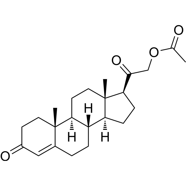
Deoxycorticosterone acetate (DOCA) is an adrenocortin, acts as a precursor to aldosterone. Deoxycorticosterone acetate is a mineralocorticoid receptor agonist. Deoxycorticosterone acetate can cause severe renal injury, including inflammation, fibrosis, glomerular damage, and proteinuria.
For research use only. We do not sell to patients.
- Purity : 99.87%
- CAS No.: 56-47-3
- Formula: C23H32O4
- Molecular Weight:372.50
-
Storage:Powder -20°C, 3 years , 4°C, 2 years ; In solvent -80°C, 1 year , -20°C, 6 months

To place orders, for customer services and technical support, please contact: MedChemExpress USA
Tel: 609-228-6898 E-mail: [email protected] [email protected]


-
 Biological Activity
-
Chemical Information
-
Solvent & Solubility
- Protocols
- Purity & Documentation
- References
-
 Help & FAQs


-
HY-B0251
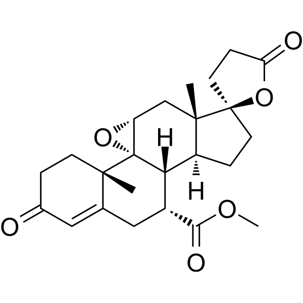
EplerenoneAldosterone Blocker -
HY-B1618
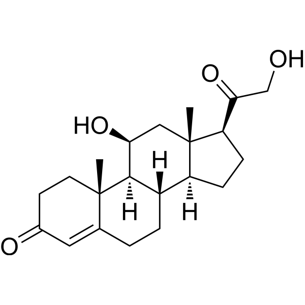
CorticosteroneEndogenous Glucocorticoids -
HY-13683
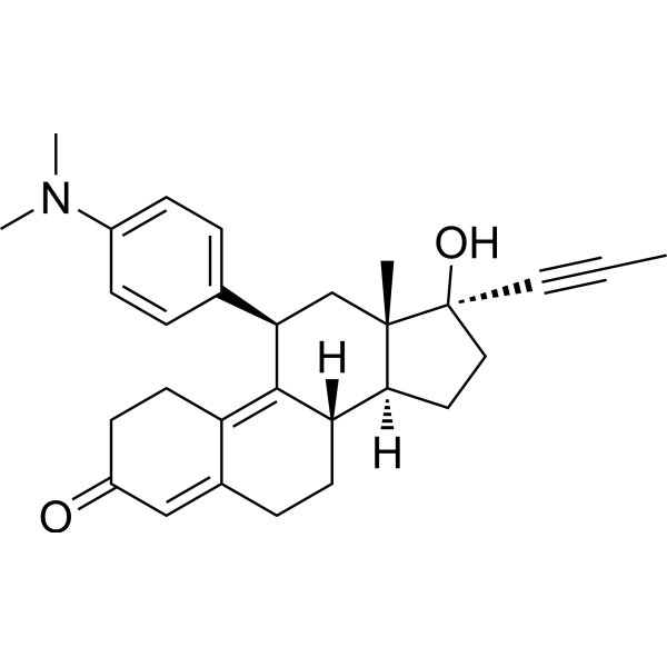
MifepristoneProgesterone Receptor Antagonist -
HY-113414
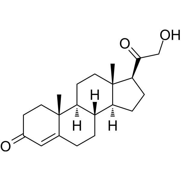
DeoxycorticosteroneMineralocorticoid Receptor Agonist -
HY-N0437
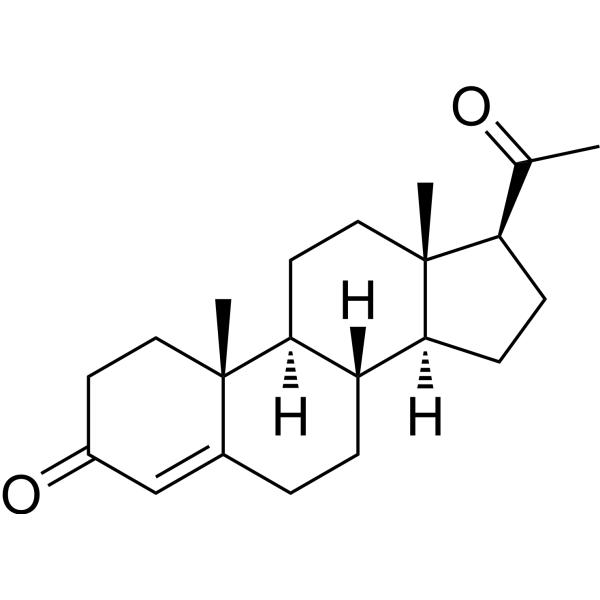
ProgesteroneEndogenous Metabolite -
HY-17461
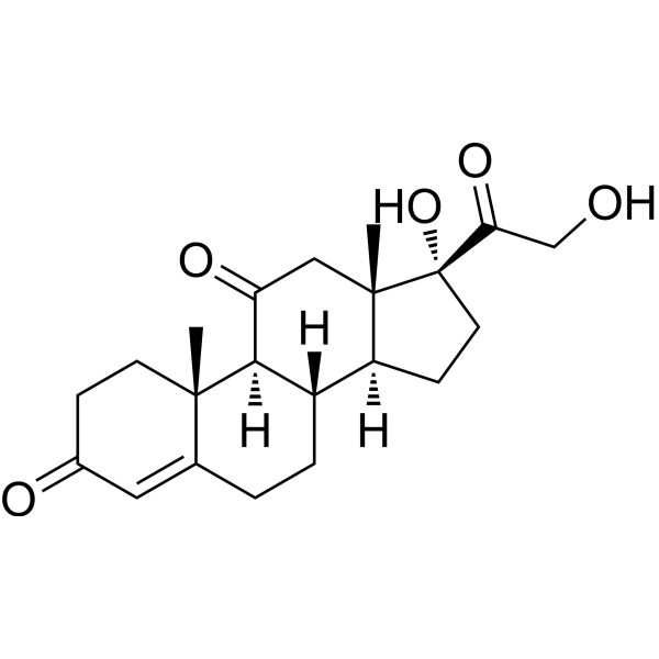
CortisoneGlucocorticoid Receptor Agonist -

HY-Y0320
Dimethyl sulfoxide, meets analytical specification of Ch.P.Aprotic Solvent -
HY-12111
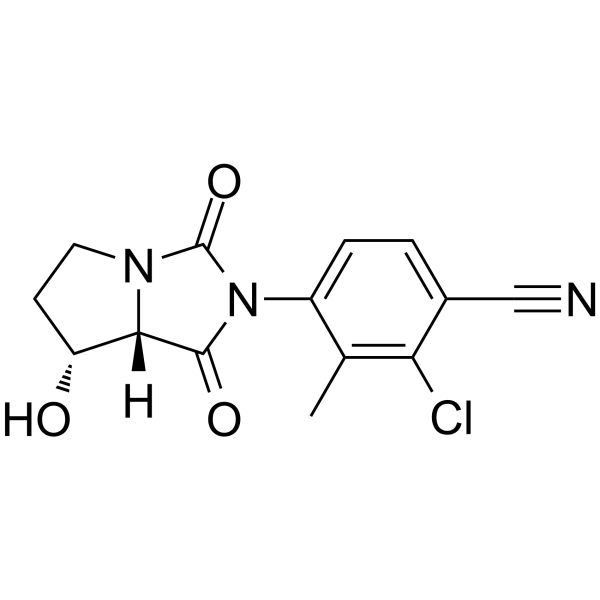
BMS-564929Androgen Receptor Agonist -
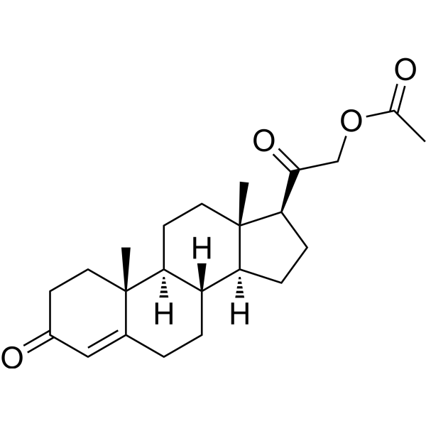
HY-B1472R
Deoxycorticosterone acetate (Standard) -
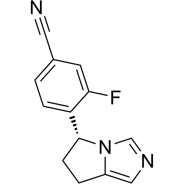
HY-16276
OsilodrostatCYP11B1/CYP11B2 Inhibitor -
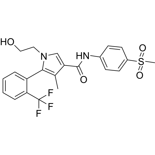
HY-100471
EsaxerenoneMineralocorticoid Antagonist -
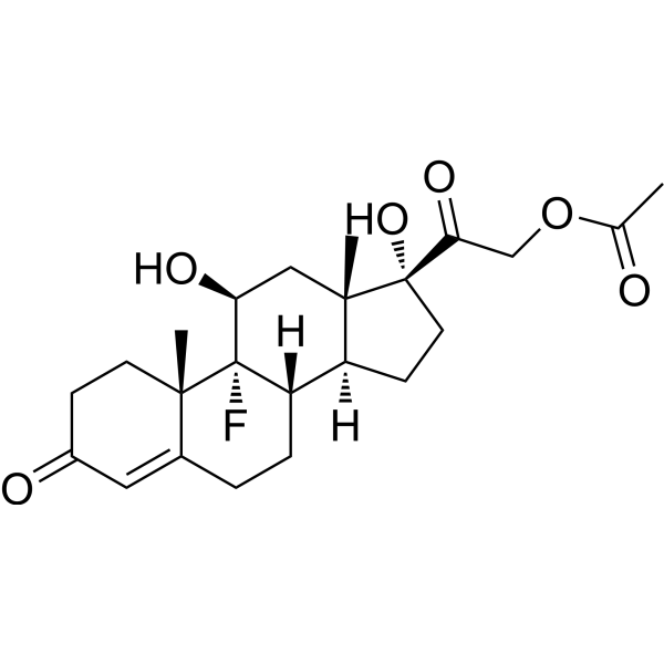
HY-B1203A
Fludrocortisone acetateMineralocorticoid
Publications Citing Use of MedChemExpress (MCE) Deoxycorticosterone acetate
More Customer Validation & Images

-
In Vivo Efficacy Study

-
Histological Imaging/Staining

-
IF

-
RT-PCR

-
WB


Biological Activity
Description
IC50 & Target
Mineralocorticoid receptor[1]
In Vivo
-
Animal Model:MR (mineralocorticoid receptor) mutant mice (MRCdh5Cre) and MR wild-type mice (MRwild-type) treated with unilateral nephrectomy (12-week-old)[1]
-
Dosage:2.5 mg/d
-
Administration:Subcutaneous implantation; 42 days; treated with 2 mg Tamoxifen (HY-13757A) (20 mg/mL in sunflower oil and 10% ethanol; i.p.; once daily on 5 consecutive days) at least 4 weeks before nephrectomy
-
Result:Increased the blood pressure without differences between both genotypes (MRCdh5Cre and MRwild-type).
Resulted glomerular injury and proteinuria, renal inflammation and fibrosis.
Induced Disease Models
Please do not refer to only one article to determine the experimental conditions. It is recommended to determine the optimal experimental conditions (animal strain, age, dosage, frequency and cycle, detection time and indicators, etc.) through preliminary experiments before the formal experiment.
Deoxycorticosterone acetate (DOCA) can be used to induce hypertension models[3][4].
Administration: 2. 100 mg sustained-release DOCA capsules were implanted subcutaneously and drinking water was replaced with 1% saline; continued for 21 days. 2. 15 mg/kg • subcutaneously implanted • drinking water was replaced with 1% saline; twice a week for 2 weeks.
MedChemExpress (MCE) has not independently confirmed the accuracy of these methods. They are for reference only.


Chemical Information
-
CAS No. 56-47-3
-
Appearance Solid
-
Molecular Weight 372.50
-
Formula C23H32O4
-
Color White to off-white
-
SMILES
C[C@@]12[C@@H](C(COC(C)=O)=O)CC[C@@]1([H])[C@]3([H])CCC4=CC(CC[C@]4(C)[C@@]3([H])CC2)=O
-
Synonyms
11-Deoxycorticosterone acetate; DOC acetate; Cortexone acetate
-
Structure Classification
-
Initial Source
-
Shipping
Room temperature in continental US; may vary elsewhere.
-
Storage
Powder -20°C 3 years 4°C 2 years In solvent -80°C 1 year -20°C 6 months


Publications (2)
-
Journal Impact Factor
-
Most Recent
-
Nat Commun
Coronavirus envelope protein activates TMED10-mediated unconventional secretion of inflammatory factors. [Abstract]2024 Oct 8;15(1):8708. PMID: 39379362 -
Phytother Res
Geniposide modulates GSK3β to inhibit Th17 differentiation and mitigate endothelial damage in intracranial aneurysm. [Abstract]2024 Nov;38(11):5184-5202. PMID: 39180344
Deoxycorticosterone acetate purchased from MedChemExpress. Usage Cited in: Phytother Res. 2024 Nov;38(11):5184-5202. [Abstract]
Changes in the aorta at the base of the mouse brain observed with body-view microscopy. Model group: Intracranial aneurysm (IA) mouse model established with multiple interventions, including daily subcutaneous injection of DOCA (Deoxycorticosterone acetate) (2.4 mg/day, dissolved in olive oil; s.c.).
Deoxycorticosterone acetate purchased from MedChemExpress. Usage Cited in: Phytother Res. 2024 Nov;38(11):5184-5202. [Abstract]
HE staining of cerebral arteries and quantification of the circumference of the Circle of Willis (n = 3 per group). Model group: Intracranial aneurysm (IA) mouse model established with multiple interventions, including daily subcutaneous injection of DOCA (Deoxycorticosterone acetate) (2.4 mg/day, dissolved in olive oil; s.c.).
Deoxycorticosterone acetate purchased from MedChemExpress. Usage Cited in: Phytother Res. 2024 Nov;38(11):5184-5202. [Abstract]
Tissue immunofluorescence detection of eNOS protein expression levels in mouse cerebral aorta. Model group: Intracranial aneurysm (IA) mouse model established with multiple interventions, including daily subcutaneous injection of DOCA (Deoxycorticosterone acetate) (2.4 mg/day, dissolved in olive oil; s.c.).
Deoxycorticosterone acetate purchased from MedChemExpress. Usage Cited in: Phytother Res. 2024 Nov;38(11):5184-5202. [Abstract]
Expression levels of GSK3B, MAPK14, IL-2, and IL-5 in transcriptome sequencing. Model group: Intracranial aneurysm (IA) mouse model established with multiple interventions, including daily subcutaneous injection of DOCA (Deoxycorticosterone acetate) (2.4 mg/day, dissolved in olive oil; s.c.).
Deoxycorticosterone acetate purchased from MedChemExpress. Usage Cited in: Phytother Res. 2024 Nov;38(11):5184-5202. [Abstract]
Western blotting detecting protein expression levels of GSK3β, STAT3, and phosphorylated STAT3. Model group: Intracranial aneurysm (IA) mouse model established with multiple interventions, including daily subcutaneous injection of DOCA (Deoxycorticosterone acetate) (2.4 mg/day, dissolved in olive oil; s.c.).


Solvent & Solubility
In Vitro:
DMSO : 16.67 mg/mL (44.75 mM; Need ultrasonic; Hygroscopic DMSO has a significant impact on the solubility of product, please use newly opened DMSO)
H2O : < 0.1 mg/mL (insoluble)
Please refer to the solubility information to select the appropriate solvent. Once prepared, please aliquot and store the solution to prevent product inactivation from repeated freeze-thaw cycles.
Storage method and period of stock solution: -80°C, 1 year; -20°C, 6 months. When stored at -80°C, please use it within 1 year. When stored at -20°C, please use it within 6 months.
Please refer to the solubility information to select the appropriate solvent. Once prepared, please aliquot and store the solution to prevent product inactivation from repeated freeze-thaw cycles.
Storage method and period of stock solution: -80°C, 1 year; -20°C, 6 months. When stored at -80°C, please use it within 1 year. When stored at -20°C, please use it within 6 months.
Concentration (start) × Volume (start) = Concentration (final) × Volume (final)
In Vivo:
Select the appropriate dissolution method based on your experimental animal and administration route.
- For the following dissolution methods, please ensure to first prepare a clear stock solution using an In Vitro approach and then sequentially add co-solvents:
- To ensure reliable experimental results, the clarified stock solution can be appropriately stored based on storage conditions. As for the working solution for In Vivo experiments, it is recommended to prepare freshly and use it on the same day.
- The percentages shown for the solvents indicate their volumetric ratio in the final prepared solution. If precipitation or phase separation occurs during preparation, heat and/or sonication can be used to aid dissolution.
Add each solvent one by one: 10% DMSO 90% (20% SBE-β-CD in Saline)
Solubility: ≥ 2.08 mg/mL (5.58 mM); Clear solution
This protocol yields a clear solution of ≥ 2.08 mg/mL (saturation unknown).
Taking 1 mL working solution as an example, add 100 μL DMSO stock solution (20.8 mg/mL) to 900 μL 20% SBE-β-CD in Saline, and mix evenly.
Preparation of 20% SBE-β-CD in Saline (4°C, storage for one week): 2 g SBE-β-CD powder is dissolved in 10 mL Saline, completely dissolve until clear.
Add each solvent one by one: 10% DMSO 40% PEG300 5% Tween-80 45% Saline
Solubility: ≥ 1.67 mg/mL (4.48 mM); Clear solution
This protocol yields a clear solution of ≥ 1.67 mg/mL (saturation unknown).
Taking 1 mL working solution as an example, add 100 μL DMSO stock solution (16.7 mg/mL) to 400 μL PEG300, and mix evenly; then add 50 μL Tween-80 and mix evenly; then add 450 μL Saline to adjust the volume to 1 mL.
Preparation of Saline: Dissolve 0.9 g sodium chloride in ddH₂O and dilute to 100 mL to obtain a clear Saline solution.
In Vivo Dissolution Calculator
Please enter the basic information of animal experiments:
-
-
-
-
Recommended: Prepare an additional quantity of animals to account for potential losses during experiments.
Please enter your animal formula composition:
-
%DMSO +
Recommended: Keep the proportion of DMSO in working solution below 2% if your animal is weak.
-
%+
-
+%Tween-80 + +
-
%Saline +
The co-solvents required include: DMSO, . All of co-solvents are available by MedChemExpress (MCE). , Tween 80. All of co-solvents are available by MedChemExpress (MCE).
Working solution concentration: 0.22 mg/mL
Method for preparing stock solution: mg drug dissolved in μL DMSO. Stock solution concentration: mg/mL.
1. Take μL DMSO stock solution;
2. Add μL .
μL , mix evenly;
3. Then add μL Tween 80, mix evenly;
4. Then add μL
Please ensure that the stock solution in the first step is dissolved to a clear state, and add co-solvents in sequence. You can use ultrasonic heating (ultrasonic cleaner, recommended frequency 20-40 kHz), vortexing, etc. to assist dissolution.


Purity & Documentation
-
Data Sheet (295 KB)
-
SDS (419 KB)
- English - EN (419 KB)
- Français - FR (419 KB)
- Deutsch - DE (419 KB)
- Norwegian - NO (419 KB)
- Español - ES (419 KB)
- Swedish - SV (419 KB)
- Italian - IT (419 KB)
- Korean - KR (419 KB)
- Portuguese - PT (419 KB)
-
Handling Instructions (2659 KB)
References
[1]. Lother A, et al. Deoxycorticosterone Acetate/Salt-Induced Cardiac But Not Renal Injury Is Mediated By Endothelial Mineralocorticoid Receptors Independently From Blood Pressure. Hypertension. 2016 Jan;67(1):130-8. [Content Brief]
[2]. Lu NZ, et al. International Union of Pharmacology. LXV. The pharmacology and classification of the nuclear receptor superfamily: glucocorticoid, mineralocorticoid, progesterone, and androgen receptors. Pharmacol Rev. 2006 Dec;58(4):782-97. [Content Brief]
[3]. M J Somers, et al. Vascular Superoxide Production and Vasomotor Function in Hypertension Induced by Deoxycorticosterone Acetate–Salt. Circulation. 2000 Apr 11;101(14):1722-8. [Content Brief]
[4]. Y Matsumura, et al. Different Contributions of Endothelin-A and Endothelin-B Receptors in the Pathogenesis of Deoxycorticosterone Acetate–Salt–Induced Hypertension in Rats. Hypertension. 1999 Feb;33(2):759-65. [Content Brief]


Complete Stock Solution Preparation Table
Please refer to the solubility information to select the appropriate solvent. Once prepared, please aliquot and store the solution to prevent product inactivation from repeated freeze-thaw cycles.
Storage method and period of stock solution: -80°C, 1 year; -20°C, 6 months. When stored at -80°C, please use it within 1 year. When stored at -20°C, please use it within 6 months.
| Optional Solvent | Concentration Solvent Mass | 1 mg | 5 mg | 10 mg | 25 mg |
|---|---|---|---|---|---|
| DMSO | 1 mM | 2.6846 mL | 13.4228 mL | 26.8456 mL | 67.1141 mL |
| 5 mM | 0.5369 mL | 2.6846 mL | 5.3691 mL | 13.4228 mL | |
| 10 mM | 0.2685 mL | 1.3423 mL | 2.6846 mL | 6.7114 mL | |
| 15 mM | 0.1790 mL | 0.8949 mL | 1.7897 mL | 4.4743 mL | |
| 20 mM | 0.1342 mL | 0.6711 mL | 1.3423 mL | 3.3557 mL | |
| 25 mM | 0.1074 mL | 0.5369 mL | 1.0738 mL | 2.6846 mL | |
| 30 mM | 0.0895 mL | 0.4474 mL | 0.8949 mL | 2.2371 mL | |
| 40 mM | 0.0671 mL | 0.3356 mL | 0.6711 mL | 1.6779 mL |


Deoxycorticosterone acetate Related Classifications
HY-B1472 Related Classifications
 Powered by Bioz