



Histatin 5
Histatin 5 inhibits the activity of the host matrix metalloproteinases MMP-2 and MMP-9 with IC50s of 0.57 and 0.25 μM, respectively.
연구목적의 판매만을 진행합니다. 환자를 대상으로 한 판매는 하지 않습니다.
- CAS No.: 115966-68-2
- 화학식: C133H195N51O33
- 분자량:3036.29
-
보관:
Please store the product under the recommended conditions in the Certificate of Analysis.


To place orders, for customer services and technical support, please contact: MedChemExpress USA
Tel: 609-228-6898 E-mail: [email protected] [email protected]


-
 Biological Activity
-
Chemical Information
-
용액&용해도
- Protocol
- 순도&문서
- References
-
 Help & FAQs


-
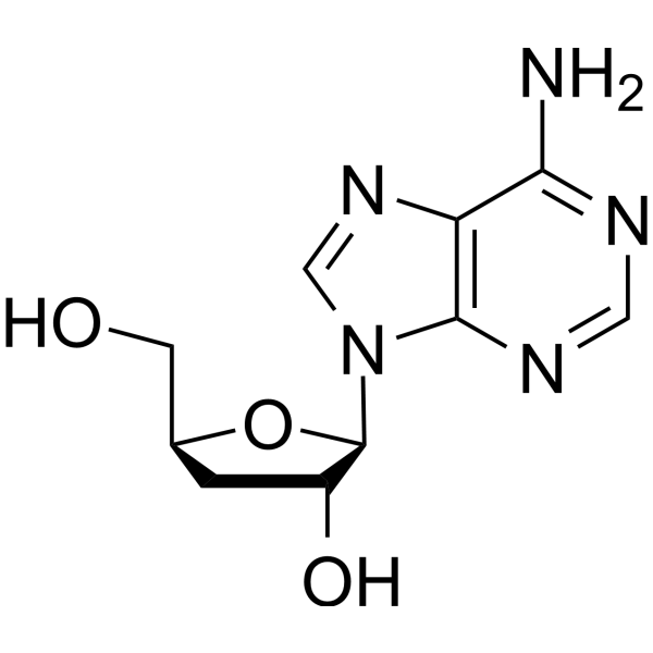
HY-N0262
CordycepinBacterial Inhibitor -
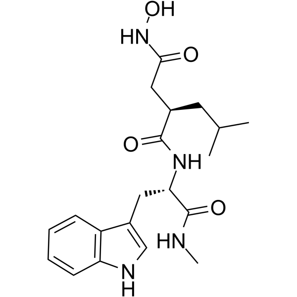
HY-15768
IlomastatMMP Inhibitor -
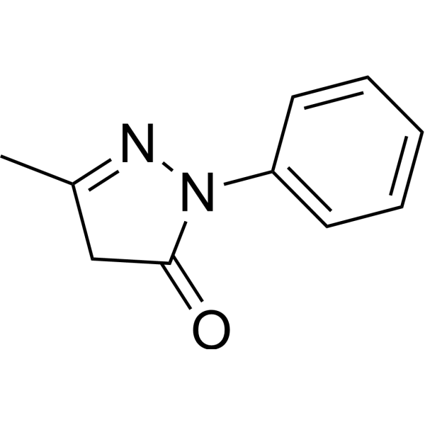
HY-B0099
EdaravoneApoptosis Inducer -
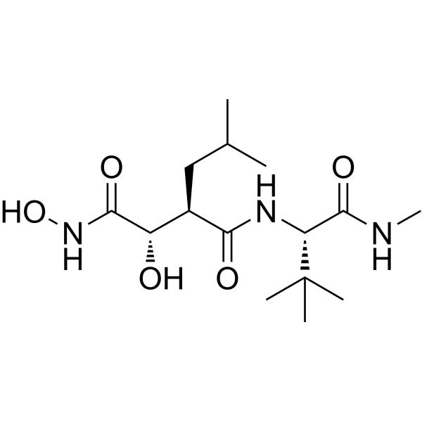
HY-12169
MarimastatMMP Inhibitor -
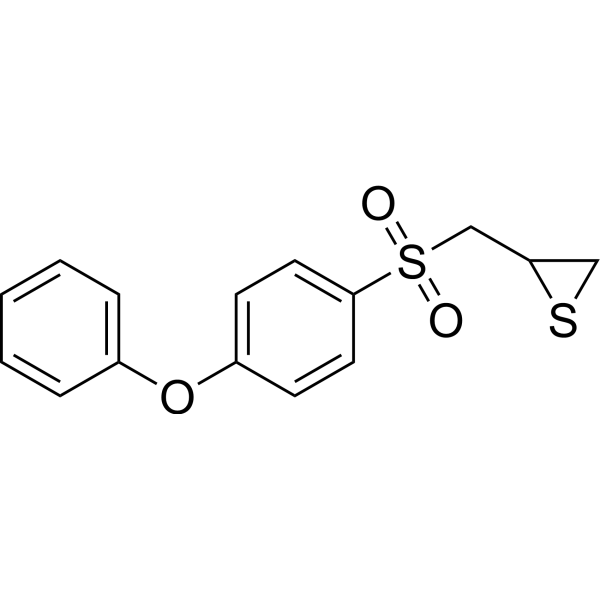
HY-12354
SB-3CTMMP2/9 Inhibitor -
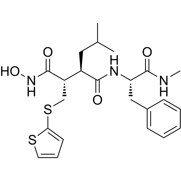
HY-13564
BatimastatMMP Inhibitor -
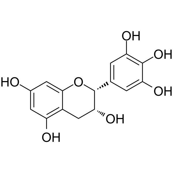
HY-N0225
(-)-EpigallocatechinAutophagy Inducer -
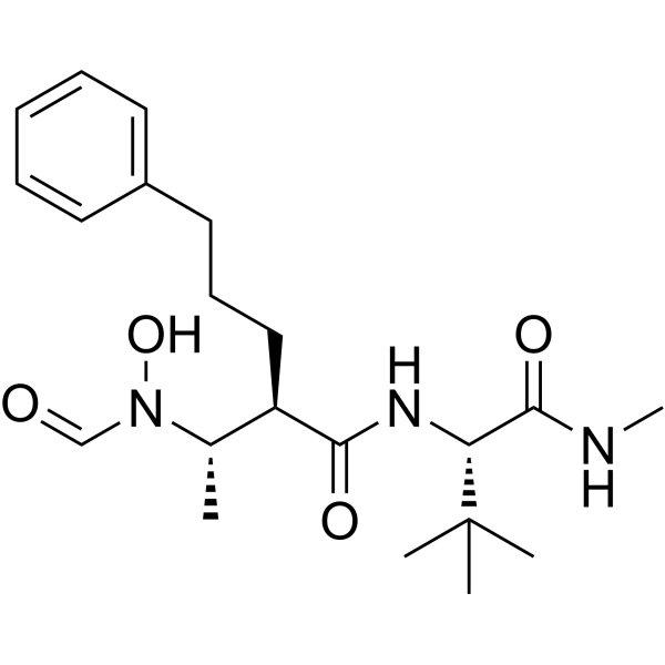
HY-19956
GI254023XADAM10/MMP9 Inhibitor -
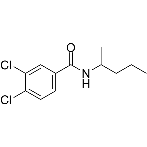
HY-15827
NSC 405020MMP14 Inhibitor -
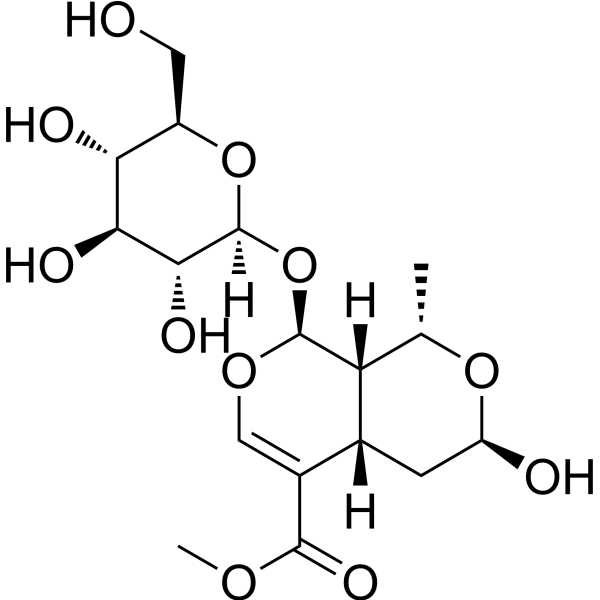
HY-N0532
MorronisidePyroptosis Inhibitor -
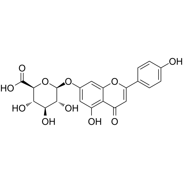
HY-N1454
Apigenin-7-glucuronideMMP Inhibitor -
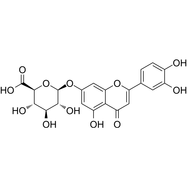
HY-N1463
Luteolin 7-O-glucuronideMMP Inhibitor


Biological Activity
제품 설명
IC50 & Target
IC50: 0.57 μM (MMP-2), 0.25 μM (MMP-9)[1]
In Vitro
Histatin 5 is a member of a family of low-molecular-weight salivary proteins secreted by parotid, submandibular, and sublingual glands. Using biotinylated gelatin as a substrate, Histatin 5 inhibits the activity of the host matrix metalloproteinases MMP-2 and MMP-9 with IC50s of 0.57 and 0.25 μM, respectively. To localize the domain responsible for this inhibition, three peptides containing different regions of Histatin 5 are synthesized and tested as inhibitors of MMP-9. Peptides comprising residues 1 to 14 and residues 4 to 15 of Histatin 5 show much lower inhibitory activities (IC50, 21.4 and 20.5 μM, respectively), while a peptide comprising residues 9 to 22 showed identical activity to Histatin 5 against MMP-9. Kinetic analysis of the inhibition of the Arg-gingipain reveals that Histatin 5 is a competitive inhibitor, affecting only the Km with a Ki of 15 μM[1]. Histatin 5 is an inhibitor of mitochondrial respiration.The human salivary antifungal peptide Histatin 5 is taken up by Candida albicans cells and associates intracellularly with mitochondria. Histatin 5 inhibits respiration of isolated C. albicans mitochondria as well as the respiration of intact blastoconidia in a dose and time-dependent manner. Histatin 5 at a concentration of 33 μM inhibits state 2 respiration[2].
MedChemExpress (MCE) has not independently confirmed the accuracy of these methods. They are for reference only. Further protocols information, click here.


Chemical Information
-
CAS No. 115966-68-2
-
분자량 3036.29
-
화학식 C133H195N51O33
-
Sequence
Asp-Ser-His-Ala-Lys-Arg-His-His-Gly-Tyr-Lys-Arg-Lys-Phe-His-Glu-Lys-His-His-Ser-His-Arg-Gly-Tyr
-
Sequence Shortening
DSHAKRHHGYKRKFHEKHHSHRGY
-
선적
Room temperature in continental US; may vary elsewhere.
-
보관
Please store the product under the recommended conditions in the Certificate of Analysis.


용액&용해도
In Vitro
H2O
Peptide Solubility and Storage Guidelines:
1. Calculate the length of the peptide.
2. Calculate the overall charge of the entire peptide according to the following table:
| Contents | Assign value | |
|---|---|---|
| Acidic amino acid | Asp (D), Glu (E), and the C-terminal -COOH. | -1 |
| Basic amino acid | Arg (R), Lys (K), His (H), and the N-terminal -NH2 | +1 |
| Neutral amino acid | Gly (G), Ala (A), Leu (L), Ile (I), Val (V), Cys (C), Met (M), Thr (T), Ser (S), Phe (F), Tyr (Y), Trp (W), Pro (P), Asn (N), Gln (Q) | 0 |
3. Recommended solution:
| Overall charge of peptide | Details |
|---|---|
| Negative (<0) |
1. Try to dissolve the peptide in water first. 2. If water fails, add NH4OH (<50 μL). 3. If the peptide still does not dissolve, add DMSO (50-100 μL) to solubilize the peptide. |
| Positive (>0) |
1. Try to dissolve the peptide in water first. 2. If water fails, try dissolving the peptide in a 10%-30% acetic acid solution. 3. If the peptide still does not dissolve, try dissolving the peptide in a small amount of DMSO. |
| Zero (=0) |
1. Try to dissolve the peptide in organic solvent (acetonitrile, methanol, etc.) first. 2. For very hydrophobic peptides, try dissolving the peptide in a small amount of DMSO, and then dilute the solution with water to the desired concentration. |

Protocol
-
Mitochondrial membrane-potential fluorescent assay
Mitochondrial membrane potential fluorescent assays estimate ΔΨm in living cells using lipophilic cationic dyes such as TMRM, TMRE, rhodamine 123, and JC-1, which accumulate in mitochondria according to membrane polarization; loss of signal after FCCP or CCCP treatment is interpreted as mitochondrial depolarization. TMRM/TMRE and rhodamine 123 are commonly used for semi-quantitative live-cell microscopy or flow cytometry, while JC-1 can report a shift from red aggregate fluorescence to green monomer fluorescence during depolarization; interpretation requires controls because dye concentration, quenching mode, cell type, dye efflux, and mitochondrial mass can affect fluorescence independently of ΔΨm.
-
Mitochondrial membrane-potential and mitochondrial mass staining
Mitochondrial membrane potential staining measures the electrochemical polarization across the mitochondrial inner membrane in live cells using lipophilic cationic fluorescent probes; early rhodamine-based work showed that selective mitochondrial dye accumulation is lost when the mitochondrial transmembrane potential is dissipated. JC-1 reports mitochondrial polarization by shifting from green monomer fluorescence to red J-aggregate fluorescence as dye concentration increases within energized mitochondria; therefore, the red/green fluorescence ratio is used as a relative readout of mitochondrial membrane potential. TMRE or TMRM staining provides a single-channel relative readout because these cationic rhodamine esters accumulate in polarized mitochondria, and lower fluorescence indicates reduced mitochondrial polarization when acquisition and dye-loading conditions are controlled. Mitochondrial mass staining is commonly performed with MitoTracker Green FM or related MitoTracker dyes as


순도&문서
References
[1]. Gusman H, et al. Salivary histatin 5 is an inhibitor of both host and bacterial enzymes implicated in periodontaldisease. Infect Immun. 2001 Mar;69(3):1402-8. [Content Brief]
[2]. Helmerhorst EJ, et al. The human salivary peptide histatin 5 exerts its antifungal activity through the formation ofreactive oxygen species. Proc Natl Acad Sci U S A. 2001 Dec 4;98(25):14637-42. [Content Brief]

Calculators
Concentration (start) × Volume (start) = Concentration (final) × Volume (final)


Histatin 5 Related Classifications
HY-P0273 Related Classifications
 Powered by Bioz