



Kisspeptin-10, human
Based on 2 publication(s) in Google Scholar
Kisspeptin-10, human is a potent vasoconstrictor and inhibitor of angiogenesis. Kisspeptin-10, human acts as a tumor metastasis suppressor via its receptor GPR54. Kisspeptin-10-GPR54 system plays an important role in embryonic kidney development. Kisspeptin-10/GPR54 signaling induces osteoblast differentiation via NFATc4-mediated BMP2 expression.
Nur für Forschungszwecke. Wir verkaufen nicht an Patienten.
- Reinheit : 99.86%
- CAS. Nr.: 374675-21-5
- Formel: C63H83N17O14
- Molecular Weight:1302.44
-
Speicherung:
Sealed storage, away from moisture and light.
Powder -80°C, 2 years , -20°C, 1 year* In solvent : -80°C, 6 months; -20°C, 1 month (sealed storage, away from moisture and light)


To place orders, for customer services and technical support, please contact: MedChemExpress USA
Tel: 609-228-6898 E-mail: [email protected] [email protected]


-
 Biologische Aktivität
-
Chemical Information
-
Lösungsmittel & Löslichkeit
- Protokoll
- Reinheit & Dokumentation
- Verweise
-
 Help & FAQs


-
HY-P1022
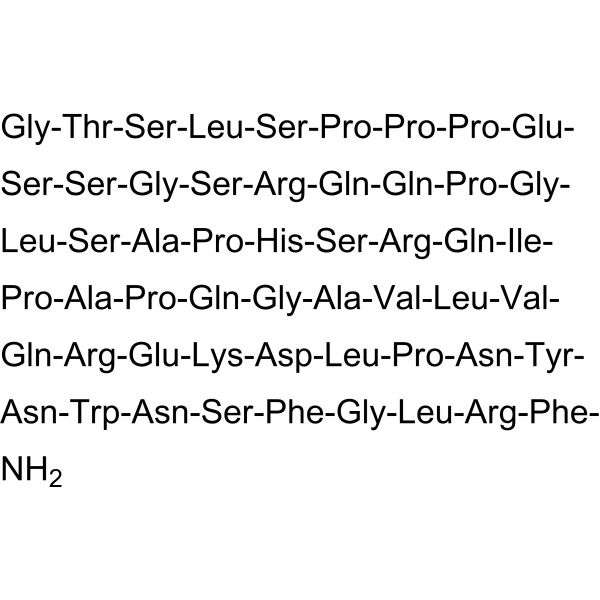
Kisspeptin-54(human)Endogenous Ligand for GPR54 -

HY-P1222
LL-37, humanAntimicrobial Peptide -
HY-19822
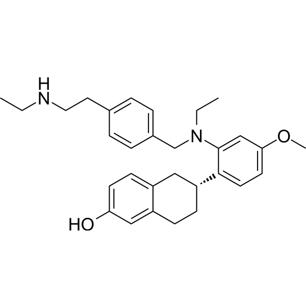
ElacestrantEstrogen Receptor Degrader -
HY-P2048
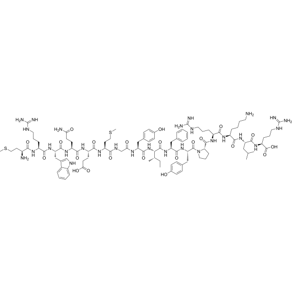
MOTS-c (human)Bioactive Peptide -
HY-18678
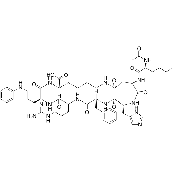
BremelanotideMelanocortin Receptor Agonist -
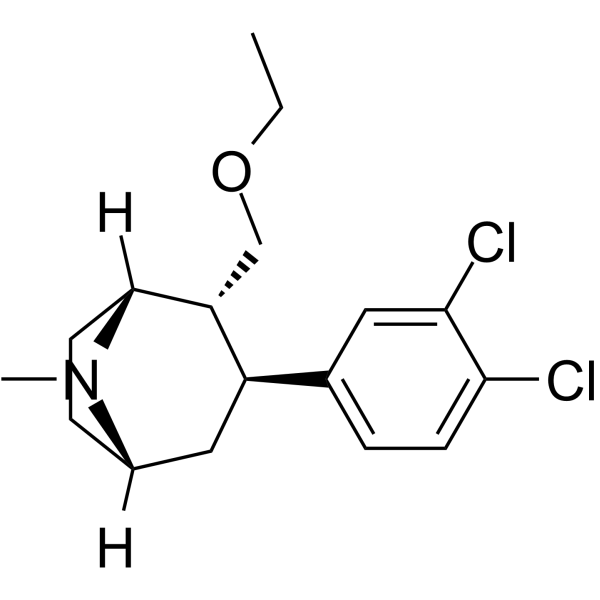
HY-14472
TesofensineDA/NE/5-HT Inhibitor -
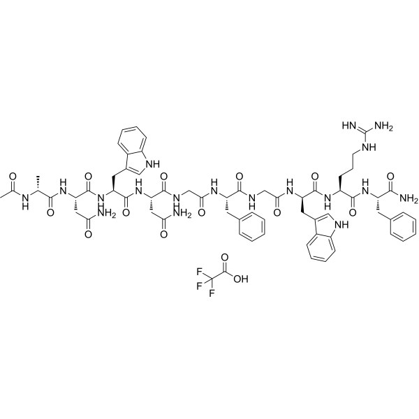
HY-P1196A
Kisspeptin 234 TFAKISS1/GPR54 Antagonist -
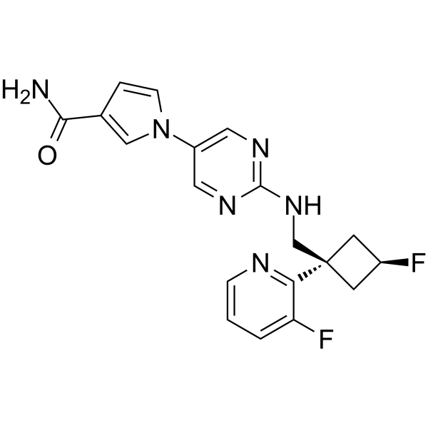
HY-109121
ReldesemtivFast Skeletal Muscle Troponin Activator -
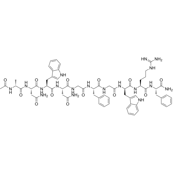
HY-P1196
Kisspeptin 234KISS1/GPR54 Antagonist -
HY-P1197
Kisspeptin-10 (mouse, rat)GPR54 Ligand
Publications Citing Use of MedChemExpress (MCE) Kisspeptin-10, human
More

Biologische Aktivität
Beschreibung
IC50 & Target
Cellular Effect
|
Cell Line

|
Type | Value | Description | References |
|---|---|---|---|---|
| CHO | EC50 |
0.087 nM
Compound: Metastin
|
Agonist activity at human KISS1R expressed in CHO cells assessed as increase in calcium mobilization measured at 2 secs interval for 2 mins by fluo-4 NW dye based FLIPR assay
Agonist activity at human KISS1R expressed in CHO cells assessed as increase in calcium mobilization measured at 2 secs interval for 2 mins by fluo-4 NW dye based FLIPR assay
|
[PMID: 30598349] |
| CHO | EC50 |
0.24 nM
Compound: kisspeptin-10
|
Agonist activity at human GPR54 expressed in CHO cells assessed as effect on kisspeptin-10-induced response by FLIPR assay
Agonist activity at human GPR54 expressed in CHO cells assessed as effect on kisspeptin-10-induced response by FLIPR assay
|
[PMID: 19007202] |
| CHO | EC50 |
0.96 nM
Compound: Metastin(45-54)
|
Agonist activity at human KISS1R transfected in CHO cells assessed as intracellular calcium flux after 24 hrs by functional FLIPR assay
Agonist activity at human KISS1R transfected in CHO cells assessed as intracellular calcium flux after 24 hrs by functional FLIPR assay
|
[PMID: 24918545] |
| CHO | EC50 |
4.2 nM
Compound: Metastin(45-54)
|
Agonist activity at rat KISS1R transfected in CHO cells assessed as intracellular calcium flux after 24 hrs by functional FLIPR assay
Agonist activity at rat KISS1R transfected in CHO cells assessed as intracellular calcium flux after 24 hrs by functional FLIPR assay
|
[PMID: 24918545] |
| CHO | IC50 |
0.07 nM
Compound: kisspeptin-10
|
Displacement of [125I]kisspeptin-15 from human GPR54 expressed in CHO cell membrane
Displacement of [125I]kisspeptin-15 from human GPR54 expressed in CHO cell membrane
|
[PMID: 19007202] |
| CHO | IC50 |
8510 nM
Compound: Metastin(45-54)
|
Inhibition of human KISS1R expressed in CHO cells assessed as inhibition of KISS1R-mediated cell growth after 4 days by cell counting kit-8 assay
Inhibition of human KISS1R expressed in CHO cells assessed as inhibition of KISS1R-mediated cell growth after 4 days by cell counting kit-8 assay
|
[PMID: 24047141] |
| HEK293 | EC50 |
17.3 nM
Compound: 10-mer, metastin-10
|
Activity at metastin receptor expressed in HEK293 cells by measuring calcium release
Activity at metastin receptor expressed in HEK293 cells by measuring calcium release
|
[PMID: 17266198] |
In Vitro
Kisspeptin-10 (KP-10) and its receptor GPR54 are key components in the regulation of GnRH secretion in humans and other mammals. Kisspeptin-10 protein binds to GPR54. Activation of Kisspeptin-10 suppresses pulmonary human melanoma and Kisspeptin-10 is a metastasis suppressor in breast cancer cells[1].
MedChemExpress (MCE) has not independently confirmed the accuracy of these methods. They are for reference only.


Chemical Information
-
CAS. Nr. 374675-21-5
-
Appearance Solid
-
Molecular Weight 1302.44
-
Formel C63H83N17O14
-
Color White to off-white
-
Sequence
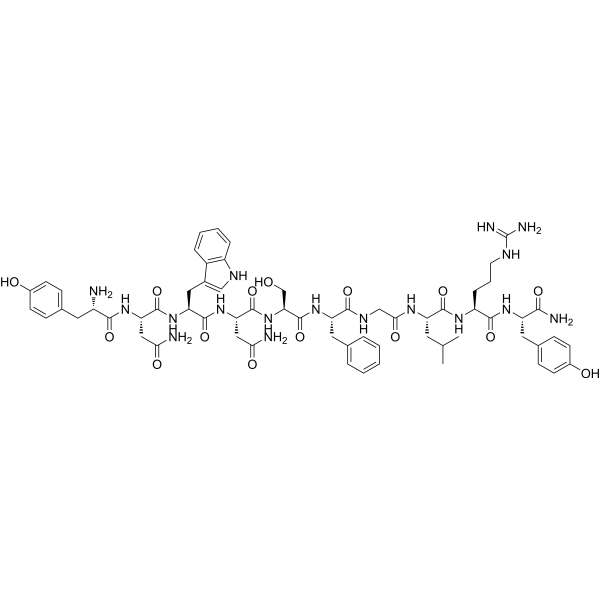
Tyr-Asn-Trp-Asn-Ser-Phe-Gly-Leu-Arg-Phe-NH2
-
Sequence Shortening
YNWNSFGLRF-NH2
-
Versand
Room temperature in continental US; may vary elsewhere.
-
Speicherung
Sealed storage, away from moisture and light
Powder -80°C 2 years -20°C 1 year * In solvent : -80°C, 6 months; -20°C, 1 month (sealed storage, away from moisture and light)


Publications (2)
-
Journal Impact Factor
-
Most Recent
-
ACS Pharmacol Transl Sci
Critical Residues in the Gβ Subunit Mediate Selective Recruitment of the Gαq Heterotrimer to Activated G Protein-Coupled Receptors. [Abstract]2025 Jun 6;8(7):2048-2060. PMID: 40672669 -
Drug Test Anal
Analysis and Characterization of Kisspeptin and Its Analogues in Serum and Urine Samples by Liquid Chromatography-High-Resolution Mass Spectrometry for Doping Control Purposes. [Abstract]2026 Apr 29. PMID: 42057309


Lösungsmittel & Löslichkeit
In Vitro:
H2O : 10 mg/mL (7.68 mM; Need ultrasonic)
Please refer to the solubility information to select the appropriate solvent. Once prepared, please aliquot and store the solution to prevent product inactivation from repeated freeze-thaw cycles.
Storage method and period of stock solution: -80°C, 6 months; -20°C, 1 month (sealed storage, away from moisture and light). When stored at -80°C, please use it within 6 months. When stored at -20°C, please use it within 1 month.
* Note: If you choose water as the stock solution, please dilute it to the working solution, then filter and sterilize it with a 0.22 μm filter before use.
Please refer to the solubility information to select the appropriate solvent. Once prepared, please aliquot and store the solution to prevent product inactivation from repeated freeze-thaw cycles.
Storage method and period of stock solution: -80°C, 6 months; -20°C, 1 month (sealed storage, away from moisture and light). When stored at -80°C, please use it within 6 months. When stored at -20°C, please use it within 1 month.
* Note: If you choose water as the stock solution, please dilute it to the working solution, then filter and sterilize it with a 0.22 μm filter before use.
Konzentration (Stammlösung) × Volumen (Stammlösung) = Konzentration (Ziellösung) × Volumen (Ziellösung)


Reinheit & Dokumentation
-
Data Sheet (270 KB)
-
SDS (252 KB)
- English - EN (252 KB)
- Français - FR (252 KB)
- Deutsch - DE (252 KB)
- Norwegian - NO (252 KB)
- Español - ES (252 KB)
- Swedish - SV (252 KB)
- Italian - IT (252 KB)
- Korean - KR (252 KB)
- Portuguese - PT (252 KB)
-
Handling Instructions (2659 KB)
Verweise


Complete Stock Solution Preparation Table
Please refer to the solubility information to select the appropriate solvent. Once prepared, please aliquot and store the solution to prevent product inactivation from repeated freeze-thaw cycles.
Storage method and period of stock solution: -80°C, 6 months; -20°C, 1 month (sealed storage, away from moisture and light). When stored at -80°C, please use it within 6 months. When stored at -20°C, please use it within 1 month.
| Optional Solvent | Concentration Solvent Mass | 1 mg | 5 mg | 10 mg | 25 mg |
|---|---|---|---|---|---|
| H2O | 1 mM | 0.7678 mL | 3.8389 mL | 7.6779 mL | 19.1947 mL |
| 5 mM | 0.1536 mL | 0.7678 mL | 1.5356 mL | 3.8389 mL |
* Note: If you choose water as the stock solution, please dilute it to the working solution, then filter and sterilize it with a 0.22 μm filter before use.


Kisspeptin-10, human Related Classifications
HY-P0254 Related Classifications
 Powered by Bioz