



Kisspeptin-10, human TFA
Based on 2 publication(s) in Google Scholar
Kisspeptin-10, human TFA is a potent vasoconstrictor and inhibitor of angiogenesis. Kisspeptin-10, human TFA acts as a tumor metastasis suppressor via its receptor GPR54. Kisspeptin-10-GPR54 system plays an important role in embryonic kidney development. Kisspeptin-10/GPR54 signaling induces osteoblast differentiation via NFATc4-mediated BMP2 expression.
For research use only. We do not sell to patients.
- Purity : 99.56%
- Formula: C63H83N17O14.C2HF3O2
- Molecular Weight:1416.46
-
Storage:
Sealed storage, away from moisture.
Powder -80°C, 2 years , -20°C, 1 year* In solvent : -80°C, 6 months; -20°C, 1 month (sealed storage, away from moisture)

To place orders, for customer services and technical support, please contact: MedChemExpress USA
Tel: 609-228-6898 E-mail: [email protected] [email protected]


-
 Biological Activity
-
Chemical Information
-
Solvent & Solubility
- Protocols
- Purity & Documentation
- References
-
 Help & FAQs



Publications Citing Use of MedChemExpress (MCE) Kisspeptin-10, human TFA
More

Biological Activity
Description
IC50 & Target
In Vitro
Kisspeptin-10 (KP-10) and its receptor GPR54 are key components in the regulation of GnRH secretion in humans and other mammals. Kisspeptin-10 protein binds to GPR54. Activation of Kisspeptin-10 suppresses pulmonary human melanoma and Kisspeptin-10 is a metastasis suppressor in breast cancer cells[1].
MedChemExpress (MCE) has not independently confirmed the accuracy of these methods. They are for reference only. Further protocols information, click here.
In Vivo
MedChemExpress (MCE) has not independently confirmed the accuracy of these methods. They are for reference only.


Chemical Information
-
Appearance Solid
-
Molecular Weight 1416.46
-
Formula C63H83N17O14.C2HF3O2
-
Color White to off-white
-
Sequence Shortening
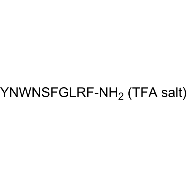
YNWNSFGLRF-NH2
-
Shipping
Room temperature in continental US; may vary elsewhere.
-
Storage
Sealed storage, away from moisture
Powder -80°C 2 years -20°C 1 year * In solvent : -80°C, 6 months; -20°C, 1 month (sealed storage, away from moisture)


Publications (2)
-
Journal Impact Factor
-
Most Recent
-
ACS Pharmacol Transl Sci
Critical Residues in the Gβ Subunit Mediate Selective Recruitment of the Gαq Heterotrimer to Activated G Protein-Coupled Receptors. [Abstract]2025 Jun 6;8(7):2048-2060. PMID: 40672669 -
Drug Test Anal
Analysis and Characterization of Kisspeptin and Its Analogues in Serum and Urine Samples by Liquid Chromatography-High-Resolution Mass Spectrometry for Doping Control Purposes. [Abstract]2026 Apr 29. PMID: 42057309


Solvent & Solubility
In Vitro:
DMSO : 100 mg/mL (70.60 mM; Need ultrasonic; Hygroscopic DMSO has a significant impact on the solubility of product, please use newly opened DMSO)
Please refer to the solubility information to select the appropriate solvent. Once prepared, please aliquot and store the solution to prevent product inactivation from repeated freeze-thaw cycles.
Storage method and period of stock solution: -80°C, 6 months; -20°C, 1 month (sealed storage, away from moisture). When stored at -80°C, please use it within 6 months. When stored at -20°C, please use it within 1 month.
Please refer to the solubility information to select the appropriate solvent. Once prepared, please aliquot and store the solution to prevent product inactivation from repeated freeze-thaw cycles.
Storage method and period of stock solution: -80°C, 6 months; -20°C, 1 month (sealed storage, away from moisture). When stored at -80°C, please use it within 6 months. When stored at -20°C, please use it within 1 month.
Concentration (start) × Volume (start) = Concentration (final) × Volume (final)

Protocols
-
RNA extraction experimental
By lysing cells, releasing RNA, and removing impurities such as proteins and DNA, high-purity RNA products are finally obtained. The commonly used traditional method is the guanidine isothiocyanate/phenol/chloroform method (Trizol), which is suitable for a variety of animal materials including animal tissues, microorganisms, cultured cells, etc., and most plant materials.
-
Human pluripotent stem cell endothelial-cell differentiation
Human pluripotent stem cell endothelial differentiation is based on stepwise developmental patterning: early activation of WNT/GSK3β inhibition promotes mesodermal or vascular progenitor entry, followed by endothelial specification using VEGF-related signaling, BMP4, FGF2, Notch modulation, or cAMP depending on the published protocol. Endothelial differentiation is read out by acquisition of CD31, CD34, VE-cadherin/CD144, KDR/VEGFR2, vWF, Tie2, NOS3, acetylated LDL uptake, tube/network formation, barrier function, and in vivo vessel-forming capacity where tested.
-
Vascular/Branching Fractal Analysis
Vascular/branching fractal analysis quantifies the geometric complexity of vessel trees or vascular networks from segmented 2D images, commonly by converting vessels into binary and/or skeletonized maps and estimating fractal dimension using box-counting or related approaches. Fractal dimension is interpreted as an image-derived readout of vascular branching complexity, space filling, or density, and has been applied to retinal photographs, fluorescein angiography, OCT angiography, capillary perfusion maps, and in vitro Matrigel angiogenesis networks. The assay readout is generated from vessel-positive pixels after image preprocessing, vessel segmentation, binarization, and optional skeletonization; reported outputs include fractal dimension, vessel density, branchpoint density, endpoint density, vessel length density, tortuosity, and generation-based branching metrics when VESGEN-style analysis is used. The biological interpretation is limited to quantitative vascular patterning and s
-
Endothelial Tube Formation Assay
Endothelial tube formation assay evaluates the ability of endothelial cells to attach, migrate, align, and organize into capillary-like networks when cultured on gelled basement membrane extract or Matrigel; the readout is the morphology and quantity of tube-like networks, which reflects an in vitro endothelial morphogenesis step related to angiogenesis. Basement membrane extract/Matrigel provides laminin-rich extracellular matrix cues that support endothelial differentiation into capillary-like structures, but it can contain biologically active growth factors, so growth-factor-reduced matrix is preferred when testing defined angiogenic stimulators or inhibitors.
-
Endothelial Cell Migration/Angiogenic Sprouting Assay
Endothelial cell migration and angiogenic sprouting assays are in vitro (and partially ex vivo-adapted) functional models that quantify the ability of endothelial cells to undergo coordinated migration, extracellular matrix invasion, and multicellular organization into capillary-like sprouts in response to pro-angiogenic stimuli such as VEGF, bFGF, or conditioned microenvironments. These assays are used to model early angiogenic events including tip-cell formation, directional migration, and lumen-like sprout extension, which collectively reflect angiogenic activation and vascular morphogenesis processes observed in vivo.
-
Orthotopic Cell-Line Xenograft
Orthotopic cell-line xenograft models involve implantation of human cancer cell lines into the anatomically corresponding organ of immunodeficient mice to reproduce tumor growth within a native microenvironment, enabling more clinically relevant tumor behavior compared with subcutaneous models. These models are widely used because orthotopic placement better recapitulates tumor progression, including invasion and metastatic spread, which are often underrepresented in heterotopic implantation systems. Compared with conventional xenografts, orthotopic implantation is described as more technically complex but provides improved simulation of tumor-microenvironment interactions and metastatic behavior, making it particularly valuable for translational oncology research. Surgical orthotopic implantation approaches have been emphasized as enabling faithful reproduction of clinical cancer features, including metastasis and disease progression patterns that align with the tumor’s organ of origi


Purity & Documentation
-
Data Sheet (270 KB)
-
SDS (252 KB)
- English - EN (252 KB)
- Français - FR (252 KB)
- Deutsch - DE (252 KB)
- Norwegian - NO (252 KB)
- Español - ES (252 KB)
- Swedish - SV (252 KB)
- Italian - IT (252 KB)
- Korean - KR (252 KB)
- Portuguese - PT (252 KB)
-
Handling Instructions (2659 KB)
References


Complete Stock Solution Preparation Table
Please refer to the solubility information to select the appropriate solvent. Once prepared, please aliquot and store the solution to prevent product inactivation from repeated freeze-thaw cycles.
Storage method and period of stock solution: -80°C, 6 months; -20°C, 1 month (sealed storage, away from moisture). When stored at -80°C, please use it within 6 months. When stored at -20°C, please use it within 1 month.
| Optional Solvent | Concentration Solvent Mass | 1 mg | 5 mg | 10 mg | 25 mg |
|---|---|---|---|---|---|
| DMSO | 1 mM | 0.7060 mL | 3.5299 mL | 7.0599 mL | 17.6496 mL |
| 5 mM | 0.1412 mL | 0.7060 mL | 1.4120 mL | 3.5299 mL | |
| 10 mM | 0.0706 mL | 0.3530 mL | 0.7060 mL | 1.7650 mL | |
| 15 mM | 0.0471 mL | 0.2353 mL | 0.4707 mL | 1.1766 mL | |
| 20 mM | 0.0353 mL | 0.1765 mL | 0.3530 mL | 0.8825 mL | |
| 25 mM | 0.0282 mL | 0.1412 mL | 0.2824 mL | 0.7060 mL | |
| 30 mM | 0.0235 mL | 0.1177 mL | 0.2353 mL | 0.5883 mL | |
| 40 mM | 0.0176 mL | 0.0882 mL | 0.1765 mL | 0.4412 mL | |
| 50 mM | 0.0141 mL | 0.0706 mL | 0.1412 mL | 0.3530 mL | |
| 60 mM | 0.0118 mL | 0.0588 mL | 0.1177 mL | 0.2942 mL |


Kisspeptin-10, human TFA Related Classifications
HY-P0254A Related Classifications
 Powered by Bioz