



Lexibulin
Based on 2 publication(s) in Google Scholar
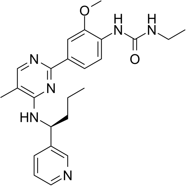
Lexibulin (CYT-997) is a potent and orally active tubulin polymerisation inhibitor with IC50s of 10-100 nM in cancer cell lines; with potent cytotoxic and vascular disrupting activity in vitro and in vivo. Lexibulin induces cell apoptosis and induces mitochondrial ROS generation in GC cells.
For research use only. We do not sell to patients.
- Purity : 99.10%
- CAS No.: 917111-44-5
- Formula: C24H30N6O2
- Molecular Weight:434.53
-
Storage:Powder -20°C, 3 years , 4°C, 2 years ; In solvent -80°C, 2 years , -20°C, 1 year

To place orders, for customer services and technical support, please contact: MedChemExpress USA
Tel: 609-228-6898 E-mail: [email protected] [email protected]


-
 Biological Activity
-
Chemical Information
-
Solvent & Solubility
- Protocols
- Purity & Documentation
- References
-
 Help & FAQs


-
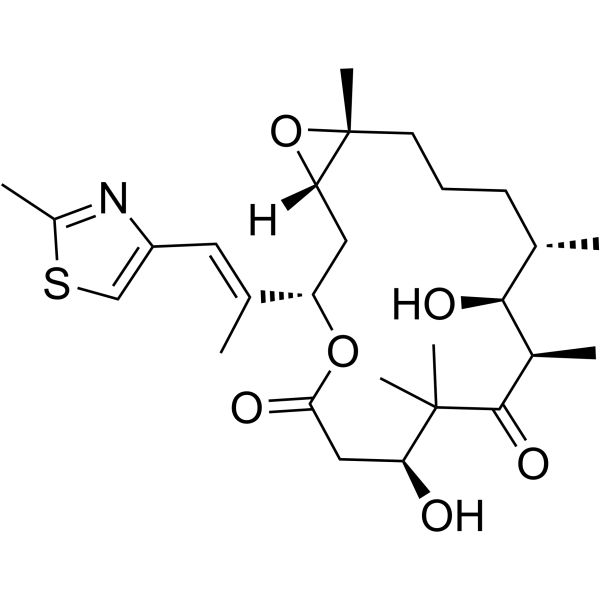
HY-17029
Epothilone BMicrotubule Stabilizer -
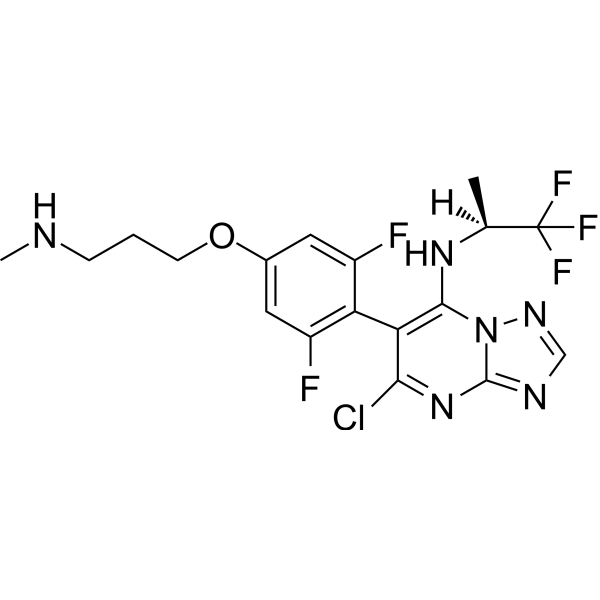
HY-14949
Cevipabulin -
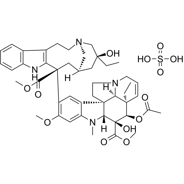
HY-13780
Vinblastine sulfatenAChR Inhibitor -

HY-B0215
AcetylcysteineMucolytic Agent -

HY-B0015
PaclitaxelAntitumor Agent -
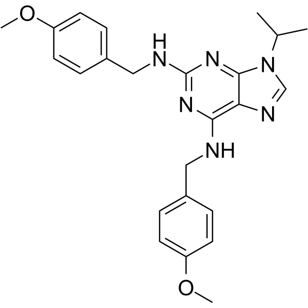
HY-W008956
MyoseverinMicrotubule-Binding Molecule -
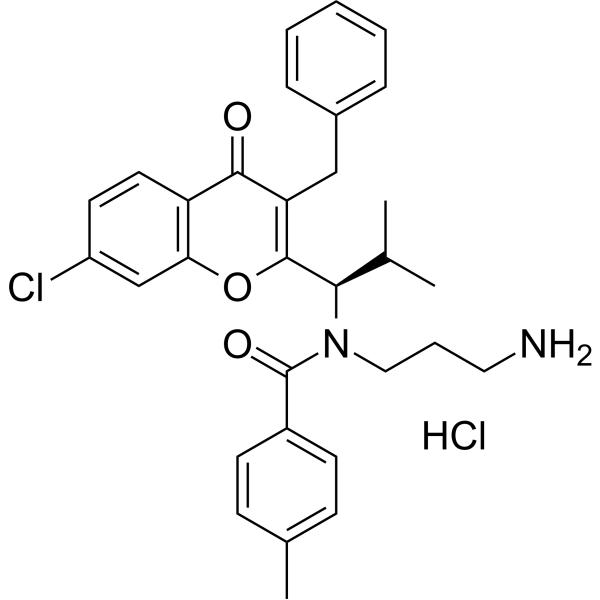
HY-12069
SB-743921 hydrochlorideEg5 Inhibitor -
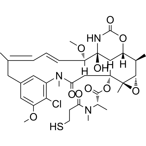
HY-19792
MertansineMicrotubule-Targeted Cytotoxin -
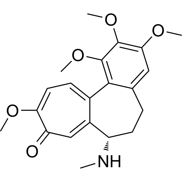
HY-N0282
DemecolcineTubulin Inhibitor -
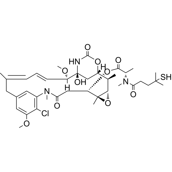
HY-12454
DM4Tubulin Inhibitor -
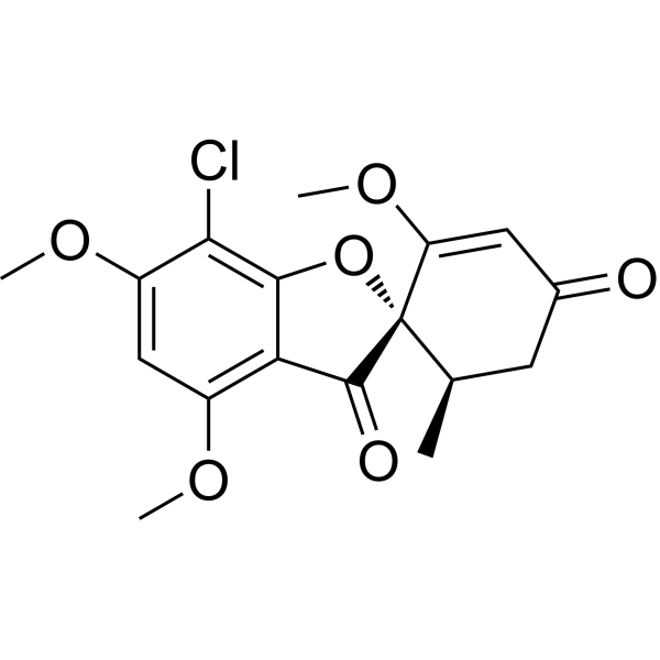
HY-17583
GriseofulvinAntifungal Antibiotic -
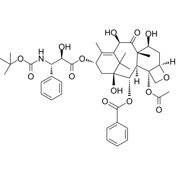
HY-B0011
DocetaxelMicrotubule Depolymerization Inhibitor
Publications Citing Use of MedChemExpress (MCE) Lexibulin
More

Biological Activity
Description
IC50 & Target
IC50 value: 10-100 nM(cell assay)[1]
Cellular Effect
|
Cell Line

|
Type | Value | Description | References |
|---|---|---|---|---|
| A549 | IC50 |
338 nM
Compound: 12, CYT997
|
Cytotoxicity against human A549 cells assessed as growth inhibition after 72 hrs by MTT assay
Cytotoxicity against human A549 cells assessed as growth inhibition after 72 hrs by MTT assay
|
[PMID: 24160376] |
| DU-145 | IC50 |
0.06 μM
Compound: 24, CYT997
|
Cytotoxicity against human DU145 cells after 72 hrs by MTT assay
Cytotoxicity against human DU145 cells after 72 hrs by MTT assay
|
[PMID: 19616947] |
| HCT-116 | IC50 |
356 nM
Compound: 12, CYT997
|
Cytotoxicity against human HCT116 cells assessed as growth inhibition after 72 hrs by MTT assay
Cytotoxicity against human HCT116 cells assessed as growth inhibition after 72 hrs by MTT assay
|
[PMID: 24160376] |
| HL-60 | IC50 |
112.3 nM
Compound: 12, CYT997
|
Cytotoxicity against human HL60 cells assessed as growth inhibition after 72 hrs by MTT assay
Cytotoxicity against human HL60 cells assessed as growth inhibition after 72 hrs by MTT assay
|
[PMID: 24160376] |
| HUVEC | GI50 |
2098 ng/mL
Compound: 12, CYT997
|
Toxicity in HUVEC assessed as growth inhibition
Toxicity in HUVEC assessed as growth inhibition
|
[PMID: 24160376] |
| NCI-H460 | IC50 |
272 nM
Compound: 12, CYT997
|
Cytotoxicity against human H460 cells assessed as growth inhibition after 72 hrs by MTT assay
Cytotoxicity against human H460 cells assessed as growth inhibition after 72 hrs by MTT assay
|
[PMID: 24160376] |
| PC-3 | IC50 |
0.06 μM
Compound: 24, CYT997
|
Cytotoxicity against human PC3 cells after 72 hrs by MTT assay
Cytotoxicity against human PC3 cells after 72 hrs by MTT assay
|
[PMID: 19616947] |
In Vitro
Lexibulin (CYT-997) prevents the in vitro polymerization of tubulin with an IC50 of ~3 μmol/L (compared with the half-maximal inhibitory concentration of 2 μmol/L for colchicine under identical conditions) as determined using the conventional turbidimetric assay for tubulin polymerization. Lexibulin is also capable of reversibly disrupting the microtubule network in cells, visualized using fluorescence microscopy. Thus, treatment of A549 cells with Lexibulin (1 μM) lead to the rapid reorganization of microtubules, including the destruction of the existing microtubule network and accumulation of tubulin in plaques within the cytoplasm of some cells. After 24 hours, major alterations in cell morphology are evident, including loss of adhesion and cell rounding. The effect of 1 hour of treatment with Lexibulin is reversible and cells rapidly recovered their normal microtubule architecture. Taken together, the data indicates that Lexibulin belongs to the class of anticancer agents that disrupt, rather than stabilize, tubulin-containing structures. Although vehicle-treated cells show 15% and 19% in G2-M phase at 15 and 24 hours (respectively), cells treated with Lexibulin (1 μM) had 38% and 43% of cells in G2-M at the same time points. Furthermore, at 24 hours post-Lexibulin treatment, only 66% of total cells are in the G1, S, and G2-M phases, which suggests that cells blocked at the G2-M boundary do not exit back to G1, as in the normal cell cycle, but most likely are driven towards apoptosis and cell death[1]. Consistent with the disruption of cellular tubulin, Lexibulin potently inhibits proliferation, induces cell cycle arrest and most importantly apoptosis of both human myeloma cell lines (HMCLs) and primary MM cells[2].
MedChemExpress (MCE) has not independently confirmed the accuracy of these methods. They are for reference only. Further protocols information, click here.
In Vivo
MedChemExpress (MCE) has not independently confirmed the accuracy of these methods. They are for reference only.


Chemical Information
-
CAS No. 917111-44-5
-
Appearance Solid
-
Molecular Weight 434.53
-
Formula C24H30N6O2
-
Color White to off-white
-
SMILES
CCC[C@H](NC1=C(C)C=NC(C2=CC=C(NC(NCC)=O)C(OC)=C2)=N1)C3=CN=CC=C3
-
Synonyms
CYT-997
-
Shipping
Room temperature in continental US; may vary elsewhere.
-
Storage
Powder -20°C 3 years 4°C 2 years In solvent -80°C 2 years -20°C 1 year


Publications (2)
-
Journal Impact Factor
-
Most Recent
-
J Exp Clin Cancer Res
Mitochondrial ROS accumulation inhibiting JAK2/STAT3 pathway is a critical modulator of CYT997-induced autophagy and apoptosis in gastric cancer. [Abstract]2020 Jun 23;39(1):119. PMID: 32576206 -


Solvent & Solubility
In Vitro:
DMSO : ≥ 100 mg/mL (230.13 mM; Hygroscopic DMSO has a significant impact on the solubility of product, please use newly opened DMSO)
* "≥" means soluble, but saturation unknown.
Please refer to the solubility information to select the appropriate solvent. Once prepared, please aliquot and store the solution to prevent product inactivation from repeated freeze-thaw cycles.
Storage method and period of stock solution: -80°C, 2 years; -20°C, 1 year. When stored at -80°C, please use it within 2 years. When stored at -20°C, please use it within 1 year.
Please refer to the solubility information to select the appropriate solvent. Once prepared, please aliquot and store the solution to prevent product inactivation from repeated freeze-thaw cycles.
Storage method and period of stock solution: -80°C, 2 years; -20°C, 1 year. When stored at -80°C, please use it within 2 years. When stored at -20°C, please use it within 1 year.
Concentration (start) × Volume (start) = Concentration (final) × Volume (final)
In Vivo:
Select the appropriate dissolution method based on your experimental animal and administration route.
- For the following dissolution methods, please ensure to first prepare a clear stock solution using an In Vitro approach and then sequentially add co-solvents:
- To ensure reliable experimental results, the clarified stock solution can be appropriately stored based on storage conditions. As for the working solution for In Vivo experiments, it is recommended to prepare freshly and use it on the same day.
- The percentages shown for the solvents indicate their volumetric ratio in the final prepared solution. If precipitation or phase separation occurs during preparation, heat and/or sonication can be used to aid dissolution.
Add each solvent one by one: 10% DMSO 40% PEG300 5% Tween-80 45% Saline
Solubility: ≥ 2.5 mg/mL (5.75 mM); Clear solution
This protocol yields a clear solution of ≥ 2.5 mg/mL (saturation unknown).
Taking 1 mL working solution as an example, add 100 μL DMSO stock solution (25.0 mg/mL) to 400 μL PEG300, and mix evenly; then add 50 μL Tween-80 and mix evenly; then add 450 μL Saline to adjust the volume to 1 mL.
Preparation of Saline: Dissolve 0.9 g sodium chloride in ddH₂O and dilute to 100 mL to obtain a clear Saline solution.
In Vivo Dissolution Calculator
Please enter the basic information of animal experiments:
-
-
-
-
Recommended: Prepare an additional quantity of animals to account for potential losses during experiments.
Please enter your animal formula composition:
-
%DMSO +
Recommended: Keep the proportion of DMSO in working solution below 2% if your animal is weak.
-
%+
-
+%Tween-80 + +
-
%Saline +
The co-solvents required include: DMSO, . All of co-solvents are available by MedChemExpress (MCE). , Tween 80. All of co-solvents are available by MedChemExpress (MCE).
Working solution concentration: 0.22 mg/mL
Method for preparing stock solution: mg drug dissolved in μL DMSO. Stock solution concentration: mg/mL.
1. Take μL DMSO stock solution;
2. Add μL .
μL , mix evenly;
3. Then add μL Tween 80, mix evenly;
4. Then add μL
Please ensure that the stock solution in the first step is dissolved to a clear state, and add co-solvents in sequence. You can use ultrasonic heating (ultrasonic cleaner, recommended frequency 20-40 kHz), vortexing, etc. to assist dissolution.

Protocols
-
Apoptosis
Apoptosis, also called programmed cell death, is generally characterized by distinct morphological characteristics.
-
TUNEL staining for apoptotic DNA fragmentation
TUNEL staining detects DNA strand breaks by using terminal deoxynucleotidyl transferase to add labeled nucleotides to exposed 3′-OH DNA termini, generating either microscopic staining in fixed cells or tissue sections, or fluorescence/cytometric signal in cell suspensions. TUNEL positivity reflects DNA fragmentation but should not be interpreted alone as definitive apoptosis, because TUNEL can also label necrotic, autolytic, mechanically damaged, or DNA-repair-associated DNA breaks.
-
Annexin V plus membrane-impermeant dye apoptosis staining
Annexin V-based apoptosis assays rely on the detection of phosphatidylserine (PS) externalization from the inner leaflet of the plasma membrane to the outer leaflet, an early biochemical hallmark of apoptosis. Fluorescently labeled Annexin V binds PS in a calcium-dependent manner, enabling identification of early apoptotic cells by flow cytometry or fluorescence microscopy. When combined with a membrane-impermeant DNA-binding dye (e. g. , propidium iodide), this approach allows discrimination between viable (Annexin V−/dye−), early apoptotic (Annexin V+/dye−), and late apoptotic or necrotic (Annexin V+/dye+) cell populations by assessing membrane integrity and PS exposure.
-
ROS/oxidative-stress fluorescent staining
ROS/oxidative-stress fluorescent staining uses cell-permeant fluorogenic probes that become fluorescent after oxidation inside cells or tissues; commonly used examples include DCFH-DA/DCFDA for broad cellular oxidant detection, DHE for superoxide-related signal detection, MitoSOX for mitochondrial superoxide-related signal detection, and CellROX probes for oxidative-stress-associated fluorescence readouts. The assay detects probe oxidation rather than a single ROS species unless the probe and analysis method have been chemically validated for that species. DCFH-DA enters cells, is deacetylated by intracellular esterases to DCFH, and produces fluorescent DCF after oxidation, so the readout is used as an operational measure of total cellular oxidative stress rather than a species-specific ROS measurement. DHE and MitoSOX can report superoxide-related oxidation, but red fluorescence alone can include non-specific ethidium-like oxidation products; HPLC or optimized spectral approaches are
-
Apoptosis Solutions
Apoptosis is a regulated, generally non-lytic cell-death pathway that removes unwanted, damaged, infected, or abnormal cells through coordinated morphological changes, caspase activation, DNA fragmentation, and membrane remodeling. The intrinsic apoptosis pathway is controlled mainly by mitochondrial outer membrane permeabilization, BCL-2 family proteins, cytochrome c release, apoptosome formation, caspase-9 activation, and downstream executioner caspase-3/7 activation. The extrinsic apoptosis pathway is initiated by death receptors such as Fas, TNFR, and TRAIL receptors, which recruit adaptor proteins and activate caspase-8 before engaging executioner caspases or mitochondrial amplification through BID cleavage. Apoptosis is linked to many phenotypes, including cancer cell killing, tissue homeostasis, immune regulation, neurodegeneration, infection response, and treatment-induced cytotoxicity; unresolved questions include how apoptosis interacts with necroptosis, pyroptosis, ferroptos
-
Cell Viability Determination by MTT Colorimetric Assay
The following protocol uses the MTT colorimetric assay as a classic literature-established method for assessing cell viability/metabolic activity in cultured mammalian cells. MTT[3-(4,5-dimethylthiazol-2-yl)-2,5-diphenyltetrazolium bromide] is reduced by metabolically active cells to a colored formazan product; the amount of formazan is quantified spectrophotometrically and provides an indirect measure of metabolically active viable cells. Importantly, MTT reduction reflects cellular oxidoreductase/metabolic activity rather than an absolute direct count of living cells, so changes in cellular metabolism can alter the signal independently of cell number.


Purity & Documentation
-
Data Sheet (290 KB)
-
SDS (393 KB)
- English - EN (393 KB)
- Français - FR (393 KB)
- Deutsch - DE (393 KB)
- Norwegian - NO (393 KB)
- Español - ES (393 KB)
- Swedish - SV (393 KB)
- Italian - IT (393 KB)
- Korean - KR (393 KB)
- Portuguese - PT (393 KB)
-
Handling Instructions (2659 KB)
References
[1]. Burns CJ, et al. CYT997: a novel orally active tubulin polymerization inhibitor with potent cytotoxic and vascular disrupting activity in vitro and in vivo. Mol Cancer Ther. 2009 Nov;8(11):3036-45. [Content Brief]
[2]. Monaghan K, et al. CYT997 causes apoptosis in human multiple myeloma. Invest New Drugs. 2011 Apr;29(2):232-8. [Content Brief]
[3]. Ya Cao, wt al. Mitochondrial ROS Accumulation Inhibiting JAK2/STAT3 Pathway Is a Critical Modulator of CYT997-induced Autophagy and Apoptosis in Gastric Cancer. J Exp Clin Cancer Res. 2020 Jun 23;39(1):119. [Content Brief]


Complete Stock Solution Preparation Table
Please refer to the solubility information to select the appropriate solvent. Once prepared, please aliquot and store the solution to prevent product inactivation from repeated freeze-thaw cycles.
Storage method and period of stock solution: -80°C, 2 years; -20°C, 1 year. When stored at -80°C, please use it within 2 years. When stored at -20°C, please use it within 1 year.
| Optional Solvent | Concentration Solvent Mass | 1 mg | 5 mg | 10 mg | 25 mg |
|---|---|---|---|---|---|
| DMSO | 1 mM | 2.3013 mL | 11.5067 mL | 23.0134 mL | 57.5334 mL |
| 5 mM | 0.4603 mL | 2.3013 mL | 4.6027 mL | 11.5067 mL | |
| 10 mM | 0.2301 mL | 1.1507 mL | 2.3013 mL | 5.7533 mL | |
| 15 mM | 0.1534 mL | 0.7671 mL | 1.5342 mL | 3.8356 mL | |
| 20 mM | 0.1151 mL | 0.5753 mL | 1.1507 mL | 2.8767 mL | |
| 25 mM | 0.0921 mL | 0.4603 mL | 0.9205 mL | 2.3013 mL | |
| 30 mM | 0.0767 mL | 0.3836 mL | 0.7671 mL | 1.9178 mL | |
| 40 mM | 0.0575 mL | 0.2877 mL | 0.5753 mL | 1.4383 mL | |
| 50 mM | 0.0460 mL | 0.2301 mL | 0.4603 mL | 1.1507 mL | |
| 60 mM | 0.0384 mL | 0.1918 mL | 0.3836 mL | 0.9589 mL | |
| 80 mM | 0.0288 mL | 0.1438 mL | 0.2877 mL | 0.7192 mL | |
| 100 mM | 0.0230 mL | 0.1151 mL | 0.2301 mL | 0.5753 mL |


Lexibulin Related Classifications
HY-10498 Related Classifications
 Powered by Bioz