



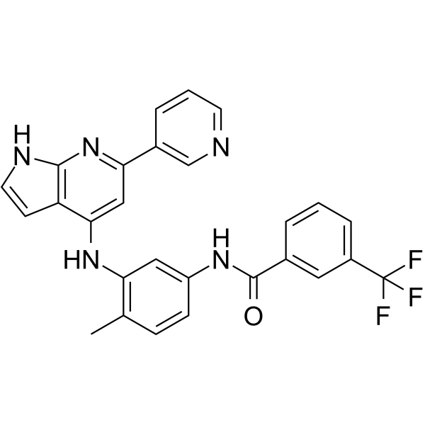
HZX-02-059

Based on 1 Customer Validation
HZX-02-059 is an allosteric inhibitor of PIKFYVE, and a methuosis inducer. HZX-02-059 disrupts the PIKfyve/TFEB axis, suppresses tubulin polymerization, reduces phosphorylated mTOR levels, downregulates p53, PI3K/AKT, c-Myc, and NF-κB pathways. HZX-02-059 induces G2/M cell cycle arrest, apoptosis, and inhibits cancer cell proliferation. HZX-02-059 can be used for the research of lymphoma, double-hit lymphoma, and B-cell acute lymphoblastic leukemia.
For research use only. We do not sell to patients.
- Purity : 99.40%
- CAS No.: 2240205-30-3
- Formula: C27H20F3N5O
- Molecular Weight:487.48
-
Storage:Powder -20°C, 3 years ; In solvent -80°C, 6 months , -20°C, 1 month

To place orders, for customer services and technical support, please contact: MedChemExpress USA
Tel: 609-228-6898 E-mail: [email protected] [email protected]


-
 Biological Activity
-
Chemical Information
-
Solvent & Solubility
- Protocols
- Purity & Documentation
- References
-
 Help & FAQs




Biological Activity
Description
IC50 & Target
Methuosis[1]
Cellular Effect
|
Cell Line

|
Type | Value | Description | References |
|---|---|---|---|---|
| A-375 | IC50 |
0.326 μM
Compound: 13
|
Antiproliferative activity against human A375 cells after 48 hrs by MTS assay
Antiproliferative activity against human A375 cells after 48 hrs by MTS assay
|
[PMID: 29878764] |
| A549 | IC50 |
1.892 μM
Compound: 13
|
Antiproliferative activity against human A549 cells after 48 hrs by MTS assay
Antiproliferative activity against human A549 cells after 48 hrs by MTS assay
|
[PMID: 29878764] |
| BGC-823 | IC50 |
1.566 μM
Compound: 13
|
Antiproliferative activity against human BGC823 cells after 48 hrs by MTS assay
Antiproliferative activity against human BGC823 cells after 48 hrs by MTS assay
|
[PMID: 29878764] |
| HCT-116 | IC50 |
1.008 μM
Compound: 13
|
Antiproliferative activity against human HCT116 cells after 48 hrs by MTS assay
Antiproliferative activity against human HCT116 cells after 48 hrs by MTS assay
|
[PMID: 29878764] |
| HeLa | IC50 |
1 μM
Compound: D-13
|
Antiproliferative activity against human HeLa cells assessed as inhibition of cell viability incubated for 24 hrs by CCK8 assay
Antiproliferative activity against human HeLa cells assessed as inhibition of cell viability incubated for 24 hrs by CCK8 assay
|
[PMID: 38117948] |
| HepG2 | IC50 |
1.197 μM
Compound: 13
|
Antiproliferative activity against human HepG2 cells after 48 hrs by MTS assay
Antiproliferative activity against human HepG2 cells after 48 hrs by MTS assay
|
[PMID: 29878764] |
| MCF-10A | IC50 |
>10 μM
Compound: 13
|
Cytotoxicity against human MCF10A cells assessed as reduction in cell viability after 48 hrs by MTS assay
Cytotoxicity against human MCF10A cells assessed as reduction in cell viability after 48 hrs by MTS assay
|
[PMID: 29878764] |
| MCF7 | IC50 |
2.611 μM
Compound: 13
|
Antiproliferative activity against human MCF7 cells after 48 hrs by MTS assay
Antiproliferative activity against human MCF7 cells after 48 hrs by MTS assay
|
[PMID: 29878764] |
| MDA-MB-231 | IC50 |
0.78 μM
Compound: D-13
|
Antiproliferative activity against human MDA-MB-231 cells assessed as inhibition of cell viability incubated for 24 hrs by CCK8 assay
Antiproliferative activity against human MDA-MB-231 cells assessed as inhibition of cell viability incubated for 24 hrs by CCK8 assay
|
[PMID: 38117948] |
| MDA-MB-231 | IC50 |
1.571 μM
Compound: 13
|
Antiproliferative activity against human MDA-MB-231 cells after 48 hrs by MTS assay
Antiproliferative activity against human MDA-MB-231 cells after 48 hrs by MTS assay
|
[PMID: 29878764] |
| MDA-MB-435S | IC50 |
2.268 μM
Compound: 13
|
Antiproliferative activity against human MDA-MB-435S cells after 48 hrs by MTS assay
Antiproliferative activity against human MDA-MB-435S cells after 48 hrs by MTS assay
|
[PMID: 29878764] |
| SK-MEL-28 | IC50 |
1.37 μM
Compound: 13
|
Antiproliferative activity against human SK-MEL-28 cells after 48 hrs by MTS assay
Antiproliferative activity against human SK-MEL-28 cells after 48 hrs by MTS assay
|
[PMID: 29878764] |
| SK-MEL-30 | IC50 |
1.662 μM
Compound: 13
|
Antiproliferative activity against human SK-MEL-30 cells after 48 hrs by MTS assay
Antiproliferative activity against human SK-MEL-30 cells after 48 hrs by MTS assay
|
[PMID: 29878764] |
In Vitro
HZX-02-059 (1 µM) binds potently to PIKFYVE (Kd = 10 nM) and strongly inhibits PIKFYVE and seven additional kinases by 99%, while inhibiting PIP4K2C by 60%[1].
HZX-02-059 reduces the viability of lymphoma cell lines in vitro with a median IC50 of 0.19 µM[1].
HZX-02-059 (1 μM) binds potently to purified PIKfyve kinase with a Kd of 10 nM[2].
HZX-02-059 (100 μM) inhibits purified tubulin polymerization[2].
HZX-02-059 (2 μM; 8 h) inhibits PIKfyve activity in 293T cells, reducing PI(3,5)P2 levels and altering late endosome/lysosome localization[2].
HZX-02-059 (48-72 h) potently inhibits proliferation of Will-2, LR, and TMD8 DHL cell lines in a dose- and time-dependent manner, with IC50 values ranging from 107 nM (Will-2, 72 h) to 344 nM (TMD8, 48 h)[2].
HZX-02-059 (0.25-4 μM; 24-72 h) induces caspase-independent cell death in Will-2, LR, and TMD8 DHL cell lines in a dose- and time-dependent manner, primarily via late apoptotic-like cell death[2].
HZX-02-059 (10 nM-2 μM; 6 h-5 days) triggers methuosis in Will-2 DHL cells via inhibition of the PIKfyve/TFEB axis, inducing characteristic vacuolation, reducing TFEB expression, and disrupting autophagy[2].
HZX-02-059 (0.25-1 μM; 24 h) induces G2/M phase cell cycle arrest in Will-2, LR, and TMD8 DHL cell lines, via inhibition of tubulin and suppression of the mTOR/Myc axis[2].
HZX-02-059 (0-4 μM; 72 h) does not induce apoptosis in primary PBMCs from healthy donors, indicating low toxicity toward normal hematopoietic cells[2].
HZX-02-059 (48-72 h) potently inhibits proliferation of Nalm6 and Sup-B15 B-ALL cells with IC50 values ranging from 376.3 nmol/L to 1068.0 nmol/L, showing greater potency with longer 72 h incubation compared to 48 h incubation[3].
HZX-02-059 (1 μmol/L; 12-24 h) induces prominent cytoplasmic vacuolization in Nalm6 and Sup-B15 B-ALL cells[3].
HZX-02-059 (0.5-1.5 μmol/L; 24-48 h) induces dose- and time-dependent late apoptosis in Nalm6 and Sup-B15 B-ALL cells[3].
HZX-02-059 (1 μmol/L; 24 h) alters transcriptome profiles in Nalm6 B-ALL cells, downregulating p53 and NF-κB pathways and upregulating apoptosis and cell cycle-related pathways[3].
HZX-02-059 (0.5-1.5 μmol/L; 24 h) downregulates p53 signaling, PI3K/AKT signaling, and downstream c-Myc and NF-κB pathways in Nalm6 and Sup-B15 B-ALL cells, with concurrent changes in cell cycle-related protein expression[3].
HZX-02-059 induces methuosis, inhibits proliferation, blocks cell cycle at G2/M phase, and dysregulates the PI3K/AKT axis in B-cell acute lymphoblastic leukemia cell lines[4].
MedChemExpress (MCE) has not independently confirmed the accuracy of these methods. They are for reference only.
-
Cell Line:Will-2, LR, and TMD8 DHL cell lines
-
Concentration:0.25; 0.5; 1; 2; 4 μM
-
Incubation Time:24; 48; 72 h
-
Result:Induced a significant dose-dependent increase in dead (Annexin V+) cells, predominantly late apoptotic (Annexin V+/PI+) cells, and a corresponding decrease in live (Annexin V−/PI−) cells across all three DHL cell lines.
-
Cell Line:Will-2, LR, and TMD8 DHL cell lines
-
Concentration:0.5 μM
-
Incubation Time:24 h
-
Result:Caused a significant decrease in the percentage of cells in the S and G0/G1 phases, and a marked increase in the G2/M phase across all three DHL cell lines at 0.5 μM for 24 h.
-
Cell Line:Will-2, LR, and TMD8 DHL cell lines
-
Concentration:0.25; 0.5; 1 μM
-
Incubation Time:24 h
-
Result:Showed reduced α-tubulin, β-tubulin, phosphorylated mTOR, phosphorylated 4EBP1, and c-MYC expression, along with altered Cyclin B1 and CDC2 levels via western blot analysis, consistent with tubulin inhibition and mTOR/Myc axis suppression.
-
Cell Line:human B-cell acute lymphoblastic leukemia (B-ALL) cell lines Nalm6 and Sup-B15
-
Concentration:0.5; 1; 1.5 μmol/L
-
Incubation Time:24; 48 h
-
Result:Triggered comparable levels of apoptosis in both cell lines after 24 h treatment with 1 or 1.5 μmol/L.
Induced substantial apoptosis in both cell lines after 48 h treatment with all tested concentrations.
Increased late apoptosis robustly, with no prominent increase in early apoptosis.
-
Cell Line:human B-cell acute lymphoblastic leukemia (B-ALL) cell lines Nalm6 and Sup-B15
-
Concentration:0.5; 1; 1.5 μmol/L
-
Incubation Time:24 h
-
Result:Decreased TP53, p27, CDC2, PI3K, phosphorylated AKT, c-Myc, and phosphorylated NF-κB p65 expression in a dose-dependent manner.
Increased cyclin B1 expression.
Decreased phosphorylated CDK2 expression.
Left cyclin D1, cyclin B2, and total CDK2 expression unchanged.
In Vivo
MedChemExpress (MCE) has not independently confirmed the accuracy of these methods. They are for reference only.
-
Animal Model:NOD-Prkdc−/−IL2rg−/− (NPI) mice (female, 6 weeks old, double-hit lymphoma model via luciferase-labeled LR cells injection after 1 Gy irradiation)[2]
-
Dosage:20 mg/kg
-
Administration:i.p.; daily; 7 consecutive days
-
Result:Significantly inhibited tumor growth at day 12 compared to the vehicle group.
Showed no statistically significant difference in average body mass compared to the vehicle group.
- Annexin V plus membrane-impermeant dye apoptosis staining
- BrdU Incorporation Assay
- Somatic Cell Culture
- CFSE Dye Dilution Proliferation Assay
- Dye-dilution cell tracking and proliferation staining
- Protocol for Cell Cycle
- CCK-8/WST-8 Cell Proliferation Assay
- Research Protocol for Inflammation-related Diseases
- Cell Counting-Based Growth Curve Assay
- Colony Formation (Clonogenic) Assay
- Cell Viability Determination by MTT Colorimetric Assay
- EdU Incorporation Assay (Click Chemistry-Based DNA Synthesis Measurement)
- Detection of Apoptosis
- TUNEL staining for apoptotic DNA fragmentation
- Ki-67 Immunostaining Proliferation Assay
- Flow cytometric DNA-content cell-cycle staining
- PCNA Immunodetection Proliferation Assay
- Protocol for Cell Counting and Cell Density Analysis
- Apoptosis Solutions
- MTT Cell Proliferation Assay


Chemical Information
-
CAS No. 2240205-30-3
-
Appearance Solid
-
Molecular Weight 487.48
-
Formula C27H20F3N5O
-
Color White to off-white
-
SMILES
CC(C=CC(NC(C1=CC(C(F)(F)F)=CC=C1)=O)=C2)=C2NC3=CC(C4=CN=CC=C4)=NC5=C3C=CN5
-
Synonyms
Methuosis inducer 1
-
Shipping
Room temperature in continental US; may vary elsewhere.
-
Storage
Powder -20°C 3 years In solvent -80°C 6 months -20°C 1 month


Solvent & Solubility
In Vitro:
DMSO : 25 mg/mL (51.28 mM; ultrasonic and warming and adjust pH to 3 with HCl and heat to 60°C; Hygroscopic DMSO has a significant impact on the solubility of product, please use newly opened DMSO)
Please refer to the solubility information to select the appropriate solvent. Once prepared, please aliquot and store the solution to prevent product inactivation from repeated freeze-thaw cycles.
Storage method and period of stock solution: -80°C, 6 months; -20°C, 1 month. When stored at -80°C, please use it within 6 months. When stored at -20°C, please use it within 1 month.
Please refer to the solubility information to select the appropriate solvent. Once prepared, please aliquot and store the solution to prevent product inactivation from repeated freeze-thaw cycles.
Storage method and period of stock solution: -80°C, 6 months; -20°C, 1 month. When stored at -80°C, please use it within 6 months. When stored at -20°C, please use it within 1 month.
Concentration (start) × Volume (start) = Concentration (final) × Volume (final)
In Vivo:
Select the appropriate dissolution method based on your experimental animal and administration route.
- For the following dissolution methods, please ensure to first prepare a clear stock solution using an In Vitro approach and then sequentially add co-solvents:
- To ensure reliable experimental results, the clarified stock solution can be appropriately stored based on storage conditions. As for the working solution for In Vivo experiments, it is recommended to prepare freshly and use it on the same day.
- The percentages shown for the solvents indicate their volumetric ratio in the final prepared solution. If precipitation or phase separation occurs during preparation, heat and/or sonication can be used to aid dissolution.
Add each solvent one by one: 10% DMSO 90% (20% SBE-β-CD in Saline)
Solubility: 2.5 mg/mL (5.13 mM); Suspended solution; Need ultrasonic
This protocol yields a suspended solution of 2.5 mg/mL. Suspended solution can be used for oral and intraperitoneal injection.
Taking 1 mL working solution as an example, add 100 μL DMSO stock solution (25.0 mg/mL) to 900 μL 20% SBE-β-CD in Saline, and mix evenly.
Preparation of 20% SBE-β-CD in Saline (4°C, storage for one week): 2 g SBE-β-CD powder is dissolved in 10 mL Saline, completely dissolve until clear.
Add each solvent one by one: 10% DMSO 40% PEG300 5% Tween-80 45% Saline
Solubility: ≥ 2.17 mg/mL (4.45 mM); Clear solution
This protocol yields a clear solution of ≥ 2.17 mg/mL (saturation unknown).
Taking 1 mL working solution as an example, add 100 μL DMSO stock solution (21.7 mg/mL) to 400 μL PEG300, and mix evenly; then add 50 μL Tween-80 and mix evenly; then add 450 μL Saline to adjust the volume to 1 mL.
Preparation of Saline: Dissolve 0.9 g sodium chloride in ddH₂O and dilute to 100 mL to obtain a clear Saline solution.
In Vivo Dissolution Calculator
Please enter the basic information of animal experiments:
-
-
-
-
Recommended: Prepare an additional quantity of animals to account for potential losses during experiments.
Please enter your animal formula composition:
-
%DMSO +
Recommended: Keep the proportion of DMSO in working solution below 2% if your animal is weak.
-
%+
-
+%Tween-80 + +
-
%Saline +
The co-solvents required include: DMSO, . All of co-solvents are available by MedChemExpress (MCE). , Tween 80. All of co-solvents are available by MedChemExpress (MCE).
Working solution concentration: 0.22 mg/mL
Method for preparing stock solution: mg drug dissolved in μL DMSO. Stock solution concentration: mg/mL.
1. Take μL DMSO stock solution;
2. Add μL .
μL , mix evenly;
3. Then add μL Tween 80, mix evenly;
4. Then add μL
Please ensure that the stock solution in the first step is dissolved to a clear state, and add co-solvents in sequence. You can use ultrasonic heating (ultrasonic cleaner, recommended frequency 20-40 kHz), vortexing, etc. to assist dissolution.


Purity & Documentation
-
Data Sheet (286 KB)
-
SDS (251 KB)
- English - EN (251 KB)
- Français - FR (251 KB)
- Deutsch - DE (251 KB)
- Norwegian - NO (251 KB)
- Español - ES (251 KB)
- Swedish - SV (251 KB)
- Italian - IT (251 KB)
- Korean - KR (251 KB)
- Portuguese - PT (251 KB)
-
Handling Instructions (2659 KB)
References
[1]. Roy A, et al. Selective Termination of Autophagy-Dependent Cancers. Cells. 2024 Jun 25;13(13):1096. [Content Brief]
[2]. Feng L, et al. Pharmacological targeting PIKfyve and tubulin as an effective treatment strategy for double-hit lymphoma. Cell Death Discov. 2022 Jan 28;8(1):39. [Content Brief]
[3]. Lu Z, et al. Novel PIKfyve/Tubulin Dual-target Inhibitor as a Promising Therapeutic Strategy for B-cell Acute Lymphoblastic Leukemia. Curr Med Sci. 2024 Apr;44(2):298-308. [Content Brief]


Complete Stock Solution Preparation Table
Please refer to the solubility information to select the appropriate solvent. Once prepared, please aliquot and store the solution to prevent product inactivation from repeated freeze-thaw cycles.
Storage method and period of stock solution: -80°C, 6 months; -20°C, 1 month. When stored at -80°C, please use it within 6 months. When stored at -20°C, please use it within 1 month.
| Optional Solvent | Concentration Solvent Mass | 1 mg | 5 mg | 10 mg | 25 mg |
|---|---|---|---|---|---|
| DMSO | 1 mM | 2.0514 mL | 10.2568 mL | 20.5137 mL | 51.2842 mL |
| 5 mM | 0.4103 mL | 2.0514 mL | 4.1027 mL | 10.2568 mL | |
| 10 mM | 0.2051 mL | 1.0257 mL | 2.0514 mL | 5.1284 mL | |
| 15 mM | 0.1368 mL | 0.6838 mL | 1.3676 mL | 3.4189 mL | |
| 20 mM | 0.1026 mL | 0.5128 mL | 1.0257 mL | 2.5642 mL | |
| 25 mM | 0.0821 mL | 0.4103 mL | 0.8205 mL | 2.0514 mL | |
| 30 mM | 0.0684 mL | 0.3419 mL | 0.6838 mL | 1.7095 mL | |
| 40 mM | 0.0513 mL | 0.2564 mL | 0.5128 mL | 1.2821 mL | |
| 50 mM | 0.0410 mL | 0.2051 mL | 0.4103 mL | 1.0257 mL |


HZX-02-059 Related Classifications
HY-112440 Related Classifications
 Powered by Bioz