



PRT-060318
Based on 4 publication(s) in Google Scholar
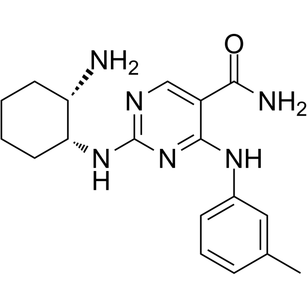
PRT-060318 (PRT318) s a potent, selective and orally active tyrosine kinase Syk inhibitor with an IC50 of 3 nM. PRT-060318 suppresses chronic lymphocytic leukemia (CLL) B cell activation and migration, and induces apoptosis. PRT-060318 prevents Heparin (HY-17567)-induced thrombocytopenia and thrombosis in a transgenic mouse model. PRT-060318 dihydrochloride can be used for CLL and thrombus research.
For research use only. We do not sell to patients.
- Purity : 99.59%
- CAS No.: 1194961-19-7
- Formula: C18H24N6O
- Molecular Weight:340.42
-
Storage:Powder -20°C, 3 years , 4°C, 2 years ; In solvent -80°C, 2 years , -20°C, 1 year

To place orders, for customer services and technical support, please contact: MedChemExpress USA
Tel: 609-228-6898 E-mail: [email protected] [email protected]


-
 Biological Activity
-
Chemical Information
-
Solvent & Solubility
- Protocols
- Purity & Documentation
- References
-
 Help & FAQs


-
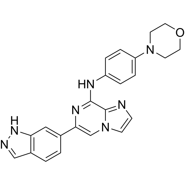
HY-15968
EntospletinibSyk Inhibitor -
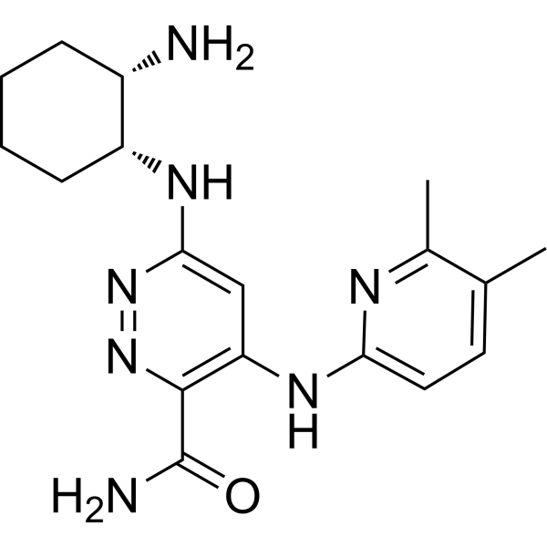
HY-16902
RO9021SYK Inhibitor -
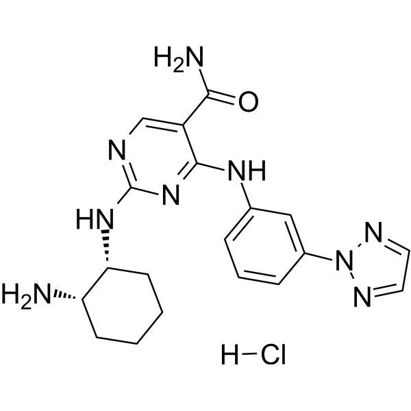
HY-15323
PRT062607 HydrochlorideSyk Inhibitor -
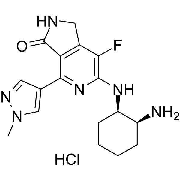
HY-100867A
Mivavotinib monohydrochlorideSYK/FLT3 Inhibitor -
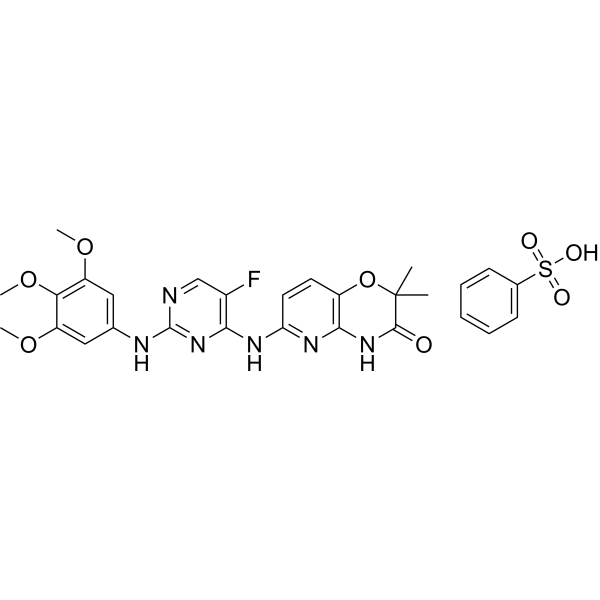
HY-12067
R406Syk/FLT3 Inhibitor -
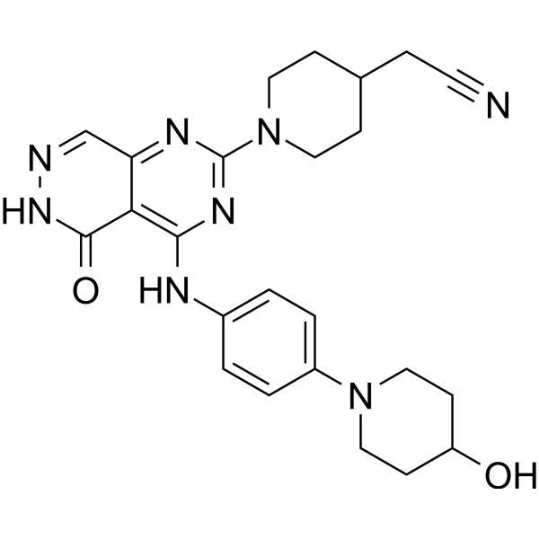
HY-103018
GusacitinibSYK/JAK Inhibitor -
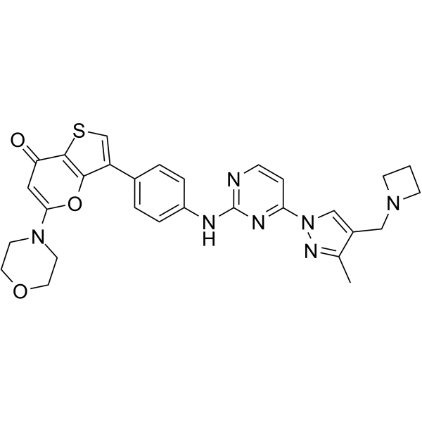
HY-136198
SRX3207Syk/PI3K Inhibitor -
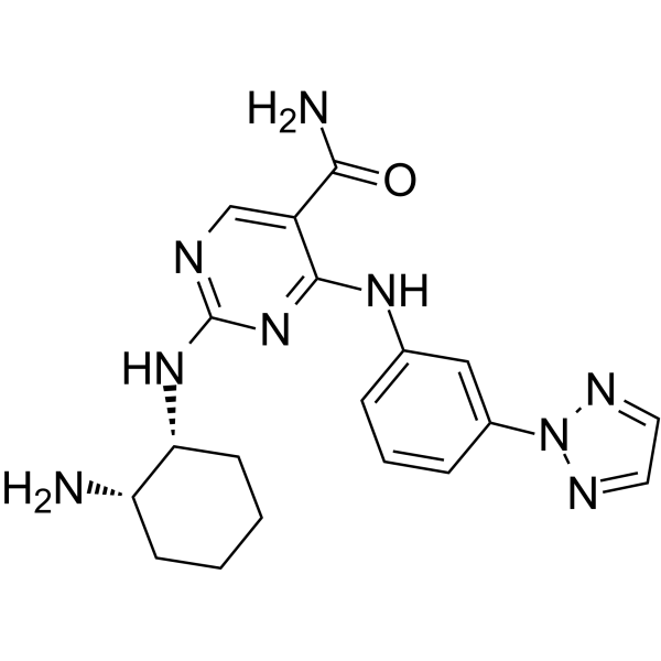
HY-15322
PRT062607Syk Inhibitor -
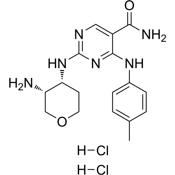
HY-12736A
GSK143 dihydrochlorideSyk Inhibitor -
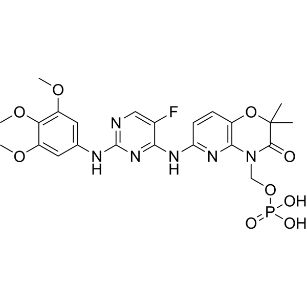
HY-13038A
FostamatinibSyk/FLT3 Inhibitor -
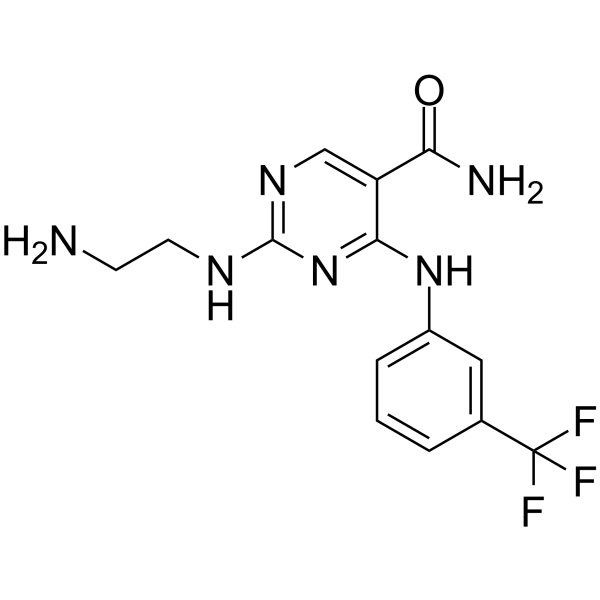
HY-112390A
Syk Inhibitor IISyk Inhibitor
Publications Citing Use of MedChemExpress (MCE) PRT-060318
More

Biological Activity
Description
In Vitro
PRT-060318 (PRT318) (0-3 µM, 15 min) completely inhibits Heparin-induced thrombocytopenia (HIT) immune complex-induced aggregation of both human and transgenic HIT mouse platelets[1].
MedChemExpress (MCE) has not independently confirmed the accuracy of these methods. They are for reference only. Further protocols information, click here.
In Vivo
MedChemExpress (MCE) has not independently confirmed the accuracy of these methods. They are for reference only.
-
Animal Model:C57Bl/6 mice treated with KKO (20 mg/kg, i.p.)[1]
-
Dosage:10 and 30 mg/kg
-
Administration:i.g., b.i.d. for 7 days
-
Result:Significantly inhibited platelet deposition without affecting bleeding time.
Markedly reduced HIT immune complex-induced thrombosis in the lungs.
Significantly reduced the thrombosis score compared to vehicle.


Chemical Information
-
CAS No. 1194961-19-7
-
Appearance Solid
-
Molecular Weight 340.42
-
Formula C18H24N6O
-
Color Light yellow to yellow
-
SMILES
O=C(C1=CN=C(N[C@H]2[C@@H](N)CCCC2)N=C1NC3=CC=CC(C)=C3)N
-
Synonyms
PRT318
-
Shipping
Room temperature in continental US; may vary elsewhere.
-
Storage
Powder -20°C 3 years 4°C 2 years In solvent -80°C 2 years -20°C 1 year


Publications (4)
-
Journal Impact Factor
-
Most Recent
-
Environ Pollut
Investigating the role and mechanism of methionine in different types of skeletal fluorosis based on Siglec-15 methylation. [Abstract]2026 Feb 1:390:127432. PMID: 41349947 -
FASEB J
Receptor-Dependent and -Independent Effects of Hemin on Platelet Plasma Membrane Disintegration. [Abstract]2026 Jan 31;40(2):e71463. PMID: 41524235 -
-


Solvent & Solubility
In Vitro:
H2O : 25 mg/mL (73.44 mM; Need ultrasonic)
DMSO : 10 mg/mL (29.38 mM; ultrasonic and warming and adjust pH to 3 with 1 M HCl and heat to 60°C; Hygroscopic DMSO has a significant impact on the solubility of product, please use newly opened DMSO)
Please refer to the solubility information to select the appropriate solvent. Once prepared, please aliquot and store the solution to prevent product inactivation from repeated freeze-thaw cycles.
Storage method and period of stock solution: -80°C, 2 years; -20°C, 1 year. When stored at -80°C, please use it within 2 years. When stored at -20°C, please use it within 1 year.
* Note: If you choose water as the stock solution, please dilute it to the working solution, then filter and sterilize it with a 0.22 μm filter before use.
Please refer to the solubility information to select the appropriate solvent. Once prepared, please aliquot and store the solution to prevent product inactivation from repeated freeze-thaw cycles.
Storage method and period of stock solution: -80°C, 2 years; -20°C, 1 year. When stored at -80°C, please use it within 2 years. When stored at -20°C, please use it within 1 year.
* Note: If you choose water as the stock solution, please dilute it to the working solution, then filter and sterilize it with a 0.22 μm filter before use.
Concentration (start) × Volume (start) = Concentration (final) × Volume (final)
In Vivo:
For the following dissolution methods, please prepare the working solution directly:
It is recommended to prepare fresh solutions and use them promptly within a short period of time.
The percentages shown for the solvents indicate their volumetric ratio in the final prepared solution. If precipitation or phase separation occurs during preparation, heat and/or sonication can be used to aid dissolution.
Add each solvent one by one: PBS
Solubility: 25 mg/mL (73.44 mM); Clear solution; Need ultrasonic

Protocols
-
Apoptosis
Apoptosis, also called programmed cell death, is generally characterized by distinct morphological characteristics.
-
TUNEL staining for apoptotic DNA fragmentation
TUNEL staining detects DNA strand breaks by using terminal deoxynucleotidyl transferase to add labeled nucleotides to exposed 3′-OH DNA termini, generating either microscopic staining in fixed cells or tissue sections, or fluorescence/cytometric signal in cell suspensions. TUNEL positivity reflects DNA fragmentation but should not be interpreted alone as definitive apoptosis, because TUNEL can also label necrotic, autolytic, mechanically damaged, or DNA-repair-associated DNA breaks.
-
Cell Cytotoxicity Assay
Cytotoxicity assays are usually based on the assessment of cell membrane damage, which can also be indirectly detected by measuring cell viability. Detection methods include MTT assay, CKK-8 assay, LDH assay and ATP assay, etc.
-
Annexin V plus membrane-impermeant dye apoptosis staining
Annexin V-based apoptosis assays rely on the detection of phosphatidylserine (PS) externalization from the inner leaflet of the plasma membrane to the outer leaflet, an early biochemical hallmark of apoptosis. Fluorescently labeled Annexin V binds PS in a calcium-dependent manner, enabling identification of early apoptotic cells by flow cytometry or fluorescence microscopy. When combined with a membrane-impermeant DNA-binding dye (e. g. , propidium iodide), this approach allows discrimination between viable (Annexin V−/dye−), early apoptotic (Annexin V+/dye−), and late apoptotic or necrotic (Annexin V+/dye+) cell populations by assessing membrane integrity and PS exposure.
-
Cell migration
Cell migration is a method that plays an important role in wound healing, cell differentiation, embryonic development, etc.
-
Apoptosis Solutions
Apoptosis is a regulated, generally non-lytic cell-death pathway that removes unwanted, damaged, infected, or abnormal cells through coordinated morphological changes, caspase activation, DNA fragmentation, and membrane remodeling. The intrinsic apoptosis pathway is controlled mainly by mitochondrial outer membrane permeabilization, BCL-2 family proteins, cytochrome c release, apoptosome formation, caspase-9 activation, and downstream executioner caspase-3/7 activation. The extrinsic apoptosis pathway is initiated by death receptors such as Fas, TNFR, and TRAIL receptors, which recruit adaptor proteins and activate caspase-8 before engaging executioner caspases or mitochondrial amplification through BID cleavage. Apoptosis is linked to many phenotypes, including cancer cell killing, tissue homeostasis, immune regulation, neurodegeneration, infection response, and treatment-induced cytotoxicity; unresolved questions include how apoptosis interacts with necroptosis, pyroptosis, ferroptos
-
How to Choose the Right Model Animal
Choosing the right model animal is a validity-driven decision in which the species, strain, sex, age, genetic background, disease-induction method, outcome measures, and welfare burden must match the scientific question rather than laboratory tradition or convenience. A model should be selected by judging face validity, construct validity, and predictive validity: whether it resembles the human phenotype, whether it reproduces relevant mechanisms, and whether results are likely to predict human biology or treatment response. Animal studies often fail to translate because of species differences, weak disease resemblance, poor experimental design, inadequate reporting, publication bias, and underuse of randomization, blinding, and sample-size justification. Unresolved questions include how to rank competing models objectively, how much human-disease complexity must be reproduced for a given objective, and when non-animal systems such as organoids, ex vivo tissue, or computational models


Purity & Documentation
-
Data Sheet (277 KB)
-
SDS (406 KB)
- English - EN (406 KB)
- Français - FR (406 KB)
- Deutsch - DE (406 KB)
- Norwegian - NO (406 KB)
- Español - ES (406 KB)
- Swedish - SV (406 KB)
- Italian - IT (406 KB)
- Korean - KR (406 KB)
- Portuguese - PT (406 KB)
-
Handling Instructions (2659 KB)
References
[1]. Reilly MP, et al. PRT-060318, a novel Syk inhibitor, prevents heparin-induced thrombocytopenia and thrombosis in a transgenic mouse model. Blood. 2011 Feb 17;117(7):2241-6. [Content Brief]
[2]. Hoellenriegel J, et al. Selective, novel spleen tyrosine kinase (Syk) inhibitors suppress chronic lymphocytic leukemia B-cell activation and migration. Leukemia. 2012 Jul;26(7):1576-83. [Content Brief]


Complete Stock Solution Preparation Table
Please refer to the solubility information to select the appropriate solvent. Once prepared, please aliquot and store the solution to prevent product inactivation from repeated freeze-thaw cycles.
Storage method and period of stock solution: -80°C, 2 years; -20°C, 1 year. When stored at -80°C, please use it within 2 years. When stored at -20°C, please use it within 1 year.
| Optional Solvent | Concentration Solvent Mass | 1 mg | 5 mg | 10 mg | 25 mg |
|---|---|---|---|---|---|
| DMSO / H2O | 1 mM | 2.9375 mL | 14.6877 mL | 29.3755 mL | 73.4387 mL |
| 5 mM | 0.5875 mL | 2.9375 mL | 5.8751 mL | 14.6877 mL | |
| 10 mM | 0.2938 mL | 1.4688 mL | 2.9375 mL | 7.3439 mL | |
| 15 mM | 0.1958 mL | 0.9792 mL | 1.9584 mL | 4.8959 mL | |
| 20 mM | 0.1469 mL | 0.7344 mL | 1.4688 mL | 3.6719 mL | |
| 25 mM | 0.1175 mL | 0.5875 mL | 1.1750 mL | 2.9375 mL | |
| H2O | 30 mM | 0.0979 mL | 0.4896 mL | 0.9792 mL | 2.4480 mL |
| 40 mM | 0.0734 mL | 0.3672 mL | 0.7344 mL | 1.8360 mL | |
| 50 mM | 0.0588 mL | 0.2938 mL | 0.5875 mL | 1.4688 mL | |
| 60 mM | 0.0490 mL | 0.2448 mL | 0.4896 mL | 1.2240 mL |
* Note: If you choose water as the stock solution, please dilute it to the working solution, then filter and sterilize it with a 0.22 μm filter before use.


PRT-060318 Related Classifications
HY-12974 Related Classifications
 Powered by Bioz