



SMER28
Based on 2 publication(s) in Google Scholar
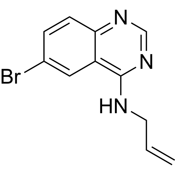
SMER28 is a positive regulator of autophagy acting via an mTOR-independent mechanism. SMER28 prevents the accumulation of amyloid beta peptide.
For research use only. We do not sell to patients.
- Purity : 99.92%
- CAS No.: 307538-42-7
- Formula: C11H10BrN3
- Molecular Weight:264.12
-
Storage:Powder -20°C, 3 years , 4°C, 2 years ; In solvent -80°C, 2 years , -20°C, 1 year

To place orders, for customer services and technical support, please contact: MedChemExpress USA
Tel: 609-228-6898 E-mail: [email protected] [email protected]


-
 Biological Activity
-
Chemical Information
-
Solvent & Solubility
- Protocols
- Purity & Documentation
- References
-
 Help & FAQs


-

HY-10219
RapamycinmTOR Inhibitor -
HY-15945
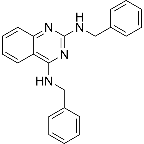
DBeQp97 Inhibitor -
HY-19312
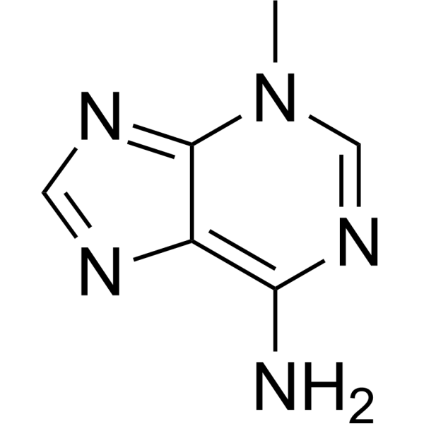
3-MethyladenineAutophagy/PI3K Inhibitor -

HY-100558
Bafilomycin A1V-ATPase Inhibitor -
HY-15713
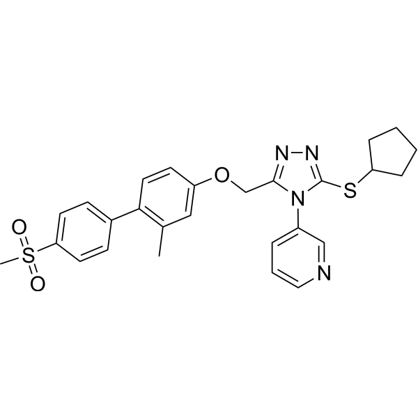
NMS-873VCP/p97 Inhibitor -
HY-B0627
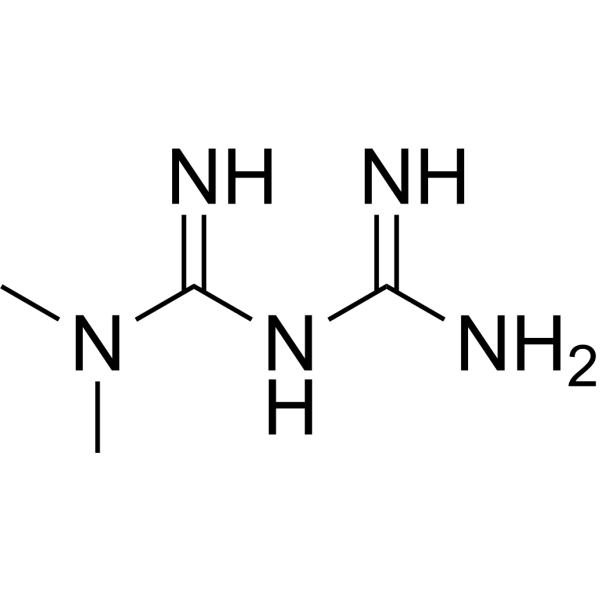
MetforminAntihyperglycemic Agent -
HY-17589A
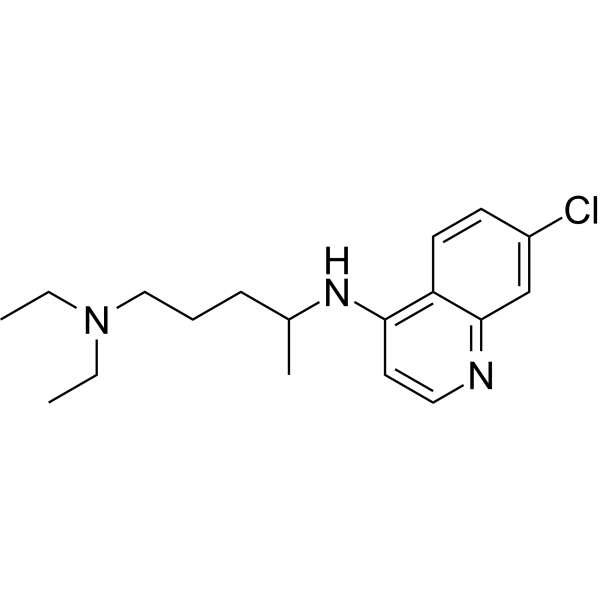
ChloroquineAntimalarial Agent -

HY-13259
MG-132Proteasome Inhibitor -
HY-100941
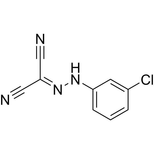
CCCPOXPHOS Uncoupler -
HY-10002
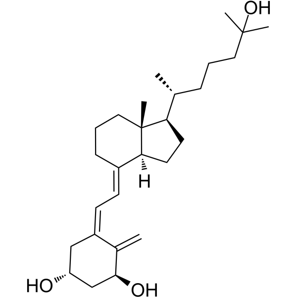
CalcitriolVDR Agonist -
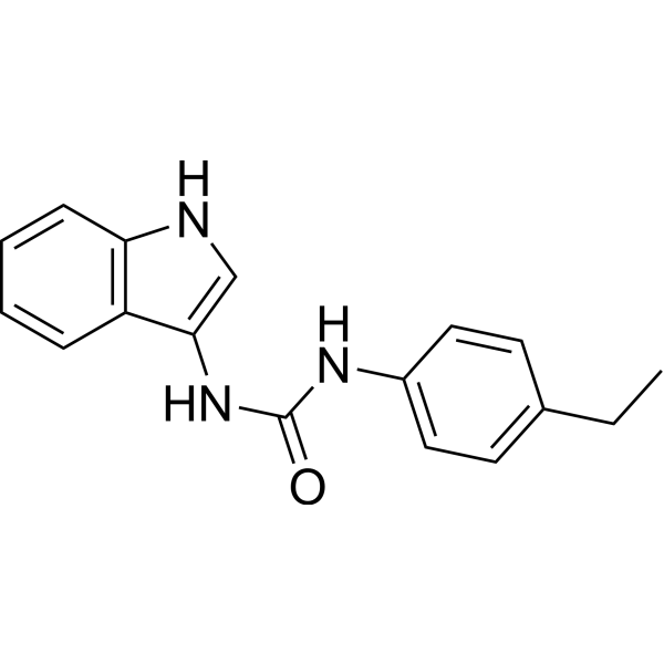
HY-112693
H-151STING Antagonist -
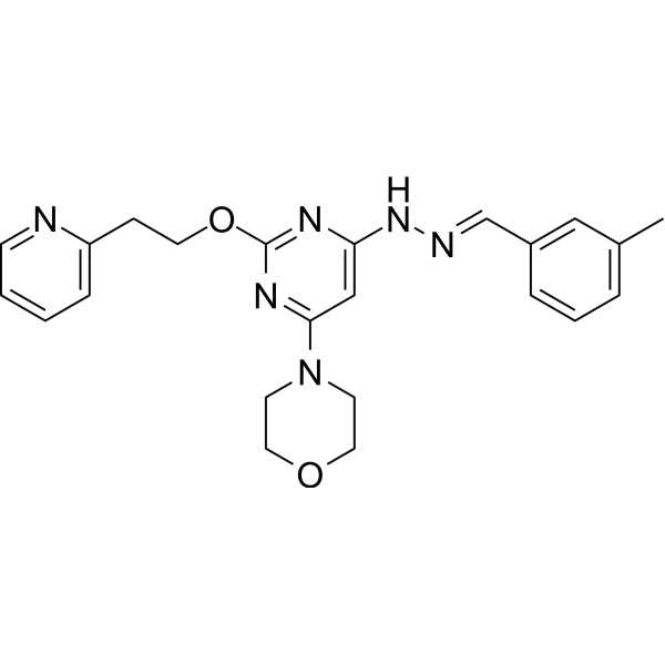
HY-14644
ApilimodIL-12/IL-23 Inhibitor
Publications Citing Use of MedChemExpress (MCE) SMER28
More

Biological Activity
Description
In Vitro
SMER28 (5-200 μM; 24 hours) shows a dose dependent decline of cell viability[4].
MedChemExpress (MCE) has not independently confirmed the accuracy of these methods. They are for reference only. Further protocols information, click here.
-
Cell Line:MMS1 cells
-
Concentration:5, 25, 50, 75, 100, 150, 200 μM
-
Incubation Time:24 hours
-
Result:Showed a dose dependent decline of cell viability.
In Vivo
MedChemExpress (MCE) has not independently confirmed the accuracy of these methods. They are for reference only.
-
Animal Model:14 to 16 weeks male mice (Balb/c)[5]
-
Dosage:15, 65 mg/kg
-
Administration:Subcutaneous injection; two days before irradiation and during the three days of irradiation (total 5 days)
-
Result:Significantly protected against post-irradiation weight loss and enhanced survival of mice at 65 mg/kg.


Chemical Information
-
CAS No. 307538-42-7
-
Appearance Solid
-
Molecular Weight 264.12
-
Formula C11H10BrN3
-
Color White to off-white
-
SMILES
C=CCNC1=C2C=C(Br)C=CC2=NC=N1
-
Shipping
Room temperature in continental US; may vary elsewhere.
-
Storage
Powder -20°C 3 years 4°C 2 years In solvent -80°C 2 years -20°C 1 year


Publications (2)
-
Journal Impact Factor
-
Most Recent
-
Neuropsychiatr Dis Treat
(2R,6R)-Hydroxynorketamine Alleviates Electroconvulsive Shock-Induced Learning Impairment by Inhibiting Autophagy. [Abstract]2021 Feb 3:17:297-304. PMID: 33568909 -


Solvent & Solubility
In Vitro:
DMSO : ≥ 32 mg/mL (121.16 mM; Hygroscopic DMSO has a significant impact on the solubility of product, please use newly opened DMSO)
* "≥" means soluble, but saturation unknown.
Please refer to the solubility information to select the appropriate solvent. Once prepared, please aliquot and store the solution to prevent product inactivation from repeated freeze-thaw cycles.
Storage method and period of stock solution: -80°C, 2 years; -20°C, 1 year. When stored at -80°C, please use it within 2 years. When stored at -20°C, please use it within 1 year.
Please refer to the solubility information to select the appropriate solvent. Once prepared, please aliquot and store the solution to prevent product inactivation from repeated freeze-thaw cycles.
Storage method and period of stock solution: -80°C, 2 years; -20°C, 1 year. When stored at -80°C, please use it within 2 years. When stored at -20°C, please use it within 1 year.
Concentration (start) × Volume (start) = Concentration (final) × Volume (final)
In Vivo:
Select the appropriate dissolution method based on your experimental animal and administration route.
- For the following dissolution methods, please ensure to first prepare a clear stock solution using an In Vitro approach and then sequentially add co-solvents:
- To ensure reliable experimental results, the clarified stock solution can be appropriately stored based on storage conditions. As for the working solution for In Vivo experiments, it is recommended to prepare freshly and use it on the same day.
- The percentages shown for the solvents indicate their volumetric ratio in the final prepared solution. If precipitation or phase separation occurs during preparation, heat and/or sonication can be used to aid dissolution.
Add each solvent one by one: 10% DMSO 40% PEG300 5% Tween-80 45% Saline
Solubility: ≥ 2.5 mg/mL (9.47 mM); Clear solution
This protocol yields a clear solution of ≥ 2.5 mg/mL (saturation unknown).
Taking 1 mL working solution as an example, add 100 μL DMSO stock solution (25.0 mg/mL) to 400 μL PEG300, and mix evenly; then add 50 μL Tween-80 and mix evenly; then add 450 μL Saline to adjust the volume to 1 mL.
Preparation of Saline: Dissolve 0.9 g sodium chloride in ddH₂O and dilute to 100 mL to obtain a clear Saline solution.
In Vivo Dissolution Calculator
Please enter the basic information of animal experiments:
-
-
-
-
Recommended: Prepare an additional quantity of animals to account for potential losses during experiments.
Please enter your animal formula composition:
-
%DMSO +
Recommended: Keep the proportion of DMSO in working solution below 2% if your animal is weak.
-
%+
-
+%Tween-80 + +
-
%Saline +
The co-solvents required include: DMSO, . All of co-solvents are available by MedChemExpress (MCE). , Tween 80. All of co-solvents are available by MedChemExpress (MCE).
Working solution concentration: 0.22 mg/mL
Method for preparing stock solution: mg drug dissolved in μL DMSO. Stock solution concentration: mg/mL.
1. Take μL DMSO stock solution;
2. Add μL .
μL , mix evenly;
3. Then add μL Tween 80, mix evenly;
4. Then add μL
Please ensure that the stock solution in the first step is dissolved to a clear state, and add co-solvents in sequence. You can use ultrasonic heating (ultrasonic cleaner, recommended frequency 20-40 kHz), vortexing, etc. to assist dissolution.

Protocols
-
Autophagy
Autophagy is a process in which eukaryotic cells use lysosomes to degrade their own cytoplasmic proteins and damaged organelles under the regulation of autophagy related gene (Atg). Microtubule-associated proteins light chain 3 (LC3) is recognized as autophagy marker, which transfers from cytoplasmic LC3 (LC3-I) to membrane type (LC3-II). LC3-II/I ratio could be detected by Western Blot and fluorescence microscopy.
-
Lysosome and acidic-vesicle live-cell staining
Lysosome and acidic-vesicle live-cell staining detects acidic intracellular compartments by using membrane-permeant acidotropic probes that accumulate in low-pH vesicles, including lysosomes, late endosomes, autolysosomes, and acidic phagosomes. LysoTracker staining is commonly used as an intensity-based readout of acidic lysosomal compartment abundance or enlargement, while acridine orange produces green fluorescence in less concentrated compartments and red fluorescence after concentration-dependent accumulation in acidic vesicular organelles. Loss or reduction of acridine-orange red signal can be used as a readout of lysosomal membrane permeabilization or reduced acidic-vesicle integrity. This protocol is designed for live cultured cells and can be adapted for fluorescence microscopy, high-content imaging, plate-reader readout, or flow cytometry when the selected literature supports the readout. Because these dyes report acidotropic accumulation rather than lysosome identity alone,
-
Macroautophagy Solutions
Macroautophagy is a conserved lysosome-dependent degradation pathway in which cytoplasmic material is sequestered into double-membrane autophagosomes and delivered to lysosomes for degradation and recycling. The pathway supports cellular homeostasis during nutrient limitation, organelle stress, protein-aggregate accumulation, infection, differentiation, and tissue remodeling by coupling cargo sequestration, autophagosome maturation, lysosomal fusion, and degradation of cargo-derived macromolecules. The core molecular sequence includes initiation by nutrient- and stress-regulated autophagy machinery, autophagosome nucleation, LC3/ATG8-family conjugation to autophagosomal membranes, cargo selection through receptors such as SQSTM1/p62, autophagosome-lysosome fusion, and lysosomal degradation. LC3 was identified as a mammalian homolog of yeast Atg8 that localizes to autophagosomal membranes after processing, and p62/SQSTM1 was shown to connect ubiquitinated cargo with autophagic degradati
-
Amyloid: Congo Red Amyloid Staining
Congo red amyloid staining is a histochemical method used to detect extracellular amyloid deposits in tissue sections based on the affinity of Congo red dye for β-pleated sheet-rich protein aggregates. When bound to amyloid, Congo red produces characteristic apple-green birefringence under polarized light microscopy, which is widely regarded as a diagnostic feature of amyloid deposition in histopathology. The diagnostic principle relies on the combination of dye binding (congophilia) and optical anisotropy under polarized illumination, which distinguishes amyloid from most non-amyloid eosinophilic extracellular deposits in routine histological evaluation. Amyloid identification by Congo red staining remains a cornerstone in diagnostic pathology despite the availability of adjunct methods such as immunohistochemistry and mass spectrometry, particularly because of its ability to localize deposits directly within tissue architecture. The specificity of Congo red-positive deposits is incre
-
Alzheimer’s Disease Modeling
Alzheimer’s Disease (AD) is a neurodegenerative disorder characterized by a progressive decline in cognitive functions and loss of specific types of neurons and synapses. Alzheimer's symptoms can be simulated in mice by injecting drugs (such as Aβ) or genetically modified.


Purity & Documentation
-
Data Sheet (277 KB)
-
SDS (476 KB)
- English - EN (476 KB)
- Français - FR (476 KB)
- Deutsch - DE (476 KB)
- Norwegian - NO (476 KB)
- Español - ES (476 KB)
- Swedish - SV (476 KB)
- Italian - IT (476 KB)
- Korean - KR (476 KB)
- Portuguese - PT (476 KB)
-
Handling Instructions (2659 KB)
References
[1]. Renna M et al. Chemical inducers of autophagy that enhance the clearance of mutant proteins in neurodegenerative diseases. J Biol Chem. 2010 Apr 9;285(15):11061-7. [Content Brief]
[2]. Nekova TS, et al. Small molecule enhancers of rapamycin induce apoptosis in myeloma cells via GSK3A/Bpreferentially within a protective bone marrow microenvironment. Br J Haematol. 2014 Oct;167(2):272-4. [Content Brief]
[3]. Shen D et al. Novel cell- and tissue-based assays for detecting misfolded and aggregated protein accumulation within aggresomes and inclusion bodies. Cell Biochem Biophys. 2011 Jul;60(3):173-85. [Content Brief]
[4]. Koukourakis MI, et al. SMER28 is a mTOR-independent small molecule enhancer of autophagy that protects mouse bone marrow and liver against radiotherapy. Invest New Drugs. 2018 Oct;36(5):773-781. [Content Brief]
[5]. Tian Y et al. A small-molecule enhancer of autophagy decreases levels of Abeta and APP-CTF via Atg5-dependent autophagy pathway. FASEB J. 2011 Jun;25(6):1934-42. [Content Brief]


Complete Stock Solution Preparation Table
Please refer to the solubility information to select the appropriate solvent. Once prepared, please aliquot and store the solution to prevent product inactivation from repeated freeze-thaw cycles.
Storage method and period of stock solution: -80°C, 2 years; -20°C, 1 year. When stored at -80°C, please use it within 2 years. When stored at -20°C, please use it within 1 year.
| Optional Solvent | Concentration Solvent Mass | 1 mg | 5 mg | 10 mg | 25 mg |
|---|---|---|---|---|---|
| DMSO | 1 mM | 3.7862 mL | 18.9308 mL | 37.8616 mL | 94.6539 mL |
| 5 mM | 0.7572 mL | 3.7862 mL | 7.5723 mL | 18.9308 mL | |
| 10 mM | 0.3786 mL | 1.8931 mL | 3.7862 mL | 9.4654 mL | |
| 15 mM | 0.2524 mL | 1.2621 mL | 2.5241 mL | 6.3103 mL | |
| 20 mM | 0.1893 mL | 0.9465 mL | 1.8931 mL | 4.7327 mL | |
| 25 mM | 0.1514 mL | 0.7572 mL | 1.5145 mL | 3.7862 mL | |
| 30 mM | 0.1262 mL | 0.6310 mL | 1.2621 mL | 3.1551 mL | |
| 40 mM | 0.0947 mL | 0.4733 mL | 0.9465 mL | 2.3663 mL | |
| 50 mM | 0.0757 mL | 0.3786 mL | 0.7572 mL | 1.8931 mL | |
| 60 mM | 0.0631 mL | 0.3155 mL | 0.6310 mL | 1.5776 mL | |
| 80 mM | 0.0473 mL | 0.2366 mL | 0.4733 mL | 1.1832 mL | |
| 100 mM | 0.0379 mL | 0.1893 mL | 0.3786 mL | 0.9465 mL |


SMER28 Related Classifications
HY-100200 Related Classifications
 Powered by Bioz