



Umbelliferone
Based on 5 publication(s) in Google Scholar
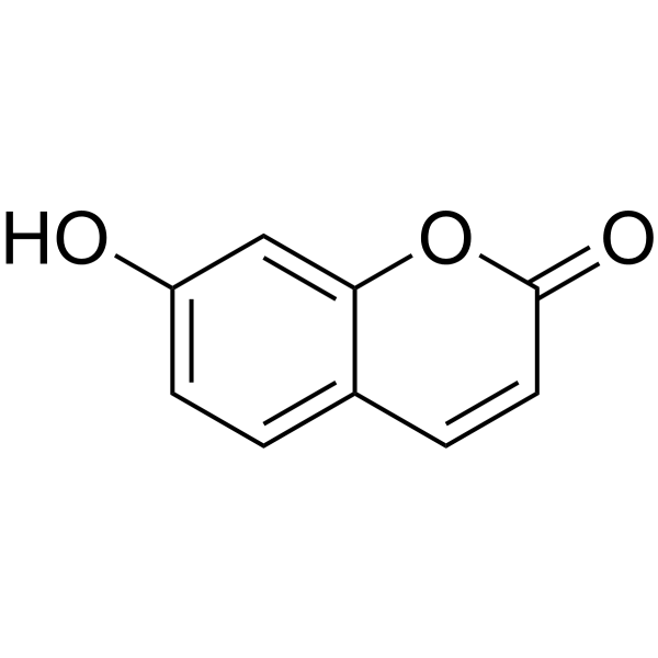
Umbelliferone (7-Hydroxycoumarin), a natural orally active product of the coumarin family, is a fluorescing compound which can be used as a sunscreen agent. Umbelliferone induces cell cycle arrest, apoptosis and DNA fragmentation in HepG2 cells. Umbelliferone exhibits significant anticancer effects. Umbelliferone attenuates the alteration characteristics of allergic airway inflammation. Umbelliferone displays the neuroprotective effects and cross the blood-brain barrier. Umbelliferone exhibits anti-inflammatory and antioxidant effects in chronic alcohol-fed rats.
For research use only. We do not sell to patients.
- Purity : 99.67%
- CAS No.: 93-35-6
- Formula: C9H6O3
- Molecular Weight:162.14
-
Storage:Powder -20°C, 3 years , 4°C, 2 years ; In solvent -80°C, 6 months , -20°C, 1 month

To place orders, for customer services and technical support, please contact: MedChemExpress USA
Tel: 609-228-6898 E-mail: [email protected] [email protected]


-
 Biological Activity
-
Chemical Information
-
Solvent & Solubility
- Protocols
- Purity & Documentation
- References
-
 Help & FAQs


-
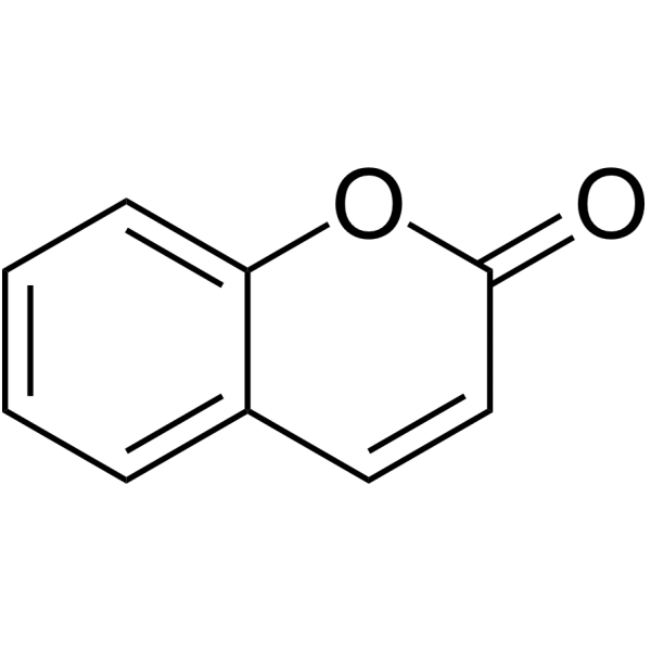
HY-N0709
CoumarinAnti-inflammatory Agent -
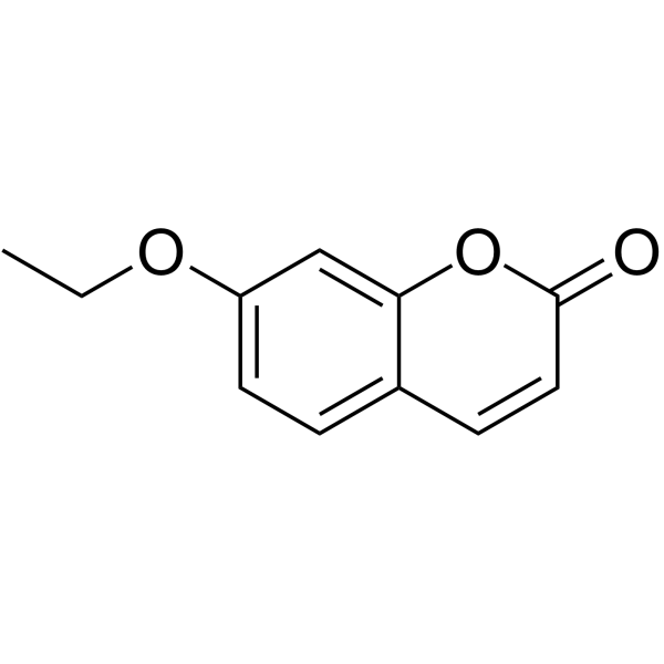
HY-133091
7-EthoxycoumarinCytochrome P451 Substrate -
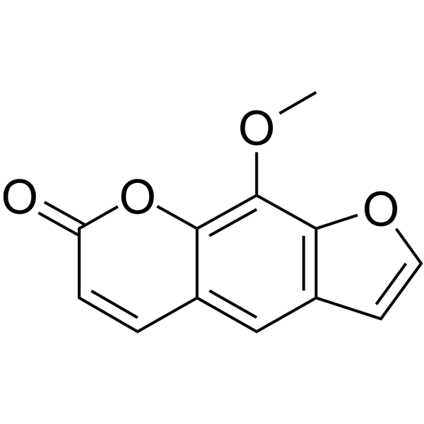
HY-30151
MethoxsalenCytochrome P450 Inhibitor -
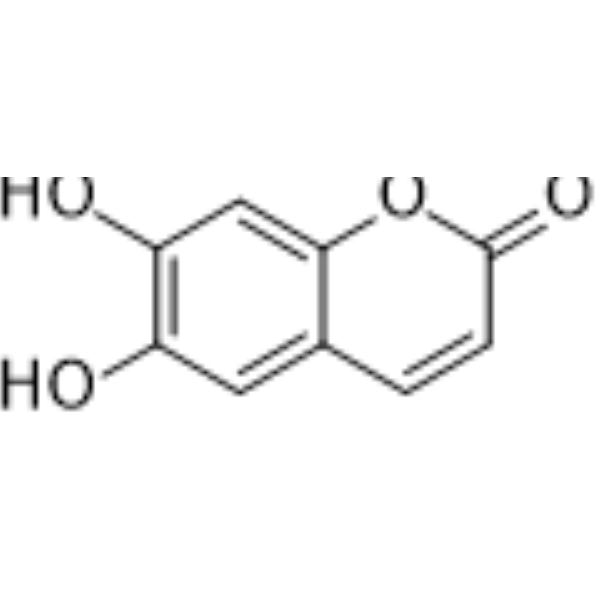
HY-N0284
EsculetinPI3K Inhibitor -
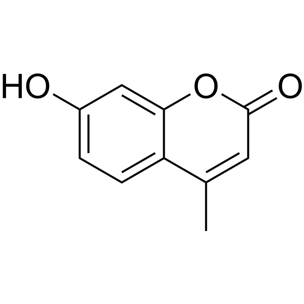
HY-N0187
4-MethylumbelliferoneHyaluronic Acid Biosynthesis Inhibitor -
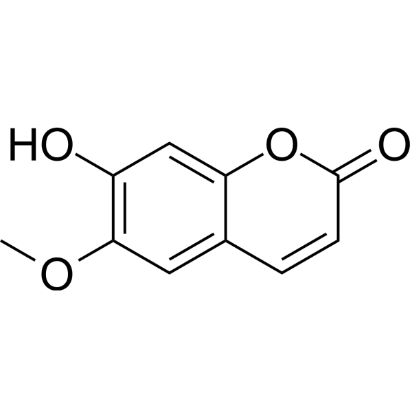
HY-N0342
ScopoletinCholinesterase (ChE) Inhibitor -
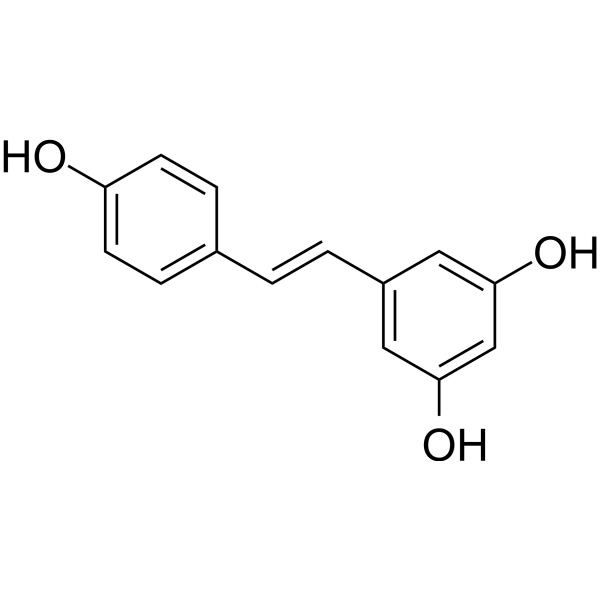
HY-16561
ResveratrolPolyphenolic Phytoalexin -

HY-N0573R
Umbelliferone (Standard)Standard of Umbelliferone -
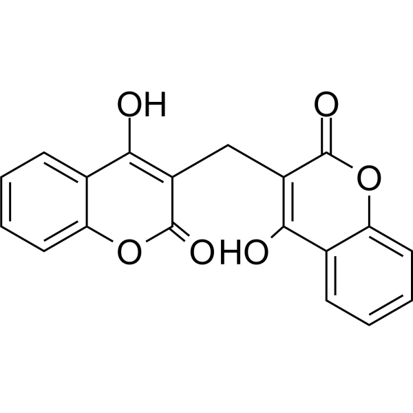
HY-N0645
DicoumarolPDHK Inhibitor -
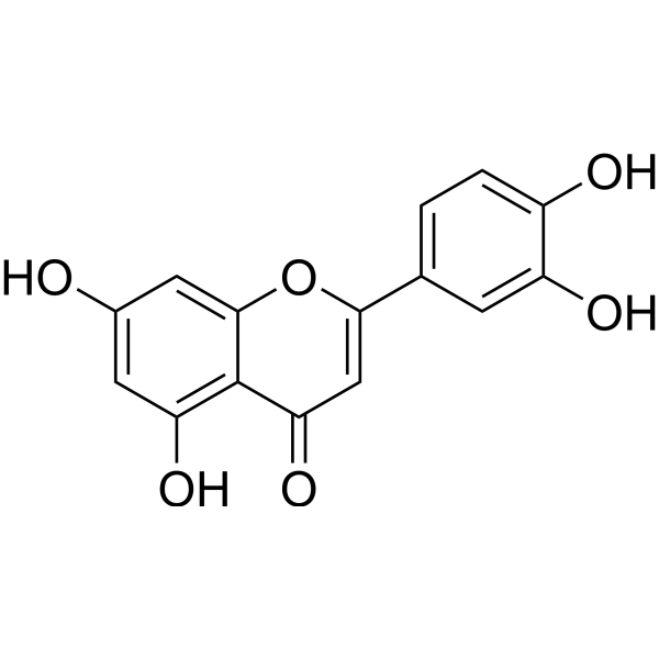
HY-N0162
LuteolinNrf2 Inhibitor -

HY-113324
NADPHReducing Agent -
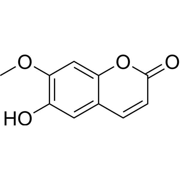
HY-N1365
IsoscopoletinHBV Inhibitor
Publications Citing Use of MedChemExpress (MCE) Umbelliferone
More Customer Validation & Images

-
RT-PCR
-
Cell Proliferation/Viability Assay
-
Cell Proliferation/Viability Assay
-
Bio/Physico-chemical Assay
-
RT-PCR


Biological Activity
Description
IC50 & Target
|
Human Endogenous Metabolite |
Cellular Effect
|
Cell Line

|
Type | Value | Description | References |
|---|---|---|---|---|
| A549 | IC50 |
>100 μM
Compound: 6
|
Cytotoxicity against human A549 cells after 72 hrs by MTT assay
Cytotoxicity against human A549 cells after 72 hrs by MTT assay
|
[PMID: 21696954] |
| A549 | IC50 |
21.11 μM
Compound: 7-C
|
Antiproliferative activity against human A549 cells after 48 hrs by MTT assay
Antiproliferative activity against human A549 cells after 48 hrs by MTT assay
|
[PMID: 28720504] |
| BT-549 | IC50 |
1 mM
Compound: 5
|
Antiproliferative activity against human BT549 cells after 24 hrs by MTT assay
Antiproliferative activity against human BT549 cells after 24 hrs by MTT assay
|
[PMID: 29144746] |
| HCT-116 | IC50 |
8.05 μM
Compound: 7
|
Cytotoxicity against human HCT116 cells after 96 hrs by MTT assay
Cytotoxicity against human HCT116 cells after 96 hrs by MTT assay
|
[PMID: 23746477] |
| HEK-293T | IC50 |
189.63 μM
Compound: 7-C
|
Antiproliferative activity against human 293T cells after 48 hrs by MTT assay
Antiproliferative activity against human 293T cells after 48 hrs by MTT assay
|
[PMID: 28720504] |
| HeLa | IC50 |
42.35 μM
Compound: 7-C
|
Antiproliferative activity against human HeLa cells after 48 hrs by MTT assay
Antiproliferative activity against human HeLa cells after 48 hrs by MTT assay
|
[PMID: 28720504] |
| HT-29 | IC50 |
22.67 μM
Compound: 7-C
|
Antiproliferative activity against human HT-29 cells after 48 hrs by MTT assay
Antiproliferative activity against human HT-29 cells after 48 hrs by MTT assay
|
[PMID: 28720504] |
| HT-29 | IC50 |
4.35 μM
Compound: 7
|
Cytotoxicity against human HT-29 cells after 96 hrs by MTT assay
Cytotoxicity against human HT-29 cells after 96 hrs by MTT assay
|
[PMID: 23746477] |
| HUVEC | ED50 |
>5 μg/mL
Compound: 7-hydroxycoumarin
|
Cytotoxicity against HUVEC
Cytotoxicity against HUVEC
|
[PMID: 15043409] |
| KB | IC50 |
200 μM
Compound: 11.1
|
Antiproliferative activity against human KB cells measured after 48 hrs by MTT assay
Antiproliferative activity against human KB cells measured after 48 hrs by MTT assay
|
[PMID: 38340509] |
| LNCaP | ED50 |
>5 μg/mL
Compound: 7-hydroxycoumarin
|
Cytotoxicity against human LNCAP cells
Cytotoxicity against human LNCAP cells
|
[PMID: 15043409] |
| LNCaP | IC50 |
>20 μg/mL
Compound: 1
|
Inhibitory activity against 5-alpha-reductase type 1 in cell culture system using LNCaP cells (androgen-sensitive human prostatic cancer cell line)
Inhibitory activity against 5-alpha-reductase type 1 in cell culture system using LNCaP cells (androgen-sensitive human prostatic cancer cell line)
|
[PMID: 11527731] |
| LoVo | IC50 |
97 μM
Compound: 6
|
Cytotoxicity against human LoVo cells after 72 hrs by MTT assay
Cytotoxicity against human LoVo cells after 72 hrs by MTT assay
|
[PMID: 21696954] |
| Lu1 | ED50 |
>5 μg/mL
Compound: 7-hydroxycoumarin
|
Cytotoxicity against human Lu1 cells
Cytotoxicity against human Lu1 cells
|
[PMID: 15043409] |
| MCF7 | ED50 |
>5 μg/mL
Compound: 7-hydroxycoumarin
|
Cytotoxicity against human MCF7 cells
Cytotoxicity against human MCF7 cells
|
[PMID: 15043409] |
| MCF7 | IC50 |
2.9 μM
Compound: 5
|
Antiproliferative activity against human MCF7 cells after 24 hrs by MTT assay
Antiproliferative activity against human MCF7 cells after 24 hrs by MTT assay
|
[PMID: 29144746] |
| MDA-MB-231 | IC50 |
3.7 μM
Compound: 5
|
Antiproliferative activity against human MDA-MB-231 cells after 24 hrs by MTT assay
Antiproliferative activity against human MDA-MB-231 cells after 24 hrs by MTT assay
|
[PMID: 29144746] |
| MT4 | IC50 |
2341 μM
Compound: 7
|
Cytotoxicity against human MT4 cells after 96 hrs by MTT assay
Cytotoxicity against human MT4 cells after 96 hrs by MTT assay
|
[PMID: 23746477] |
| P388 | ED50 |
0.4 μg/mL
Compound: umbelliferone, NSC-019790
|
Cytotoxicity against mouse P388 cells
Cytotoxicity against mouse P388 cells
|
[PMID: 6875580] |
| P388 | ED50 |
3.1 μg/mL
Compound: Umbelliferone
|
Cytotoxicity against mouse P388 cells
Cytotoxicity against mouse P388 cells
|
[PMID: 3783168] |
| PC-3 | IC50 |
>100 μM
Compound: 6
|
Cytotoxicity against human PC3 cells after 72 hrs by MTT assay
Cytotoxicity against human PC3 cells after 72 hrs by MTT assay
|
[PMID: 21696954] |
| SK-MEL-28 | IC50 |
>100 μM
Compound: 6
|
Cytotoxicity against human SK-MEL-28 cells after 72 hrs by MTT assay
Cytotoxicity against human SK-MEL-28 cells after 72 hrs by MTT assay
|
[PMID: 21696954] |
| SK-MEL-28 | IC50 |
0.48 μM
Compound: 5
|
Anticancer activity against human SK-MEL-28 cells assessed as reduction in cell viability incubated for 24 hrs by MTT assay
Anticancer activity against human SK-MEL-28 cells assessed as reduction in cell viability incubated for 24 hrs by MTT assay
|
[PMID: 37197457] |
| SK-MEL3 | IC50 |
1.6 μM
Compound: 5
|
Anticancer activity against human SK-MEL3 cells assessed as reduction in cell viability incubated for 24 hrs in presence of Artesunic acid by MTT assay
Anticancer activity against human SK-MEL3 cells assessed as reduction in cell viability incubated for 24 hrs in presence of Artesunic acid by MTT assay
|
[PMID: 37197457] |
| SK-MEL3 | IC50 |
2.1 μM
Compound: 5
|
Anticancer activity against human SK-MEL3 cells assessed as reduction in cell viability incubated for 24 hrs by MTT assay
Anticancer activity against human SK-MEL3 cells assessed as reduction in cell viability incubated for 24 hrs by MTT assay
|
[PMID: 37197457] |
| SMMC-7721 | IC50 |
19.9 μM
Compound: 7-C
|
Antiproliferative activity against human SMMC7721 cells after 48 hrs by MTT assay
Antiproliferative activity against human SMMC7721 cells after 48 hrs by MTT assay
|
[PMID: 28720504] |
| U-373MG ATCC | IC50 |
>100 μM
Compound: 6
|
Cytotoxicity against human U373 cells after 72 hrs by MTT assay
Cytotoxicity against human U373 cells after 72 hrs by MTT assay
|
[PMID: 21696954] |
| V79 | IC50 |
58 μM
Compound: 7-Hydroxy-coumarin
|
Cytotoxicity against V-79 cells
Cytotoxicity against V-79 cells
|
10.1016/0960-894X(96)00315-0 |
In Vitro
Umbelliferone (1-50 μM, 12-48 h) induces a dose‑dependent and time‑dependent reduction in HepG2 cells viability[1].
Umbelliferone (0-50 μM, 24 h) results in the appearance of HepG2 cell shrinkage, membrane blebbing, nuclear condensation and apoptotic body formation[1].
MedChemExpress (MCE) has not independently confirmed the accuracy of these methods. They are for reference only. Further protocols information, click here.
-
Cell Line:HepG2 cells
-
Concentration:0, 5, 25 50 μM
-
Incubation Time:24 h
-
Result:Resulted in the appearance of cell shrinkage and membrane blebbing, when compared with the morphology of untreated cells.
At 50 μM almost all the HepG2 cancer cells shrank significantly and no cells with normal morphological features were detected.
Reduced large nuclei.
Induced nuclear condensation and apoptotic body formation.
-
Cell Line:HepG2 cells
-
Concentration:0, 5, 25 50 μM
-
Incubation Time:24 h
-
Result:Increased in the proportion of cells in S phase (45.0, 51.3 and 62.1%, compared with 43.6% in untreated cells) and reduced in the fraction of cells in G1 phase (42.1, 45.6 and 32.2%, compared with 53.2% in the untreated cells).
In Vivo
Umbelliferone (15-30 mg/kg, i.g., daily, 21 days) shows neuroprotective effects on CUMS (chronic unpredictable mild stress)-induced rat model of depression, which is associated with the inhibition of neuronal apoptosis modulated by ROCK/Akt pathway[3].
Umbelliferone (0.05 g/L, p.o., daily, 8 weeks) protects against alcohol-induced liver damage by inhibiting the TLR4 signaling pathway and activating the antioxidant system in rats with liber-decarli liquid diet containing 5% alcohol [4].
MedChemExpress (MCE) has not independently confirmed the accuracy of these methods. They are for reference only.
-
Animal Model:Male BALB/c mice, 4-6 weeks old received systemic immunization by subcutaneous injection of 10 μg of chicken egg ovalbumin diluted in 2 mg/mL alum followed by a booster injection at day 14. A nasal challenge was performed starting at day 28, by inhalational exposure to aerosolised ovalbumin for 15 min/day, on five consecutive days[2].
-
Dosage:30, 60, 90 mg/kg
-
Administration:p.o., daily, 5 days
-
Result:Mice sensitized and challenged with ovalbumin have increased the number of inflammatory cells in bronchoalveolar lavage fluid.
Reduced the number of total cells and of neutrophils in the bronchoalveolar lavage fluid.
At 60 and 90 mg/kg, but not at 30 mg/kg, decreased significantly the number of eosinophils found in the bronchoalveolar lavage fluid of challenged mice.
At 60 mg/kg, decreased the number of mononuclear cells.
Caused a reduction in the levels of Th2-associated cytokines (IL-4, IL-5, IL-13).
-
Animal Model:While the control group of rats without receiving stress treatment had free access to food and water, other groups of rats were subjected to different types of stressors: cage tilting for 24 h, damp sawdust for 24 h, predator sounds, swimming in 4 ℃ cold water for 5min, swimming in 45 ℃ hot water for 5min, fasting for 48 h, water deprivation for 24 h, shaking for 15 min, nip trail for 1 min[3][5].
-
Dosage:15,30 mg/kg
-
Administration:i.g., daily, 21 days
-
Result:Exhibited an antidepressant-like effect, as sucrose consumption in CUMS rats significantly increased.
Decreased the expression of caspase3 and bax and increased the expression of bcl2 in CA1 region of hippocampus compared to the control group.
Remarkably increased the number of nissl-positive cells.
Down-regulated the levels of inflammatory cytokines IL-1β, IL-6 and TNF-α in hippocampus in the CUMS group versus control group.
Decreased the protein level of ROCK2 and phosphorylation expression of PTEN stimulated by CUMS.
Up-regulated the phosphorylation expressions of Akt and GSK3.
-
Animal Model:Rats were fed a Liber-Decarli liquid diet containing 5% alcohol for 8 weeks, while normal rats received an isocaloric carbohydrate liquid diet[4].
-
Dosage:0.05 g/L
-
Administration:p.o., daily, 8 weeks
-
Result:Decreased in serum TNF-α and IL-6 levels and increased IL-10 level.
Decreased the mRNA expressions of LBP, TLR4, NF-κB and TNF-α and IL-6.
Up-regulated SOD and CAT mRNA levels and activity.
Increased H2O2 and GSH levels and decreased hepatic MDA (Malondialdehyde) levels.


Chemical Information
-
CAS No. 93-35-6
-
Appearance Solid
-
Molecular Weight 162.14
-
Formula C9H6O3
-
Color Light yellow to orange
-
SMILES
O=C1C=CC2=CC=C(O)C=C2O1
-
Synonyms
7-Hydroxycoumarin; Hydrangin; NSC 19790
-
Structure Classification
-
Initial Source
-
Shipping
Room temperature in continental US; may vary elsewhere.
-
Storage
Powder -20°C 3 years 4°C 2 years In solvent -80°C 6 months -20°C 1 month


Publications (5)
-
Journal Impact Factor
-
Most Recent
-
J Agric Food Chem
Therapeutic Potential of Raspberry Extract in High-Fat Diet-Induced Liver Injury via Apoptosis and AMPK/PPARα Pathways. [Abstract]2025 Apr 1. PMID: 40168586 -
ACS Omega
2026 Mar 6;11(10):16756-16764. PMID: 41867607
Umbelliferone purchased from MedChemExpress. Usage Cited in: ACS Omega. 2026 Mar 6;11(10):16756-16764. [Abstract]
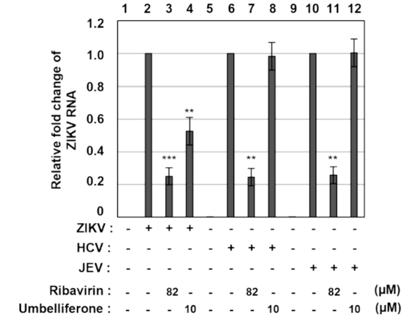
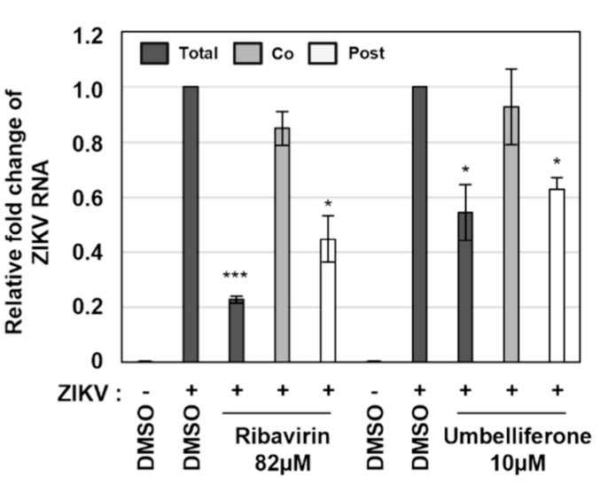
Cells were inoculated with culture medium alone (lanes 1, 5, and 9) or ZIKV (lanes 2–4), HCV (lanes 6–8), or JEV (lanes 10–12) and simultaneously treated with DMSO (vehicle, negative control), ribavirin (82 μM; positive control), or Umbelliferone (10 μM). Following the initial adsorption period (ZIKV, 6 h; HCV, 4 h; JEV, 1 h), the inoculum was replaced with fresh medium containing the same compounds. At 48 h postinfection (hpi), intracellular viral RNA was quantified via RT-qPCR using primers specific for viral NS1 and GAPDH.
Umbelliferone purchased from MedChemExpress. Usage Cited in: ACS Omega. 2026 Mar 6;11(10):16756-16764. [Abstract]
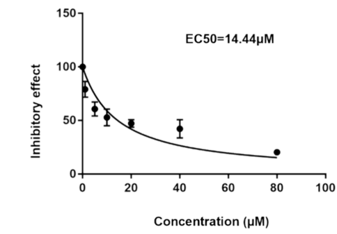
Dose-response analysis of ZIKV-infected cells treated with Umbelliferone (1, 5, 10, 20, 40, 80 μM). Data points represent mean ± SEM (n = 3). Significant inhibition was observed at concentrations ≥ 20 μM, yielding an estimated 50% inhibitory concentration (IC50) of 14.44 μM.
Umbelliferone purchased from MedChemExpress. Usage Cited in: ACS Omega. 2026 Mar 6;11(10):16756-16764. [Abstract]
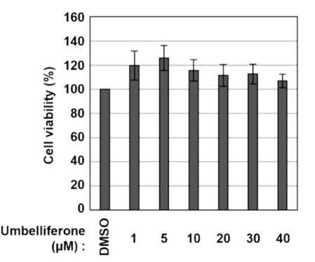
Cytotoxicity of Umbelliferone in Vero E6 cells was assessed 48 h after treatment with DMSO or Umbelliferone (1–40 μM) using the CellTiter-Glo luminescent viability assay. Cell viability was calculated from relative luminescence units (RLU) and normalized to the DMSO group (100%).
Umbelliferone purchased from MedChemExpress. Usage Cited in: ACS Omega. 2026 Mar 6;11(10):16756-16764. [Abstract]
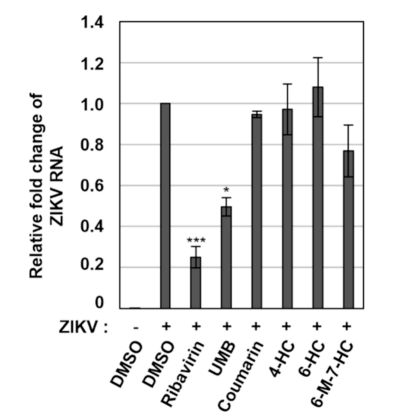
Evaluation of the anti-Zika efficacy of Umbelliferone derivatives. The four derivatives identified in Figure with no detectable cytotoxicity were selected to evaluate the antiviral activity against ZIKV in parallel with Umbelliferone. At 10 μM, a concentration at which Umbelliferone displayed robust antiviral activity, none of the derivatives exerted significant inhibitory effects.
Umbelliferone purchased from MedChemExpress. Usage Cited in: ACS Omega. 2026 Mar 6;11(10):16756-16764. [Abstract]
Vero E6 cells were infected with ZIKV at an MOI of 0.1 and treated with DMSO (negative control), ribavirin (82 μM; positive control), or Umbelliferone (10 μM). At 48 h postinfection (hpi), intracellular ZIKV RNA was quantified via RT-qPCR using primers specific for ZIKV NS1 and GAPDH.
-
J Biochem Mol Toxicol
The Protective Effects of Dose-Dependent Umbelliferone Application on CLP-Induced Acute Lung Injury (ALI) Model. [Abstract]2025 Oct;39(10):e70549. PMID: 41046538 -
J Pharm Biomed Anal
Determination of the in vitro metabolic stability and metabolites of the anticancer derivative riccardin D-N in human and mouse hepatic S9 fractions using HPLC-Q-LIT-MS. [Abstract]2019 Sep 10:174:734-743. PMID: 31299454 -
J Appl Toxicol
Umbelliferone protects against methylglyoxal-induced HUVECs dysfunction through suppression of apoptosis and oxidative stress. [Abstract]2023 Apr;43(4):490-499. PMID: 36170298


Solvent & Solubility
In Vitro:
DMSO : 100 mg/mL (616.75 mM; Need ultrasonic; Hygroscopic DMSO has a significant impact on the solubility of product, please use newly opened DMSO)
H2O : < 0.1 mg/mL (insoluble)
Please refer to the solubility information to select the appropriate solvent. Once prepared, please aliquot and store the solution to prevent product inactivation from repeated freeze-thaw cycles.
Storage method and period of stock solution: -80°C, 6 months; -20°C, 1 month. When stored at -80°C, please use it within 6 months. When stored at -20°C, please use it within 1 month.
Please refer to the solubility information to select the appropriate solvent. Once prepared, please aliquot and store the solution to prevent product inactivation from repeated freeze-thaw cycles.
Storage method and period of stock solution: -80°C, 6 months; -20°C, 1 month. When stored at -80°C, please use it within 6 months. When stored at -20°C, please use it within 1 month.
Concentration (start) × Volume (start) = Concentration (final) × Volume (final)
In Vivo:
Select the appropriate dissolution method based on your experimental animal and administration route.
- For the following dissolution methods, please ensure to first prepare a clear stock solution using an In Vitro approach and then sequentially add co-solvents:
- To ensure reliable experimental results, the clarified stock solution can be appropriately stored based on storage conditions. As for the working solution for In Vivo experiments, it is recommended to prepare freshly and use it on the same day.
- The percentages shown for the solvents indicate their volumetric ratio in the final prepared solution. If precipitation or phase separation occurs during preparation, heat and/or sonication can be used to aid dissolution.
Add each solvent one by one: 10% DMSO 40% PEG300 5% Tween-80 45% Saline
Solubility: 1.43 mg/mL (8.82 mM); Clear solution; Need ultrasonic
This protocol yields a clear solution of 1.43 mg/mL.
Taking 1 mL working solution as an example, add 100 μL DMSO stock solution (14.3 mg/mL) to 400 μL PEG300, and mix evenly; then add 50 μL Tween-80 and mix evenly; then add 450 μL Saline to adjust the volume to 1 mL.
Preparation of Saline: Dissolve 0.9 g sodium chloride in ddH₂O and dilute to 100 mL to obtain a clear Saline solution.
Add each solvent one by one: 10% DMSO 90% (20% SBE-β-CD in Saline)
Solubility: 1.43 mg/mL (8.82 mM); Clear solution; Need ultrasonic
This protocol yields a clear solution of 1.43 mg/mL.
Taking 1 mL working solution as an example, add 100 μL DMSO stock solution (14.3 mg/mL) to 900 μL 20% SBE-β-CD in Saline, and mix evenly.
Preparation of 20% SBE-β-CD in Saline (4°C, storage for one week): 2 g SBE-β-CD powder is dissolved in 10 mL Saline, completely dissolve until clear.
In Vivo Dissolution Calculator
Please enter the basic information of animal experiments:
-
-
-
-
Recommended: Prepare an additional quantity of animals to account for potential losses during experiments.
Please enter your animal formula composition:
-
%DMSO +
Recommended: Keep the proportion of DMSO in working solution below 2% if your animal is weak.
-
%+
-
+%Tween-80 + +
-
%Saline +
The co-solvents required include: DMSO, . All of co-solvents are available by MedChemExpress (MCE). , Tween 80. All of co-solvents are available by MedChemExpress (MCE).
Working solution concentration: 0.22 mg/mL
Method for preparing stock solution: mg drug dissolved in μL DMSO. Stock solution concentration: mg/mL.
1. Take μL DMSO stock solution;
2. Add μL .
μL , mix evenly;
3. Then add μL Tween 80, mix evenly;
4. Then add μL
Please ensure that the stock solution in the first step is dissolved to a clear state, and add co-solvents in sequence. You can use ultrasonic heating (ultrasonic cleaner, recommended frequency 20-40 kHz), vortexing, etc. to assist dissolution.

Protocols
-
Apoptosis
Apoptosis, also called programmed cell death, is generally characterized by distinct morphological characteristics.
-
TUNEL staining for apoptotic DNA fragmentation
TUNEL staining detects DNA strand breaks by using terminal deoxynucleotidyl transferase to add labeled nucleotides to exposed 3′-OH DNA termini, generating either microscopic staining in fixed cells or tissue sections, or fluorescence/cytometric signal in cell suspensions. TUNEL positivity reflects DNA fragmentation but should not be interpreted alone as definitive apoptosis, because TUNEL can also label necrotic, autolytic, mechanically damaged, or DNA-repair-associated DNA breaks.
-
Flow cytometric DNA-content cell-cycle staining
Flow cytometric DNA-content cell-cycle staining measures the fluorescence intensity of DNA-bound fluorochromes in single cells or nuclei to estimate DNA content distributions, allowing assignment of populations to G0/G1, S, and G2/M phases by DNA histogram deconvolution. Propidium iodide (PI) intercalates into DNA, and PI fluorescence is proportional to cellular DNA content when staining is performed under conditions that make DNA accessible and minimize non-DNA signal. Cells with G2/M DNA content are expected to show approximately twice the fluorescence intensity of G0/G1 cells, while S-phase cells occupy intermediate fluorescence values. PI-based DNA-content analysis can also detect cells with fractional DNA content, often reported as sub-G1, when DNA fragmentation and extraction during staining reduce retained DNA signal in apoptotic cells. DAPI is an alternative DNA fluorochrome for univariate DNA-content analysis, while bivariate approaches combining DNA content with proliferation
-
Annexin V plus membrane-impermeant dye apoptosis staining
Annexin V-based apoptosis assays rely on the detection of phosphatidylserine (PS) externalization from the inner leaflet of the plasma membrane to the outer leaflet, an early biochemical hallmark of apoptosis. Fluorescently labeled Annexin V binds PS in a calcium-dependent manner, enabling identification of early apoptotic cells by flow cytometry or fluorescence microscopy. When combined with a membrane-impermeant DNA-binding dye (e. g. , propidium iodide), this approach allows discrimination between viable (Annexin V−/dye−), early apoptotic (Annexin V+/dye−), and late apoptotic or necrotic (Annexin V+/dye+) cell populations by assessing membrane integrity and PS exposure.
-
BrdU Incorporation Assay
Bromodeoxyuridine (BrdU) incorporation assay is based on the principle that BrdU, a thymidine analog, is incorporated into newly synthesized DNA during the S phase of the cell cycle, thereby serving as a marker of DNA replication and cellular proliferation. Incorporated BrdU can be detected using anti-BrdU antibodies following DNA denaturation, enabling visualization or quantification of proliferating cells through immunochemical detection methods such as immunofluorescence or immunohistochemistry.
-
Ovalbumin-Induced Allergic Airway Inflammation
Ovalbumin-induced allergic airway inflammation is a mouse model in which systemic sensitization to ovalbumin, usually with aluminum hydroxide adjuvant, is followed by airway ovalbumin challenge to induce allergic airway inflammation, eosinophil recruitment, mucus production, serum antigen-specific IgE, Th2 cytokine responses, and airway hyperresponsiveness to methacholine. The model is used to study allergen-driven airway inflammation and asthma-like immune responses, but it does not reproduce every feature of human asthma. The main readouts are bronchoalveolar lavage fluid cellularity, lung histopathology, airway hyperresponsiveness, serum OVA-specific IgE, and cytokines such as IL-4, IL-5, and IL-13 in bronchoalveolar lavage fluid or lung samples. Eosinophilia and Th2 cytokines reflect allergic type 2 inflammation, while methacholine responsiveness provides a functional airway-reactivity endpoint.
-
Apoptosis Solutions
Apoptosis is a regulated, generally non-lytic cell-death pathway that removes unwanted, damaged, infected, or abnormal cells through coordinated morphological changes, caspase activation, DNA fragmentation, and membrane remodeling. The intrinsic apoptosis pathway is controlled mainly by mitochondrial outer membrane permeabilization, BCL-2 family proteins, cytochrome c release, apoptosome formation, caspase-9 activation, and downstream executioner caspase-3/7 activation. The extrinsic apoptosis pathway is initiated by death receptors such as Fas, TNFR, and TRAIL receptors, which recruit adaptor proteins and activate caspase-8 before engaging executioner caspases or mitochondrial amplification through BID cleavage. Apoptosis is linked to many phenotypes, including cancer cell killing, tissue homeostasis, immune regulation, neurodegeneration, infection response, and treatment-induced cytotoxicity; unresolved questions include how apoptosis interacts with necroptosis, pyroptosis, ferroptos
-
Protocol for Cell Cycle
Cell-cycle analysis by flow cytometry measures DNA content in single cells to estimate the fraction of cells in G0/G1, S, and G2/M phases. Propidium iodide intercalates into DNA, and after RNA removal with RNase, fluorescence intensity reflects cellular DNA content: 2N cells are assigned to G0/G1, cells between 2N and 4N to S phase, and 4N cells to G2/M. DNA-content analysis alone cannot reliably separate G0 from G1 or G2 from M. Ki-67 can distinguish quiescent G0 cells from cycling cells, EdU or BrdU incorporation marks active DNA synthesis in S phase, and phospho-histone H3 staining identifies mitotic cells within the 4N population.
-
Research Protocol for Inflammation-related Diseases
The NLRP3 inflammasome is a cytosolic innate immune signaling platform that integrates priming signals and danger-signal activation to promote caspase-1 activation, maturation of IL-1β and IL-18, and gasdermin D-mediated pyroptotic cell death. The core experimental logic is to determine whether inflammatory disease phenotypes are driven by increased NLRP3 expression, ASC-containing inflammasome assembly, caspase-1 cleavage, GSDMD cleavage, and extracellular release of IL-1β/IL-18 rather than by nonspecific cell injury alone. The pathway is strongly linked to inflammation-related disease phenotypes because monosodium urate crystals activate NALP3/NLRP3 inflammasome signaling in gout-like crystal inflammation, cholesterol crystals activate NLRP3 inflammasomes in atherogenesis models, and DSS-induced intestinal inflammation has been reported to involve NLRP3 inflammasome activity. However, experimental colitis studies also show context-dependent protective effects of NLRP3 inflammasome co


Purity & Documentation
-
Data Sheet (284 KB)
-
SDS (394 KB)
- English - EN (394 KB)
- Français - FR (394 KB)
- Deutsch - DE (394 KB)
- Norwegian - NO (394 KB)
- Español - ES (394 KB)
- Swedish - SV (394 KB)
- Italian - IT (394 KB)
- Korean - KR (394 KB)
- Portuguese - PT (394 KB)
-
Handling Instructions (2659 KB)
References
[1]. Yu SM, et al. Umbelliferone exhibits anticancer activity via the induction of apoptosis and cell cycle arrest in HepG2 hepatocellular carcinoma cells. Mol Med Rep. 2015 Sep;12(3):3869-3873. [Content Brief]
[2]. Vasconcelos JF, et al. Effects of umbelliferone in a murine model of allergic airway inflammation. Eur J Pharmacol. 2009 May 1;609(1-3):126-31. [Content Brief]
[3]. Qin T, et al. Umbelliferone reverses depression-like behavior in chronic unpredictable mild stress-induced rats by attenuating neuronal apoptosis via regulating ROCK/Akt pathway. Behav Brain Res. 2017 Jan 15;317:147-156. [Content Brief]
[4]. Sim MO, et al. Anti-inflammatory and antioxidant effects of umbelliferone in chronic alcohol-fed rats. Nutr Res Pract. 2015 Aug;9(4):364-9. [Content Brief]
[5]. Luo DD, et al. Involvement of hippocampal serotonin and neuropeptide Y in depression induced by chronic unpredicted mild stress. Brain Res Bull. 2008 Sep 5;77(1):8-12. [Content Brief]


Complete Stock Solution Preparation Table
Please refer to the solubility information to select the appropriate solvent. Once prepared, please aliquot and store the solution to prevent product inactivation from repeated freeze-thaw cycles.
Storage method and period of stock solution: -80°C, 6 months; -20°C, 1 month. When stored at -80°C, please use it within 6 months. When stored at -20°C, please use it within 1 month.
| Optional Solvent | Concentration Solvent Mass | 1 mg | 5 mg | 10 mg | 25 mg |
|---|---|---|---|---|---|
| DMSO | 1 mM | 6.1675 mL | 30.8375 mL | 61.6751 mL | 154.1877 mL |
| 5 mM | 1.2335 mL | 6.1675 mL | 12.3350 mL | 30.8375 mL | |
| 10 mM | 0.6168 mL | 3.0838 mL | 6.1675 mL | 15.4188 mL | |
| 15 mM | 0.4112 mL | 2.0558 mL | 4.1117 mL | 10.2792 mL | |
| 20 mM | 0.3084 mL | 1.5419 mL | 3.0838 mL | 7.7094 mL | |
| 25 mM | 0.2467 mL | 1.2335 mL | 2.4670 mL | 6.1675 mL | |
| 30 mM | 0.2056 mL | 1.0279 mL | 2.0558 mL | 5.1396 mL | |
| 40 mM | 0.1542 mL | 0.7709 mL | 1.5419 mL | 3.8547 mL | |
| 50 mM | 0.1234 mL | 0.6168 mL | 1.2335 mL | 3.0838 mL | |
| 60 mM | 0.1028 mL | 0.5140 mL | 1.0279 mL | 2.5698 mL | |
| 80 mM | 0.0771 mL | 0.3855 mL | 0.7709 mL | 1.9273 mL | |
| 100 mM | 0.0617 mL | 0.3084 mL | 0.6168 mL | 1.5419 mL |


Umbelliferone Related Classifications
HY-N0573 Related Classifications
 Powered by Bioz