



XEN445
Based on 1 publication(s) in Google Scholar
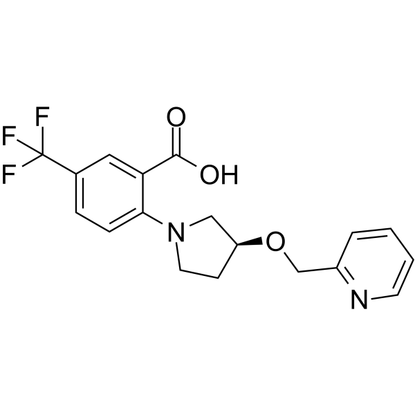
XEN445 is a potent, selective and orally active endothelial lipase (EL) inhibitor with an IC50 value of 0.237 μM. XEN445 selectively inhibits phospholipase enzymatic activity of LIPG. XEN445 raises plasma HDL and cholesterol levles. XEN445 induces G1 cell cycle arrest, reduces cell viability, suppresses cancer stem cell self-renewal, and inhibits tumor formation in LIPG-expressing triple-negative breast cancer cells, while showing no inhibitory effect on invasiveness or cancer stem cell stemness in these cells. XEN445 can be used for the research of cancer and metabolic disease, such as triple-negative breast cancer.
For research use only. We do not sell to patients.
- Purity : 99.63%
- CAS No.: 1515856-92-4
- Formula: C18H17F3N2O3
- Molecular Weight:366.33
-
Storage:Powder -20°C, 3 years , 4°C, 2 years ; In solvent -80°C, 2 years , -20°C, 1 year

To place orders, for customer services and technical support, please contact: MedChemExpress USA
Tel: 609-228-6898 E-mail: [email protected] [email protected]


-
 Biological Activity
-
Chemical Information
-
Solvent & Solubility
- Protocols
- Purity & Documentation
- References
-
 Help & FAQs


-
HY-15859
AtglistatinATGL Inhibitor -

HY-14471
CetilistatLipase Inhibitor -

HY-14471R
Cetilistat (Standard)Lipase Inhibitor -
HY-148756
NG-497ATGL Inhibitor -
HY-N1937
PristimerinMGL Inhibitor -
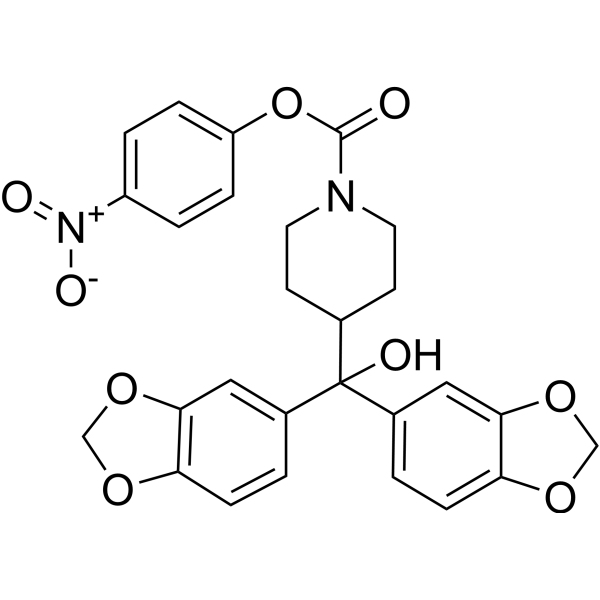
HY-15249
JZL 184MAGL Inhibitor -

HY-15859R
Atglistatin (Standard)ATGL Inhibitor -
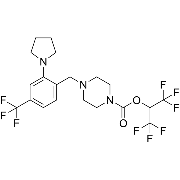
HY-117632
ElcubragistatMAGL Inhibitor -
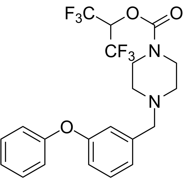
HY-12332
JW 642MAGL Inhibitor -

HY-15249R
JZL 184 (Standard)MAGL Inhibitor -

HY-N1937R
Pristimerin (Standard)MGL Inhibitor -

HY-12332R
JW 642 (Standard)MAGL Inhibitor
Publications Citing Use of MedChemExpress (MCE) XEN445
More

Biological Activity
Description
Cellular Effect
|
Cell Line

|
Type | Value | Description | References |
|---|---|---|---|---|
| HEK293 | IC50 |
0.25 μM
Compound: 13, XEN445
|
Inhibition of full length human endothelial lipase transfected in HEK293 cells using PED-A1 as substrate preincubated for 30 mins followed by substrate addition by fluorescence assay
Inhibition of full length human endothelial lipase transfected in HEK293 cells using PED-A1 as substrate preincubated for 30 mins followed by substrate addition by fluorescence assay
|
[PMID: 24211162] |
In Vitro
XEN445 potently inhibits recombinant human endothelial lipase with an IC50 of 0.237 μM, and shows selectivity over human lipoprotein lipase ( IC50 = 20 μM) and human hepatic lipase ( IC50 = 9.5 μM)[1].
XEN445 (2.5 μM; 4 h) exhibits high binding to rat, mouse, and human plasma proteins, with 97.4%, 88.8%, and 95.4% binding respectively at 2.5 μM after 4 h at 37°C[1].
XEN445 potently inhibits recombinant human endothelial lipase-mediated hydrolysis of purified human HDL particles with an IC50 of 0.11 μM[1].
XEN445 (30 min) inhibits endothelial lipase activity in EL-transfected HEK-293 cells with an IC50 of 0.25 μM[1].
XEN445 (30 min) specifically inhibits LIPG phospholipase activity in parental and LIPG-overexpressing MDA-MB-468 cells, with an IC50 of 2.172 μM[2].
XEN445 (100-250 μM; 4 days) reduces viability of LIPG-expressing TNBC cells (MCF10DCIS, MDA-MB-468) via G1 cell cycle arrest in a LIPG-dependent manner[2].
XEN445 (250 μM; 3-4 days) enhances migration and invasion of LIPG-expressing TNBC cells (MDA-MB-468, MCF10DCIS)[2].
XEN445 (1 week) suppresses CSC self-renewal in LIPG-expressing TNBC cells (MCF10DCIS, MDA-MB-468)[2].
XEN445 upregulates expression of select stemness and EMT-related genes in LIPG-expressing TNBC cells (MDA-MB-468, MCF10DCIS)[2].
MedChemExpress (MCE) has not independently confirmed the accuracy of these methods. They are for reference only. Further protocols information, click here.
-
Cell Line:MCF10DCIS, MDA-MB-468, MCF7, T47D, LIPG-overexpressing MDA-MB-468, LIPG-knockdown MDA-MB-468, LIPG-overexpressing MCF7 cells
-
Concentration:1, 2.5, 5, 10, 25, 50, 100, 200, 250 μM
-
Incubation Time:4 days
-
Result:Reduced viability of MCF10DCIS cells at ≥100 μM.
Selectively reduced viability of LIPG-expressing TNBC cell lines (MCF10DCIS, MDA-MB-468) but not LIPG-negative luminal breast cancer cell lines (MCF7, T47D) at 200 and 250 μM.
Sensitized MDA-MB-468 and MCF7 cells to viability reduction via LIPG overexpression.
Rendered MDA-MB-468 cells resistant to viability reduction via LIPG knockdown.
Parmacokinetics
In Vivo
XEN445 (50 mg/kg; i.p.; three times per week; 32 days) significantly inhibits in vivo triple-negative breast cancer xenograft tumor growth in nude mice were trans-
planted with MDA-MB-468 cells [2].
MedChemExpress (MCE) has not independently confirmed the accuracy of these methods. They are for reference only.
-
Animal Model:C57BL/6 (male, 12-16 weeks of age, wild-type)[1]
-
Dosage:30 mg/kg
-
Administration:p.o.; b.i.d.; 3 or 9 days
-
Result:Increased total plasma cholesterol by 18% (from 106.1 mg/dL to 125.2 mg/dL) at 30 mg/kg for 3 days.
Increased plasma HDLc by 16% (from 74.7 mg/dL to 86.5 mg/dL) at 30 mg/kg for 3 days.
Increased total plasma cholesterol by 21% (from 108.3 mg/dL to 131.2 mg/dL) for 9 days.
Increased plasma HDLc by 30% (from 68.2 mg/dL to 88.7 mg/dL) for 9 days.
-
Animal Model:NU/NU (8-week-old female) trans-
planted with MDA-MB-468 cells[2] -
Dosage:50 mg/kg
-
Administration:i.p.; three times per week; 32 days
-
Result:Significantly inhibited tumor growth compared to vehicle control.
Reduced Ki67-positive cells to 150 ± 18 per 1000 tumor cells, representing a decrease compared to vehicle control's 423 ± 27 per 1000 tumor cells.
Showed no significant difference in vimentin staining compared to vehicle control.


Chemical Information
-
CAS No. 1515856-92-4
-
Appearance Solid
-
Molecular Weight 366.33
-
Formula C18H17F3N2O3
-
Color Off-white to light yellow
-
SMILES
O=C(O)C1=CC(C(F)(F)F)=CC=C1N2C[C@@H](OCC3=NC=CC=C3)CC2
-
Shipping
Room temperature in continental US; may vary elsewhere.
-
Storage
Powder -20°C 3 years 4°C 2 years In solvent -80°C 2 years -20°C 1 year


Publications (1)
-
Journal Impact Factor
-
Most Recent
-
Neoplasma
Endothelial lipase promotes acute myeloid leukemia progression through metabolic reprogramming. [Abstract]2022 Jul;69(4):755-763 PMID: 35263993


Solvent & Solubility
In Vitro:
DMSO : 100 mg/mL (272.98 mM; Need ultrasonic; Hygroscopic DMSO has a significant impact on the solubility of product, please use newly opened DMSO)
Please refer to the solubility information to select the appropriate solvent. Once prepared, please aliquot and store the solution to prevent product inactivation from repeated freeze-thaw cycles.
Storage method and period of stock solution: -80°C, 2 years; -20°C, 1 year. When stored at -80°C, please use it within 2 years. When stored at -20°C, please use it within 1 year.
Please refer to the solubility information to select the appropriate solvent. Once prepared, please aliquot and store the solution to prevent product inactivation from repeated freeze-thaw cycles.
Storage method and period of stock solution: -80°C, 2 years; -20°C, 1 year. When stored at -80°C, please use it within 2 years. When stored at -20°C, please use it within 1 year.
Concentration (start) × Volume (start) = Concentration (final) × Volume (final)

Protocols
-
Cell Cytotoxicity Assay
Cytotoxicity assays are usually based on the assessment of cell membrane damage, which can also be indirectly detected by measuring cell viability. Detection methods include MTT assay, CKK-8 assay, LDH assay and ATP assay, etc.
-
Flow cytometric DNA-content cell-cycle staining
Flow cytometric DNA-content cell-cycle staining measures the fluorescence intensity of DNA-bound fluorochromes in single cells or nuclei to estimate DNA content distributions, allowing assignment of populations to G0/G1, S, and G2/M phases by DNA histogram deconvolution. Propidium iodide (PI) intercalates into DNA, and PI fluorescence is proportional to cellular DNA content when staining is performed under conditions that make DNA accessible and minimize non-DNA signal. Cells with G2/M DNA content are expected to show approximately twice the fluorescence intensity of G0/G1 cells, while S-phase cells occupy intermediate fluorescence values. PI-based DNA-content analysis can also detect cells with fractional DNA content, often reported as sub-G1, when DNA fragmentation and extraction during staining reduce retained DNA signal in apoptotic cells. DAPI is an alternative DNA fluorochrome for univariate DNA-content analysis, while bivariate approaches combining DNA content with proliferation
-
Mammalian live/dead viability and cytotoxicity staining
Live/dead viability and cytotoxicity staining assays are based on the simultaneous detection of intracellular esterase activity in metabolically active (viable) cells and membrane integrity loss in non-viable cells. In commonly used dual-staining approaches, membrane-permeant fluorogenic substrates are converted by intracellular esterases into fluorescent products in live cells, while impermeant DNA-binding dyes selectively enter cells with compromised plasma membranes and label nucleic acids in dead or dying cells, enabling discrimination between viable and non-viable populations by fluorescence microscopy or flow cytometry.
-
BrdU Incorporation Assay
Bromodeoxyuridine (BrdU) incorporation assay is based on the principle that BrdU, a thymidine analog, is incorporated into newly synthesized DNA during the S phase of the cell cycle, thereby serving as a marker of DNA replication and cellular proliferation. Incorporated BrdU can be detected using anti-BrdU antibodies following DNA denaturation, enabling visualization or quantification of proliferating cells through immunochemical detection methods such as immunofluorescence or immunohistochemistry.
-
Protocol for Cell Cycle
Cell-cycle analysis by flow cytometry measures DNA content in single cells to estimate the fraction of cells in G0/G1, S, and G2/M phases. Propidium iodide intercalates into DNA, and after RNA removal with RNase, fluorescence intensity reflects cellular DNA content: 2N cells are assigned to G0/G1, cells between 2N and 4N to S phase, and 4N cells to G2/M. DNA-content analysis alone cannot reliably separate G0 from G1 or G2 from M. Ki-67 can distinguish quiescent G0 cells from cycling cells, EdU or BrdU incorporation marks active DNA synthesis in S phase, and phospho-histone H3 staining identifies mitotic cells within the 4N population.
-
CCK-8/WST-8 Cell Proliferation Assay
The CCK-8/WST-8 assay is based on the reduction of the water-soluble tetrazolium salt WST-8 to a water-soluble formazan product by cellular dehydrogenases in metabolically active cells, where the generated formazan amount is proportional to the number of living cells and is quantified by measuring absorbance in the visible range, providing a colorimetric readout for cell viability and proliferation assessment. This class of tetrazolium-based assays improves upon earlier MTT-based systems by producing a water-soluble formazan, eliminating the need for organic solubilization steps and enabling direct spectrophotometric measurement in culture medium.
-
MTT Cell Proliferation Assay
The MTT assay is a colorimetric endpoint assay for estimating viable cell number, cell growth, cytotoxicity, or cell activation in cultured mammalian cells. Living cells reduce the yellow tetrazolium salt MTT into purple/blue formazan, while dead cells do not generate the same signal; the resulting color can be quantified with a multiwell spectrophotometer. MTT reduction is commonly interpreted as a readout of metabolic activity that often correlates with viable cell number, but it should not be treated as a direct cell-counting method unless the assay is optimized for the cell type and experimental condition. Studies show that MTT reduction can involve mitochondrial and non-mitochondrial reducing systems, and formazan may accumulate in intracellular lipid droplets rather than simply marking mitochondria.
-
hPSC maintenance and expansion
This protocol maintains and expands human pluripotent stem cells under feeder-free, chemically defined conditions using E8 medium and vitronectin-coated culture surfaces; the readout is sustained adherent colony growth with undifferentiated morphology and retained pluripotency-marker expression during serial passaging. E8-based hPSC culture relies on defined soluble factors and matrix-dependent adhesion rather than feeder cells; vitronectin supports hPSC attachment through integrin-mediated interactions, and EDTA passaging dissociates colonies as small aggregates without enzymatic digestion, centrifugation, or routine ROCK-inhibitor treatment.
-
Breast Cancer Modeling
Breast cancer is a heterogeneous cancer, and it has been distinguished into four subtypes: luminal A, luminal B, HER2-positive and basal-like. Molecular mutations, epigenetic alterations, hormone exposure and immune microenvironment are related to the progression of breast cancer.
-
Research Protocol for Metabolic Diseases
AMP-activated protein kinase, AMPK, is a conserved cellular energy sensor that responds to reduced cellular energy status and coordinates metabolism by increasing ATP-generating catabolic pathways while suppressing ATP-consuming anabolic processes. In metabolic disease research, the AMPK pathway is experimentally relevant because it regulates hepatic lipid synthesis, fatty acid oxidation, glucose production, skeletal-muscle glucose disposal, mTORC1-linked biosynthesis, autophagy, mitochondrial homeostasis, and whole-body energy balance. The central pathway logic is that energy stress, metformin, exercise-like stimulation, or direct AMPK activators increase AMPKα Thr172 phosphorylation and downstream substrate phosphorylation, including ACC and RAPTOR. Phosphorylation of ACC suppresses lipogenesis and supports fatty acid oxidation, whereas phosphorylation of RAPTOR suppresses mTORC1 signaling and links cellular energy status to growth and protein synthesis control. The pathway is linked
-
Cell Viability Determination by MTT Colorimetric Assay
The following protocol uses the MTT colorimetric assay as a classic literature-established method for assessing cell viability/metabolic activity in cultured mammalian cells. MTT[3-(4,5-dimethylthiazol-2-yl)-2,5-diphenyltetrazolium bromide] is reduced by metabolically active cells to a colored formazan product; the amount of formazan is quantified spectrophotometrically and provides an indirect measure of metabolically active viable cells. Importantly, MTT reduction reflects cellular oxidoreductase/metabolic activity rather than an absolute direct count of living cells, so changes in cellular metabolism can alter the signal independently of cell number.


Purity & Documentation
-
Data Sheet (278 KB)
-
SDS (396 KB)
- English - EN (396 KB)
- Français - FR (396 KB)
- Deutsch - DE (396 KB)
- Norwegian - NO (396 KB)
- Español - ES (396 KB)
- Swedish - SV (396 KB)
- Italian - IT (396 KB)
- Korean - KR (396 KB)
- Portuguese - PT (396 KB)
-
Handling Instructions (2659 KB)
References
[1]. Sun S, et al. Discovery of XEN445: a potent and selective endothelial lipase inhibitor raises plasma HDL-cholesterol concentration in mice. Bioorg Med Chem. 2013;21(24):7724-7734. [Content Brief]
[2]. Lo PK, et al. Inhibition of LIPG phospholipase activity suppresses tumor formation of human basal-like triple-negative breast cancer. Sci Rep. 2020;10(1):8911. Published 2020 Jun 2. [Content Brief]


Complete Stock Solution Preparation Table
Please refer to the solubility information to select the appropriate solvent. Once prepared, please aliquot and store the solution to prevent product inactivation from repeated freeze-thaw cycles.
Storage method and period of stock solution: -80°C, 2 years; -20°C, 1 year. When stored at -80°C, please use it within 2 years. When stored at -20°C, please use it within 1 year.
| Optional Solvent | Concentration Solvent Mass | 1 mg | 5 mg | 10 mg | 25 mg |
|---|---|---|---|---|---|
| DMSO | 1 mM | 2.7298 mL | 13.6489 mL | 27.2978 mL | 68.2445 mL |
| 5 mM | 0.5460 mL | 2.7298 mL | 5.4596 mL | 13.6489 mL | |
| 10 mM | 0.2730 mL | 1.3649 mL | 2.7298 mL | 6.8244 mL | |
| 15 mM | 0.1820 mL | 0.9099 mL | 1.8199 mL | 4.5496 mL | |
| 20 mM | 0.1365 mL | 0.6824 mL | 1.3649 mL | 3.4122 mL | |
| 25 mM | 0.1092 mL | 0.5460 mL | 1.0919 mL | 2.7298 mL | |
| 30 mM | 0.0910 mL | 0.4550 mL | 0.9099 mL | 2.2748 mL | |
| 40 mM | 0.0682 mL | 0.3412 mL | 0.6824 mL | 1.7061 mL | |
| 50 mM | 0.0546 mL | 0.2730 mL | 0.5460 mL | 1.3649 mL | |
| 60 mM | 0.0455 mL | 0.2275 mL | 0.4550 mL | 1.1374 mL | |
| 80 mM | 0.0341 mL | 0.1706 mL | 0.3412 mL | 0.8531 mL | |
| 100 mM | 0.0273 mL | 0.1365 mL | 0.2730 mL | 0.6824 mL |


XEN445 Related Classifications
HY-12246 Related Classifications
 Powered by Bioz