



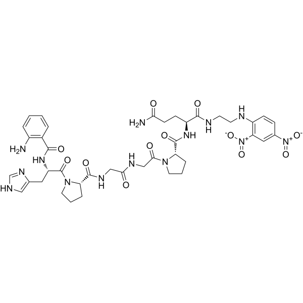
Abz-HPGGPQ-EDDnp
Abz-HPGGPQ-EDDnp (Cathepsin K substrate) is a biological active peptide. (Cathepsins are a class of globular lysosomal proteases, playing a vital role in mammalian cellular turnover. They degrade polypeptides and are distinguished by their substrate specificities. Cathepsin K is the lysosomal cysteine protease involved in bone remodeling and resorption. It has potential as a drug target in autoimmune diseases and osteoporosis.This FRET peptide can be used to monitor selectively cathepsin K activity in physiological fluids and cell lysates. Abz-HPGGPQ-EDDnp [where Abz represents o-aminobenzoic acid and EDDnp represents N -(2, 4-dinitrophenyl)-ethylenediamine], a substrate initially developed for trypanosomal enzymes, is efficiently cleaved at the Gly-Gly bond by cathepsin K. This peptide is resistant to hydrolysis by cathepsins B, F, H, L, S and V, Ex/Em=340 nm/420 nm.)
For research use only. We do not sell to patients.
- CAS No.: 221055-89-6
- Formula: C40H50N14O12
- Molecular Weight:918.91
-
Storage:
Please store the product under the recommended conditions in the Certificate of Analysis.

To place orders, for customer services and technical support, please contact: MedChemExpress USA
Tel: 609-228-6898 E-mail: [email protected] [email protected]


-
 Biological Activity
-
Chemical Information
- Protocols
- Purity & Documentation
-
 Help & FAQs




Biological Activity
Description


Chemical Information
-
CAS No. 221055-89-6
-
Molecular Weight 918.91
-
Formula C40H50N14O12
-
Synonyms
Cathepsin K substrate
-
Sequence
{Abz}-His-Pro-Gly-Gly-Pro-Gln-{EDDnp}
-
Sequence Shortening
{Abz}-HPGGPQ-{EDDnp}
-
Shipping
Room temperature in continental US; may vary elsewhere.
-
Storage
Please store the product under the recommended conditions in the Certificate of Analysis.

Protocols
-
Lysosome and acidic-vesicle live-cell staining
Lysosome and acidic-vesicle live-cell staining detects acidic intracellular compartments by using membrane-permeant acidotropic probes that accumulate in low-pH vesicles, including lysosomes, late endosomes, autolysosomes, and acidic phagosomes. LysoTracker staining is commonly used as an intensity-based readout of acidic lysosomal compartment abundance or enlargement, while acridine orange produces green fluorescence in less concentrated compartments and red fluorescence after concentration-dependent accumulation in acidic vesicular organelles. Loss or reduction of acridine-orange red signal can be used as a readout of lysosomal membrane permeabilization or reduced acidic-vesicle integrity. This protocol is designed for live cultured cells and can be adapted for fluorescence microscopy, high-content imaging, plate-reader readout, or flow cytometry when the selected literature supports the readout. Because these dyes report acidotropic accumulation rather than lysosome identity alone,


Purity & Documentation

Calculators
Concentration (start) × Volume (start) = Concentration (final) × Volume (final)


Abz-HPGGPQ-EDDnp Related Classifications
HY-P5377 Related Classifications
 Powered by Bioz