



ACA-28
Based on 1 publication(s) in Google Scholar
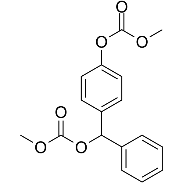
ACA-28 is a potent ERK MAPK signaling modulator. ACA-28 selectively inhibits cancer cell growth and induces apoptosis. ACA-28 can activate nuclear factor erythroid 2-related factor 2 (Nrf2) signaling via inducing ROS production. ACA-28 can be used for the research of cancer, such as melanoma .
For research use only. We do not sell to patients.
- Purity : 99.70%
- CAS No.: 948044-25-5
- Formula: C17H16O6
- Molecular Weight:316.31
-
Storage:Powder -20°C, 3 years , 4°C, 2 years ; In solvent -80°C, 6 months , -20°C, 1 month

To place orders, for customer services and technical support, please contact: MedChemExpress USA
Tel: 609-228-6898 E-mail: [email protected] [email protected]


-
 Biological Activity
-
Chemical Information
-
Solvent & Solubility
- Protocols
- Purity & Documentation
- References
-
 Help & FAQs


-
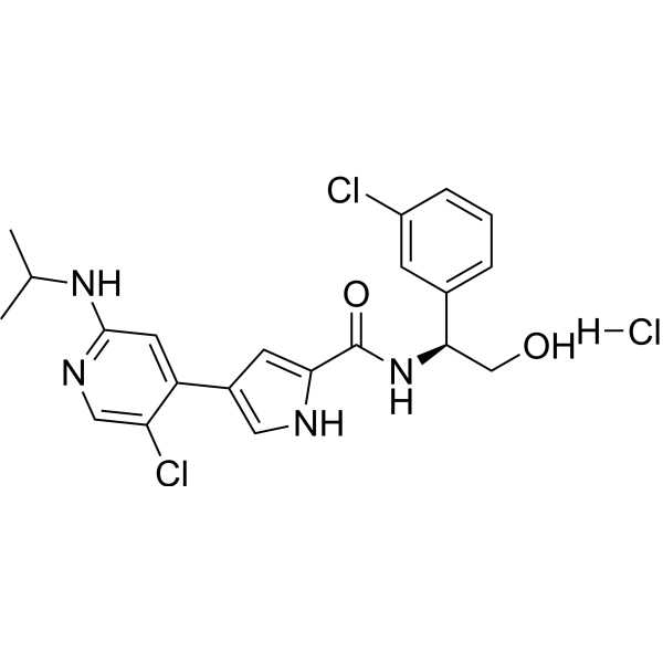
HY-15816A
Ulixertinib hydrochlorideERK1/2 Inhibitor -
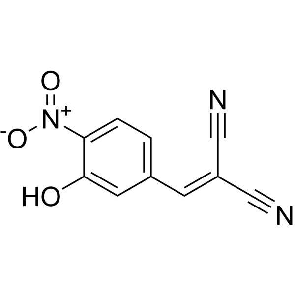
HY-108330
AG126Tyrosine Kinase Inhibitor -
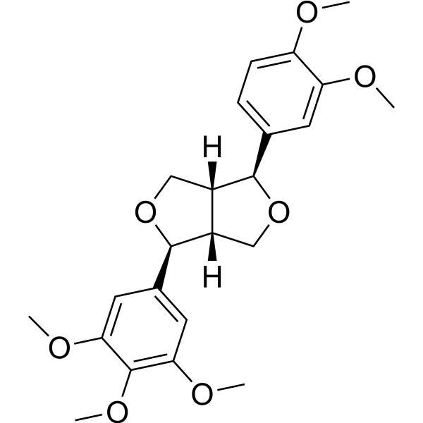
HY-N1374
MagnolinERK Inhibitor -
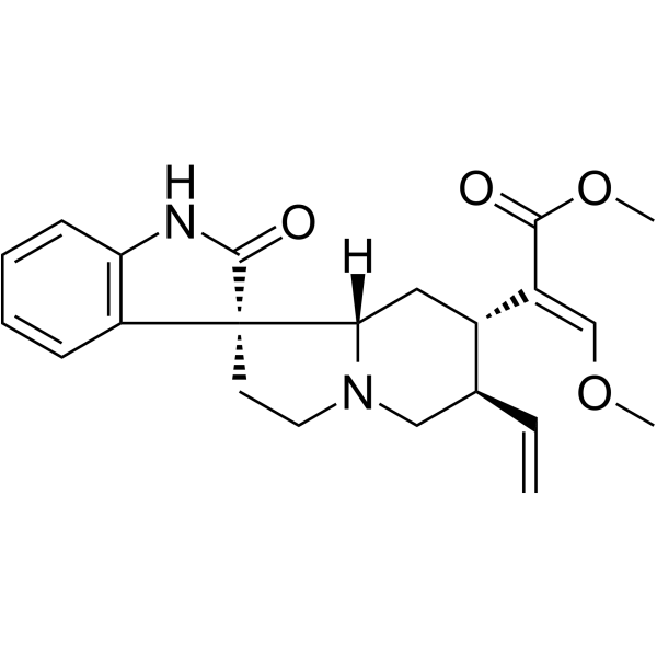
HY-N0590
CorynoxeineERK Inhibitor -
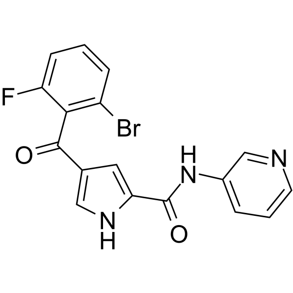
HY-128341
ERK5-IN-2ERK5 Inhibitor -
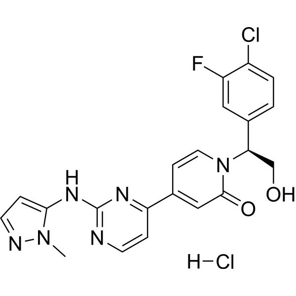
HY-15947A
Ravoxertinib hydrochlorideERK1/2 Inhibitor -
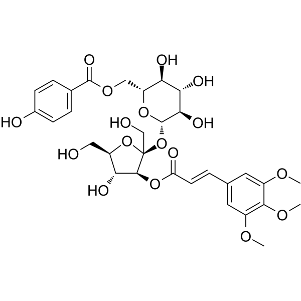
HY-N6076
Tenuifoliside AERK Inhibitor -
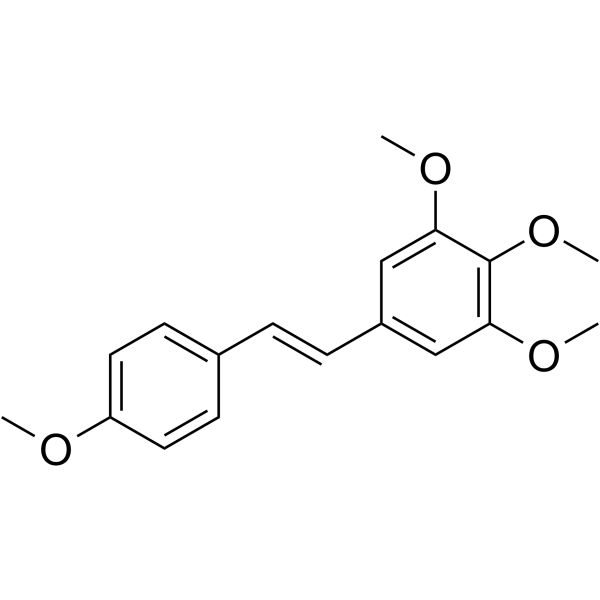
HY-137977
DMU-212Anticancer Compound -
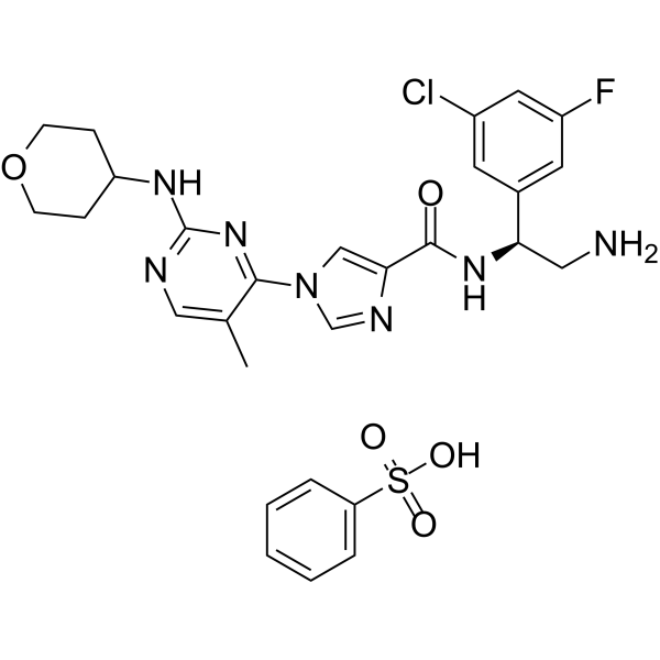
HY-136579A
ASN007 benzenesulfonateERK Inhibitor -
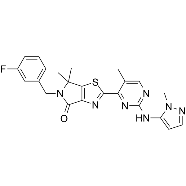
HY-142433
ERK1/2 inhibitor 7ERK Inhibitor -
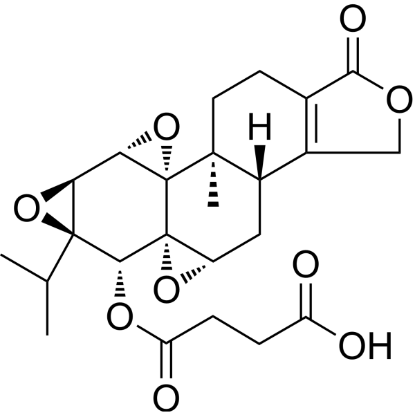
HY-16363
OmtriptolideTriptolide Derivative -
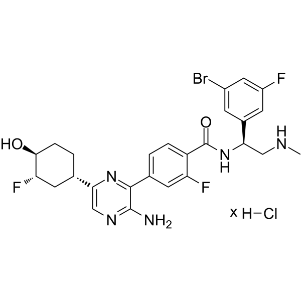
HY-114491A
Rineterkib hydrochlorideERK1/2 Inhibitor
Publications Citing Use of MedChemExpress (MCE) ACA-28
More

Biological Activity
Description
Cellular Effect
|
Cell Line

|
Type | Value | Description | References |
|---|---|---|---|---|
| RBL-2H3 | IC50 |
18 μM
Compound: 9
|
Antiallergic activity against antigen-IgE-mediated degranulation in rat RBL2H3 cells assessed as inhibition of DNP-BSA-induced beta-hexosaminidase release preincubated for 10 mins before DNP-BSA challenge measured after 10 mins
Antiallergic activity against antigen-IgE-mediated degranulation in rat RBL2H3 cells assessed as inhibition of DNP-BSA-induced beta-hexosaminidase release preincubated for 10 mins before DNP-BSA challenge measured after 10 mins
|
[PMID: 19414259] |
In Vitro
ACA-28 inhibits cell growth of melanoma cells (SK-MEL-28) and normal melanocytes (NHEM), with IC50 values of 5.3 and 10.1 μM, respectively[1].
ACA-28 increases p-ERK and p-ELK1 levels in SK-MEL-28 cells[1].
ACA-28 (10 μM, 6 h) increases Nrf2 levels in SK-MEL-28 cells[2].
ACA-28 (20-50 μM, 1 h) induces ROS production in SK-MEL-28 cells[2].
ACA-28 shows an IC50 of 27.9 μM against A549 cells[2].
MedChemExpress (MCE) has not independently confirmed the accuracy of these methods. They are for reference only.
-
Cell Line:SK-MEL-28 cells
-
Concentration:10 μM
-
Incubation Time:6 h
-
Result:Increased Nrf2 levels.
- Detection of Apoptosis
- TUNEL staining for apoptotic DNA fragmentation
- Annexin V plus membrane-impermeant dye apoptosis staining
- Protocol for Cell Counting and Cell Density Analysis
- ROS/oxidative-stress fluorescent staining
- Somatic Cell Culture
- Apoptosis Solutions
- CCK-8/WST-8 Cell Proliferation Assay
- Cell Counting-Based Growth Curve Assay
- MTT Cell Proliferation Assay
- Cell Viability Determination by MTT Colorimetric Assay


Chemical Information
-
CAS No. 948044-25-5
-
Appearance Solid
-
Molecular Weight 316.31
-
Formula C17H16O6
-
Color White to off-white
-
SMILES
O=C(OC)OC1=CC=C(C(C2=CC=CC=C2)OC(OC)=O)C=C1
-
Shipping
Room temperature in continental US; may vary elsewhere.
-
Storage
Powder -20°C 3 years 4°C 2 years In solvent -80°C 6 months -20°C 1 month


Publications (1)
-
Journal Impact Factor
-
Most Recent
-
Cell Signal
Inhibition of ERO1L induces autophagy and apoptosis via endoplasmic reticulum stress in colorectal cancer. [Abstract]2025 Mar:127:111560. PMID: 39657838


Solvent & Solubility
In Vitro:
DMSO : 100 mg/mL (316.15 mM; Need ultrasonic; Hygroscopic DMSO has a significant impact on the solubility of product, please use newly opened DMSO)
Please refer to the solubility information to select the appropriate solvent. Once prepared, please aliquot and store the solution to prevent product inactivation from repeated freeze-thaw cycles.
Storage method and period of stock solution: -80°C, 6 months; -20°C, 1 month. When stored at -80°C, please use it within 6 months. When stored at -20°C, please use it within 1 month.
Please refer to the solubility information to select the appropriate solvent. Once prepared, please aliquot and store the solution to prevent product inactivation from repeated freeze-thaw cycles.
Storage method and period of stock solution: -80°C, 6 months; -20°C, 1 month. When stored at -80°C, please use it within 6 months. When stored at -20°C, please use it within 1 month.
Concentration (start) × Volume (start) = Concentration (final) × Volume (final)
In Vivo:
Select the appropriate dissolution method based on your experimental animal and administration route.
- For the following dissolution methods, please ensure to first prepare a clear stock solution using an In Vitro approach and then sequentially add co-solvents:
- To ensure reliable experimental results, the clarified stock solution can be appropriately stored based on storage conditions. As for the working solution for In Vivo experiments, it is recommended to prepare freshly and use it on the same day.
- The percentages shown for the solvents indicate their volumetric ratio in the final prepared solution. If precipitation or phase separation occurs during preparation, heat and/or sonication can be used to aid dissolution.
Add each solvent one by one: 10% DMSO 40% PEG300 5% Tween-80 45% Saline
Solubility: ≥ 2.5 mg/mL (7.90 mM); Clear solution
This protocol yields a clear solution of ≥ 2.5 mg/mL (saturation unknown).
Taking 1 mL working solution as an example, add 100 μL DMSO stock solution (25.0 mg/mL) to 400 μL PEG300, and mix evenly; then add 50 μL Tween-80 and mix evenly; then add 450 μL Saline to adjust the volume to 1 mL.
Preparation of Saline: Dissolve 0.9 g sodium chloride in ddH₂O and dilute to 100 mL to obtain a clear Saline solution.
Add each solvent one by one: 10% DMSO 90% (20% SBE-β-CD in Saline)
Solubility: ≥ 2.5 mg/mL (7.90 mM); Clear solution
This protocol yields a clear solution of ≥ 2.5 mg/mL (saturation unknown).
Taking 1 mL working solution as an example, add 100 μL DMSO stock solution (25.0 mg/mL) to 900 μL 20% SBE-β-CD in Saline, and mix evenly.
Preparation of 20% SBE-β-CD in Saline (4°C, storage for one week): 2 g SBE-β-CD powder is dissolved in 10 mL Saline, completely dissolve until clear.
In Vivo Dissolution Calculator
Please enter the basic information of animal experiments:
-
-
-
-
Recommended: Prepare an additional quantity of animals to account for potential losses during experiments.
Please enter your animal formula composition:
-
%DMSO +
Recommended: Keep the proportion of DMSO in working solution below 2% if your animal is weak.
-
%+
-
+%Tween-80 + +
-
%Saline +
The co-solvents required include: DMSO, . All of co-solvents are available by MedChemExpress (MCE). , Tween 80. All of co-solvents are available by MedChemExpress (MCE).
Working solution concentration: 0.22 mg/mL
Method for preparing stock solution: mg drug dissolved in μL DMSO. Stock solution concentration: mg/mL.
1. Take μL DMSO stock solution;
2. Add μL .
μL , mix evenly;
3. Then add μL Tween 80, mix evenly;
4. Then add μL
Please ensure that the stock solution in the first step is dissolved to a clear state, and add co-solvents in sequence. You can use ultrasonic heating (ultrasonic cleaner, recommended frequency 20-40 kHz), vortexing, etc. to assist dissolution.


Purity & Documentation
-
Data Sheet (277 KB)
-
SDS (251 KB)
- English - EN (251 KB)
- Français - FR (251 KB)
- Deutsch - DE (251 KB)
- Norwegian - NO (251 KB)
- Español - ES (251 KB)
- Swedish - SV (251 KB)
- Italian - IT (251 KB)
- Korean - KR (251 KB)
- Portuguese - PT (251 KB)
-
Handling Instructions (2659 KB)
References
[1]. Satoh R, et al. Discovery of new benzhydrol biscarbonate esters as potent and selective apoptosis inducers of human melanomas bearing the activated ERK pathway: SAR studies on an ERK MAPK signaling modulator, ACA-28. Bioorg Chem. 2020 Oct;103:104137. [Content Brief]
[2]. Takasaki T, et al. ACA-28, an ERK MAPK Signaling Modulator, Exerts Anticancer Activity through ROS Induction in Melanoma and Pancreatic Cancer Cells. Oxid Med Cell Longev. 2024 Mar 11;2024:7683793. [Content Brief]


Complete Stock Solution Preparation Table
Please refer to the solubility information to select the appropriate solvent. Once prepared, please aliquot and store the solution to prevent product inactivation from repeated freeze-thaw cycles.
Storage method and period of stock solution: -80°C, 6 months; -20°C, 1 month. When stored at -80°C, please use it within 6 months. When stored at -20°C, please use it within 1 month.
| Optional Solvent | Concentration Solvent Mass | 1 mg | 5 mg | 10 mg | 25 mg |
|---|---|---|---|---|---|
| DMSO | 1 mM | 3.1615 mL | 15.8073 mL | 31.6146 mL | 79.0364 mL |
| 5 mM | 0.6323 mL | 3.1615 mL | 6.3229 mL | 15.8073 mL | |
| 10 mM | 0.3161 mL | 1.5807 mL | 3.1615 mL | 7.9036 mL | |
| 15 mM | 0.2108 mL | 1.0538 mL | 2.1076 mL | 5.2691 mL | |
| 20 mM | 0.1581 mL | 0.7904 mL | 1.5807 mL | 3.9518 mL | |
| 25 mM | 0.1265 mL | 0.6323 mL | 1.2646 mL | 3.1615 mL | |
| 30 mM | 0.1054 mL | 0.5269 mL | 1.0538 mL | 2.6345 mL | |
| 40 mM | 0.0790 mL | 0.3952 mL | 0.7904 mL | 1.9759 mL | |
| 50 mM | 0.0632 mL | 0.3161 mL | 0.6323 mL | 1.5807 mL | |
| 60 mM | 0.0527 mL | 0.2635 mL | 0.5269 mL | 1.3173 mL | |
| 80 mM | 0.0395 mL | 0.1976 mL | 0.3952 mL | 0.9880 mL | |
| 100 mM | 0.0316 mL | 0.1581 mL | 0.3161 mL | 0.7904 mL |


ACA-28 Related Classifications
HY-147855 Related Classifications
 Powered by Bioz