



Aducanumab

Based on 1 Customer Validation
Aducanumab (BIIB037) is a human monoclonal antibody that selectively targets aggregated amyloid-beta (Aβ). Aducanumab shows blood-brain-barrier penetration, and can be used for Alzheimer's disease (AD) research.
For research use only. We do not sell to patients.
- Purity : 99.25%
- CAS No.: 1384260-65-4
- Molecular Weight:146.2 kDa
-
Storage:
Please store the product under the recommended conditions in the Certificate of Analysis.


To place orders, for customer services and technical support, please contact: MedChemExpress USA
Tel: 609-228-6898 E-mail: [email protected] [email protected]


-
 Biological Activity
-
Chemical Information
- Protocols
- Purity & Documentation
- References
-
 Help & FAQs


-

HY-P99022
GantenerumabAnti-Aβ Monoclonal Antibody -

HY-P99859
DonanemabAnti-Aβ Antibody -

HY-P99185
BapineuzumabAmyloid Beta Binder -

HY-P99317
SolanezumabAnti-Aβ Antibody -

HY-P99835
CrenezumabAnti-Aβ Antibody -

HY-P990110
Lecanemab (Mouse IgG2a)Monoclonal Antibody -

HY-P99001
Human IgG1 kappa, Isotype ControlIgG1κ Isotype Control -

HY-P9999
TrontinemabAβ/TFRC Antibody -
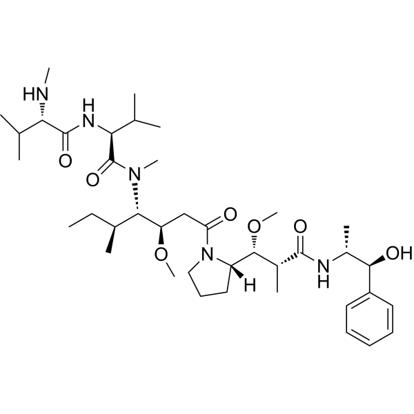
HY-15162
Monomethyl auristatin EAntimitotic Agent -

HY-P9902
PembrolizumabPD-1 Inhibitor -
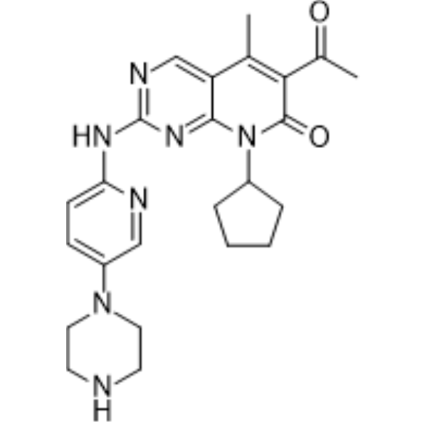
HY-50767
PalbociclibCDK4/6 Inhibitor -

HY-P9913
RituximabCD20 Inhibitor


Biological Activity
Description
Isotype
Human IgG1 kappa
Recommend Isotype Controls
Species Reactivity
Human
IC50 & Target
Amyloid Beta/Aβ
In Vivo
Aducanumab (0.3-30 mg/kg, i.p., weekly, 6 months) reduces soluble and insoluble Aβ in a dose-dependent manner in 9.5- to 15.5-month-old Tg2576 transgenic mice[1].
Aducanumab (10 mg/kg, i.p., weekly, 6 months) restores intracellular calcium to control levels in 18-month-old Tg2576 mice[2].
Aducanumab (0.4-1 mg/mL, ICV, 20 min) leads to rapid decrease in amyloid burden, plaque clearance in Tg2576 mice[2].
MedChemExpress (MCE) has not independently confirmed the accuracy of these methods. They are for reference only.
-
Animal Model:9.5- to 15.5-month-old Tg2576 transgenic mice [1]
-
Dosage:0.3-30 mg/kg
-
Administration:Intraperitoneal injection (i.p.), weekly, 6 months
-
Result:Increased recruitment of Iba-1-positive microglia to Aβ plaques.
-
Animal Model:18-month-old Tg2576 mice[2]
-
Dosage:10 mg/kg
-
Administration:Intraperitoneal injection (i.p.), weekly, 6 months
-
Result:Restored neurite baseline calcium to control levels.
Decreased the number of neurites with elevated levels of calcium after 2 weeks.
Decreased the percentage of cell bodies with calcium overload.
Restored the levels of VILIP and SERCA to control levels.
Increased the cell numbers of NR1 and NR2A.
-
Animal Model:22-month-old transgenic Tg2576 mice[2]
-
Dosage:0.4-1 mg/mL
-
Administration:Intracerebroventricular injection (ICV), 20 min
-
Result:Decreased the number of amyloid plaque.
Decreased the size of the remaining individual plaques.
Reduced amyloid plaque burden.
Clinical Trial
| NCT Number | Sponsor | Condition | Start Date |
Phase

|
|---|---|---|---|---|
| NCT01329991 | Plexxikon| | 2011-05 | PHASE1 |
Gene ID
Accession
Conjugated
Unconjugated
Reconsititution
The product can be reconstituted/diluted with sterile PBS or saline.
Format
-
Human IgG1 kappa
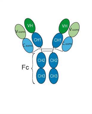
Application
ELISA, FACS, Functional assay
Verified Bioactivity
-
 Immobilized Human Beta-amyloid/Beta-APP Protein (hFc Tag) can bind Aducanumab. The EC50 for this effect is 603.66 ng/mL.
 -
 The EC50 for this effect is 603.66 ng/mL. Flow cytometric analysis of 1.5×10⁶ HUVEC cells with Aducanumab (HY-P9967, red). Cells were fixed with 4% paraformaldehyde and permeabilised with 90% methanol. Then stained with the primary antibody at 1/200 dilution for an hour at 4℃. Goat Anti-Human IgG H&L (AF488) (HY-P83776) was used as the secondary antibody at 1/1,000 dilution for 30 minutes at 4℃. Human IgG1 kappa, Isotype Control (HY-P99001, blue) was used as the isotype control.


Chemical Information
-
CAS No. 1384260-65-4
-
Appearance Liquid
-
Molecular Weight 146.2 kDa
-
Color Colorless to light yellow
-
SMILES
[Aducanumab]
-
Synonyms
BIIB037
-
Shipping
Shipping with dry ice.
-
Formulation
Please refer to the lot-specific COA for specific buffer information.
-
Storage
Please store the product under the recommended conditions in the Certificate of Analysis.

Protocols
-
Amyloid: Congo Red Amyloid Staining
Congo red amyloid staining is a histochemical method used to detect extracellular amyloid deposits in tissue sections based on the affinity of Congo red dye for β-pleated sheet-rich protein aggregates. When bound to amyloid, Congo red produces characteristic apple-green birefringence under polarized light microscopy, which is widely regarded as a diagnostic feature of amyloid deposition in histopathology. The diagnostic principle relies on the combination of dye binding (congophilia) and optical anisotropy under polarized illumination, which distinguishes amyloid from most non-amyloid eosinophilic extracellular deposits in routine histological evaluation. Amyloid identification by Congo red staining remains a cornerstone in diagnostic pathology despite the availability of adjunct methods such as immunohistochemistry and mass spectrometry, particularly because of its ability to localize deposits directly within tissue architecture. The specificity of Congo red-positive deposits is incre
-
Alzheimer’s Disease Modeling
Alzheimer’s Disease (AD) is a neurodegenerative disorder characterized by a progressive decline in cognitive functions and loss of specific types of neurons and synapses. Alzheimer's symptoms can be simulated in mice by injecting drugs (such as Aβ) or genetically modified.


Purity & Documentation
-
Data Sheet (261 KB)
-
SDS (251 KB)
- English - EN (251 KB)
- Français - FR (251 KB)
- Deutsch - DE (251 KB)
- Norwegian - NO (251 KB)
- Español - ES (251 KB)
- Swedish - SV (251 KB)
- Italian - IT (251 KB)
- Korean - KR (251 KB)
- Portuguese - PT (251 KB)
-
Inhibitory Antibodies User Guide (603 KB)
References
[1]. Sevigny J, et al. The antibody aducanumab reduces Aβ plaques in Alzheimer's disease. Nature. 2016 Sep 1;537(7618):50-6. [Content Brief]
[2]. Kastanenka KV, et al. Immunotherapy with Aducanumab Restores Calcium Homeostasis in Tg2576 Mice. J Neurosci. 2016 Dec 14;36(50):12549-12558. [Content Brief]

Calculators
Concentration (start) × Volume (start) = Concentration (final) × Volume (final)


Aducanumab Related Classifications
HY-P9967 Related Classifications
 Powered by Bioz