



AMPK alpha 2 Antibody (YA833)
(Synonyms: AMPK, AMPK2, PRKAA2, 5'-AMP-activated protein kinase catalytic subunit alpha-2, AMPK subunit alpha-2, Acetyl-CoA carboxylase kinase, Hydroxymethylglutaryl-CoA reductase kinase, ACACA kinase, HMGCR kinase)
Based on 1 Customer Validation
AMPK alpha 2 Antibody (YA833) is a Mouse-derived and non-conjugated IgG1 monoclonal antibody, targeting to AMPK alpha 2.
-
Host:
Mouse
-
Isotype:
IgG
-
Application:
WB, ICC/IF, IHC-P
-
Reactivity :
Human, Mouse, Rat
-
Formulation:
Supplied in 1*TBS (pH7.4), 0.2% BSA and 50% Glycerol. Preservative: 0.05% Sodium Azide.
-
Conjugation:
Non-conjugated


To place orders, for customer services and technical support, please contact: MedChemExpress USA
Tel: 609-228-6898 E-mail: [email protected] [email protected]





Applications
| Application |
WB

WB: Western Blot
|
ICC/IF
ICC/IF: Immunocytochemistry/
Immunofluorescence |
IHC-P
IHC-P: Immunohistochemistry-Paraffin
|
|---|---|---|---|
| Dilution Ratio | 1:500 | 1:50 | 1:100-1:500 |


Product Details
AMPK alpha 2 Antibody (YA833) is a Mouse-derived and non-conjugated IgG1 monoclonal antibody, targeting to AMPK alpha 2.
-
Host Mouse

-
Clonality Monoclonal

-
Species ReactivityHuman, Mouse, Rat
-
Observed Molecular WeightObserved band size: 62 kDa
Note: Due to possible protein modifications or aggregation, the molecular weight should be confirmed by actual measurement, and the predicted value is for reference only.

-
Calculated Molecular Weight Predicted band size: 62 kDa

Entrez Gene: 5563 Human ; 108079 Mouse ; 78975 Rat
SwissProt: P54646 Human ; Q8BRK8 Mouse ; Q09137 Rat
OMIM: 600497 Human
Synthetic peptide corresponding to Human AMPK alpha 2.AA range:231-430.
Endogenous
Protein G affinity purified.
Non-conjugated
Unmodified
IgG


Product Properties
-
Appearance
Solution
-
Formulation
Supplied in 1*TBS (pH7.4), 0.2% BSA and 50% Glycerol. Preservative: 0.05% Sodium Azide.
-
Concentration
Batch-dependent, Please check the COA for the concentration of each lot. Check Lot Concentration
-
Storage & Stability
Stored at -20°C for 1 year. Avoid repeated freeze / thaw cycles.
-
Shipping
Shipping with blue ice.


Verification Images
-
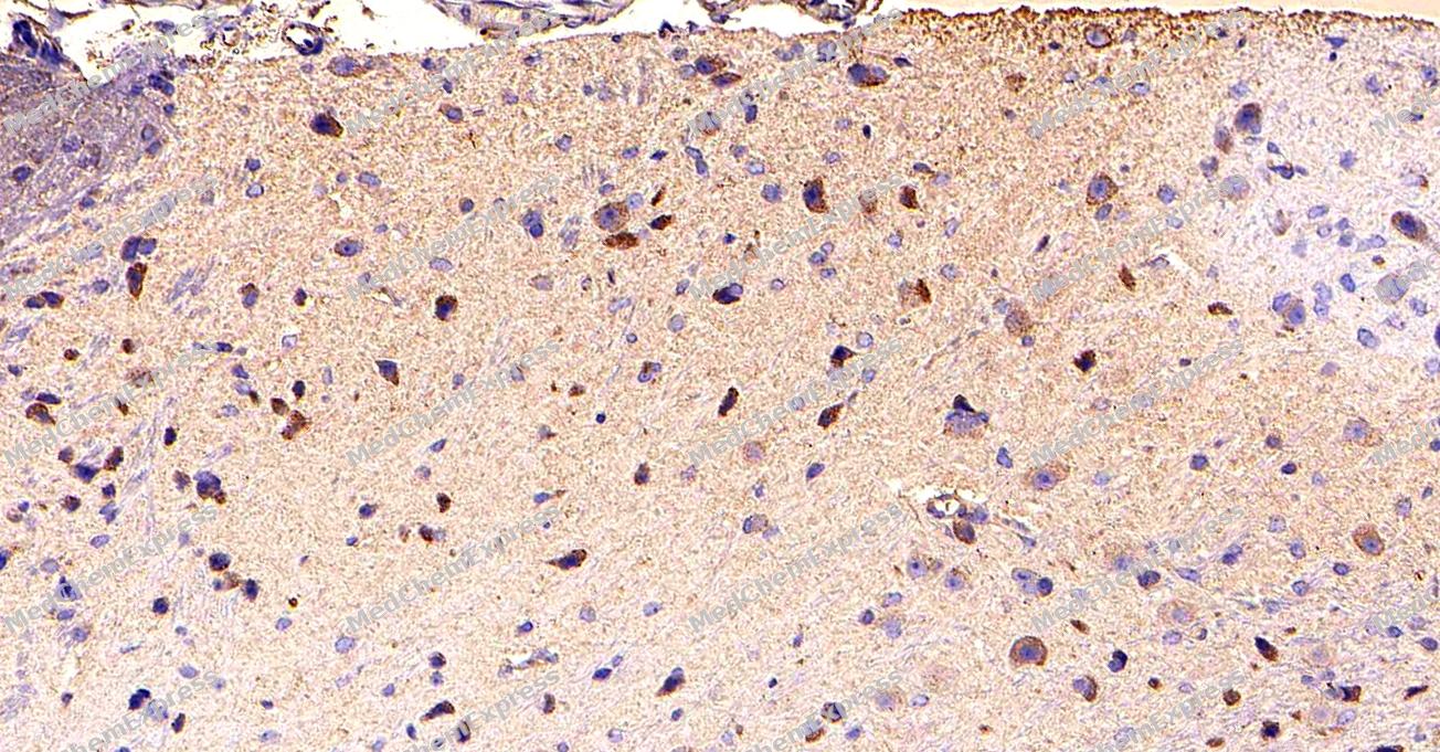 Immunohistochemical analysis of paraffin-embedded Mouse brain tissue using AMPK alpha 2 Antibody (YA833). The section was pre-treated using heat mediated antigen retrieval with sodium citrate buffer (pH 6.0) for 8 minutes. The tissues were blocked in QuickBlock for 20 minutes at room temperature, washed with ddH2O and PBS, and then probed with the primary antibody (HY-P80377, 1/200) in 4℃ overnight. The detection was performed using an HRP conjugated compact polymer system. DAB was used as the chromogen. Tissues were counterstained with hematoxylin and mounted with DPX.
 -
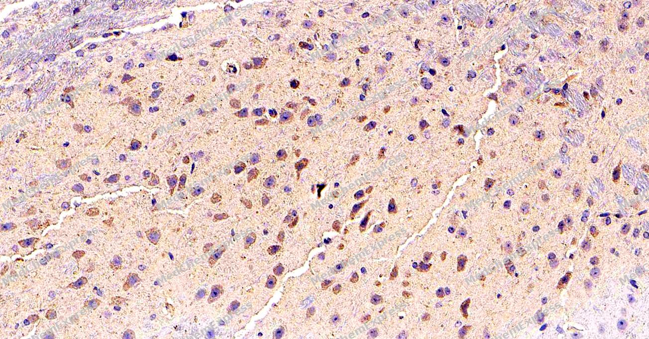 Immunohistochemical analysis of paraffin-embedded Mouse brain tissue using AMPK alpha 2 Antibody (YA833). The section was pre-treated using heat mediated antigen retrieval with sodium citrate buffer (pH 6.0) for 8 minutes. The tissues were blocked in QuickBlock for 20 minutes at room temperature, washed with ddH2O and PBS, and then probed with the primary antibody (HY-P80377, 1/200) in 4℃ overnight. The detection was performed using an HRP conjugated compact polymer system. DAB was used as the chromogen. Tissues were counterstained with hematoxylin and mounted with DPX.
 -
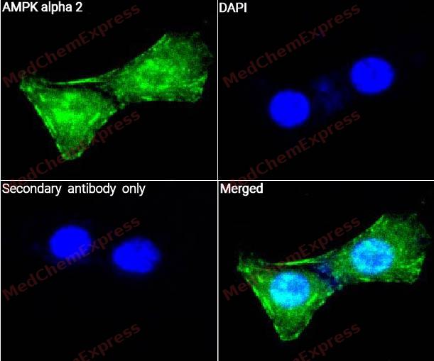 Immunocytochemistry analysis of NIH/3T3 cells labeling AMPK alpha 2 with AMPK alpha 2 (YA833)(HY-P80377) at 1/100 dilution. Cells were fixed in 4% paraformaldehyde for 15 minutes at room temperature, then blocked with QuickBlock™ Blocking Buffer for Immunol Staining for 10 min at room temperature. Cells were then incubated with AMPK alpha 2 (YA833)Antibody(HY-P80377) at 1/100 dilution in QuickBlock™ Blocking Buffer for Immunol Staining at 4 ℃. Alexa Fluor® 488-conjugated AffiniPure Goat Anti-Mouse IgG H&L(HY-P8005, Green) was used as the secondary antibody at 1/1,000 dilution. PBS instead of the primary antibody was used as the secondary antibody only control. The Nuclear counterstain was DAPI (Blue).
 -
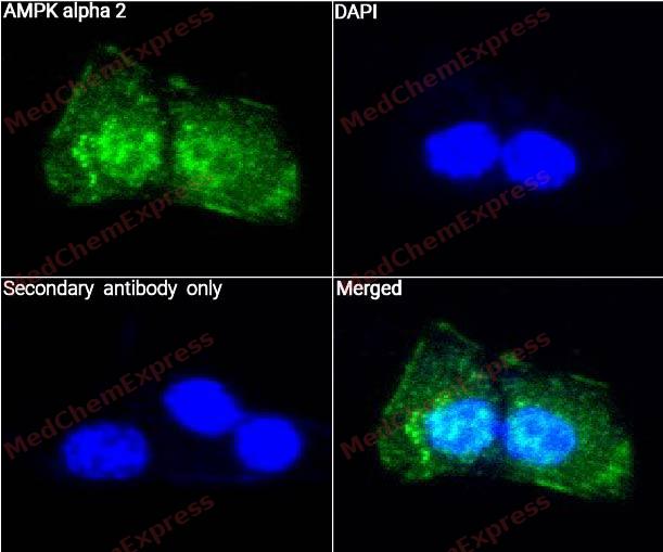 Immunocytochemistry analysis of NIH/3T3 cells labeling AMPK alpha 2 with AMPK alpha 2 Antibody (YA833)(HY-P80377) at 1/50 dilution. Cells were fixed in 4% paraformaldehyde for 15 minutes at room temperature, then blocked with QuickBlock™ Blocking Buffer for Immunol Staining for 10 min at room temperature. Cells were then incubated with AMPK alpha 2 (YA833) Antibody(HY-P80377) at 1/50 dilution in QuickBlock™ Blocking Buffer for Immunol Staining at 4 ℃. Alexa Fluor® 488-conjugated AffiniPure Goat Anti-Mouse IgG H&L(HY-P8005, Green) was used as the secondary antibody at 1/1,000 dilution. PBS instead of the primary antibody was used as the secondary antibody only control. The Nuclear counterstain was DAPI (Blue).


Background
-
Function
AMPK alpha 2 is a Catalytic subunit of AMP-activated protein kinase (AMPK), an energy sensor protein kinase that plays a key role in regulating cellular energy metabolism. In response to reduction of intracellular ATP levels, AMPK activates energy-producing pathways and inhibits energy-consuming processes: inhibits protein, carbohydrate and lipid biosynthesis, as well as cell growth and proliferation. AMPK acts via direct phosphorylation of metabolic enzymes, and by longer-term effects via phosphorylation of transcription regulators. Regulates lipid synthesis by phosphorylating and inactivating lipid metabolic enzymes such as ACACA, ACACB, GYS1, HMGCR and LIPE; regulates fatty acid and cholesterol synthesis by phosphorylating acetyl-CoA carboxylase (ACACA and ACACB) and hormone-sensitive lipase (LIPE) enzymes, respectively. Promotes lipolysis of lipid droplets by mediating phosphorylation of isoform 1 of CHKA (CHKalpha2). Regulates insulin-signaling and glycolysis by phosphorylating IRS1, PFKFB2 and PFKFB3. Involved in insulin receptor/INSR internalization. AMPK stimulates glucose uptake in muscle by increasing the translocation of the glucose transporter SLC2A4/GLUT4 to the plasma membrane, possibly by mediating phosphorylation of TBC1D4/AS160. Regulates transcription and chromatin structure by phosphorylating transcription regulators involved in energy metabolism such as CRTC2/TORC2, FOXO3, histone H2B, HDAC5, MEF2C, MLXIPL/ChREBP, EP300, HNF4A, p53/TP53, SREBF1, SREBF2 and PPARGC1A. Acts as a key regulator of glucose homeostasis in liver by phosphorylating CRTC2/TORC2, leading to CRTC2/TORC2 sequestration in the cytoplasm. In response to stress, phosphorylates 'Ser-36' of histone H2B (H2BS36ph), leading to promote transcription. Acts as a key regulator of cell growth and proliferation by phosphorylating FNIP1, TSC2, RPTOR, WDR24 and ATG1/ULK1: in response to nutrient limitation, negatively regulates the mTORC1 complex by phosphorylating RPTOR component of the mTORC1 complex and by phosphorylating and activating TSC2. Also phosphorylates and inhibits GATOR2 subunit WDR24 in response to nutrient limitation, leading to suppress glucose-mediated mTORC1 activation. In response to energetic stress, phosphorylates FNIP1, inactivating the non-canonical mTORC1 signaling, thereby promoting nuclear translocation of TFEB and TFE3, and inducing transcription of lysosomal or autophagy genes. In response to nutrient limitation, promotes autophagy by phosphorylating and activating ATG1/ULK1. In that process, it also activates WDR45/WIPI4. Phosphorylates CASP6, thereby preventing its autoprocessing and subsequent activation. AMPK also acts as a regulator of circadian rhythm by mediating phosphorylation of CRY1, leading to destabilize it. May regulate the Wnt signaling pathway by phosphorylating CTNNB1, leading to stabilize it. Also acts as a regulator of cellular polarity by remodeling the actin cytoskeleton; probably by indirectly activating myosin. Also phosphorylates CFTR, EEF2K, KLC1, NOS3 and SLC12A1. Plays an important role in the differential regulation of pro-autophagy (composed of PIK3C3, BECN1, PIK3R4 and UVRAG or ATG14) and non-autophagy (composed of PIK3C3, BECN1 and PIK3R4) complexes, in response to glucose starvation. Can inhibit the non-autophagy complex by phosphorylating PIK3C3 and can activate the pro-autophagy complex by phosphorylating BECN1. Upon glucose starvation, promotes ARF6 activation in a kinase-independent manner leading to cell migration. Upon glucose deprivation mediates the phosphorylation of ACSS2 at 'Ser-659', which exposes the nuclear localization signal of ACSS2, required for its interaction with KPNA1 and nuclear translocation. Upon stress, regulates mitochondrial fragmentation through phosphorylation of MTFR1L[1][2][3][4][5][6][7][8][9][10][11][12][13][14][15][16][17][18][19][20][21][22][23].
-
Subcellular Localization
Cytoplasm; Nucleus
-
Subunit
AMPK is a heterotrimer of an alpha catalytic subunit (PRKAA1 or PRKAA2), a beta (PRKAB1 or PRKAB2) and a gamma non-catalytic subunits (PRKAG1, PRKAG2 or PRKAG3) (PubMed:21543851).
-
SwissProt ID
-
Synonyms
AMPK, AMPK2, PRKAA2, 5'-AMP-activated protein kinase catalytic subunit alpha-2, AMPK subunit alpha-2, Acetyl-CoA carboxylase kinase, Hydroxymethylglutaryl-CoA reductase kinase, ACACA kinase, HMGCR kinase
-
Research Field
Neuroscience


Documentation
-
Data Sheet (265 KB)
-
SDS (252 KB)
- English - EN (252 KB)
- Français - FR (252 KB)
- Deutsch - DE (252 KB)
- Norwegian - NO (252 KB)
- Español - ES (252 KB)
- Swedish - SV (252 KB)
- Italian - IT (252 KB)
- Korean - KR (252 KB)
- Portuguese - PT (252 KB)
-
User Guide for Antibodies (1077 KB)
References
[1]. Towler MC, et al. AMP-activated protein kinase in metabolic control and insulin signaling. Circ Res. 2007 Feb 16;100(3):328-41. [Content Brief]
[2]. Hardie DG, et al. AMP-activated/SNF1 protein kinases: conserved guardians of cellular energy. Nat Rev Mol Cell Biol. 2007 Oct;8(10):774-85. [Content Brief]
[3]. Aguan K, et al. Characterization and chromosomal localization of the human homologue of a rat AMP-activated protein kinase-encoding gene: a major regulator of lipid metabolism in mammals. Gene. 1994 Nov 18;149(2):345-50. [Content Brief]
[4]. Liu R, et al. Choline kinase alpha 2 acts as a protein kinase to promote lipolysis of lipid droplets. Mol Cell. 2021 Jul 1;81(13):2722-2735.e9. [Content Brief]
[5]. Boutchueng-Djidjou M, et al. The last enzyme of the de novo purine synthesis pathway 5-aminoimidazole-4-carboxamide ribonucleotide formyltransferase/IMP cyclohydrolase (ATIC) plays a central role in insulin signaling and the Golgi/endosomes protein network. Mol Cell Proteomics. 2015 Apr;14(4):1079-92. [Content Brief]
[6]. Yang W, et al. Regulation of transcription by AMP-activated protein kinase: phosphorylation of p300 blocks its interaction with nuclear receptors. J Biol Chem. 2001 Oct 19;276(42):38341-4. [Content Brief]
[7]. Imamura K, et al. Cell cycle regulation via p53 phosphorylation by a 5'-AMP activated protein kinase activator, 5-aminoimidazole- 4-carboxamide-1-beta-D-ribofuranoside, in a human hepatocellular carcinoma cell line. Biochem Biophys Res Commun. 2001 Sep 21;287(2):562-7. [Content Brief]
[8]. Jones RG, et al. AMP-activated protein kinase induces a p53-dependent metabolic checkpoint. Mol Cell. 2005 Apr 29;18(3):283-93. [Content Brief]
[9]. Greer EL, et al. The energy sensor AMP-activated protein kinase directly regulates the mammalian FOXO3 transcription factor. J Biol Chem. 2007 Oct 12;282(41):30107-19. [Content Brief]
[10]. McGee SL, et al. AMP-activated protein kinase regulates GLUT4 transcription by phosphorylating histone deacetylase 5. Diabetes. 2008 Apr;57(4):860-7. [Content Brief]
[11]. Inoki K, et al. TSC2 mediates cellular energy response to control cell growth and survival. Cell. 2003 Nov 26;115(5):577-90. [Content Brief]
[12]. Alexander A, et al. ATM signals to TSC2 in the cytoplasm to regulate mTORC1 in response to ROS. Proc Natl Acad Sci U S A. 2010 Mar 2;107(9):4153-8. [Content Brief]
[13]. Egan DF, et al. Phosphorylation of ULK1 (hATG1) by AMP-activated protein kinase connects energy sensing to mitophagy. Science. 2011 Jan 28;331(6016):456-61. [Content Brief]
[14]. Dai X, et al. AMPK-dependent phosphorylation of the GATOR2 component WDR24 suppresses glucose-mediated mTORC1 activation. Nat Metab. 2023 Feb;5(2):265-276. [Content Brief]
[15]. Malik N, et al. Induction of lysosomal and mitochondrial biogenesis by AMPK phosphorylation of FNIP1. Science. 2023 Apr 21;380(6642):eabj5559. [Content Brief]
[16]. Bakula D, et al. WIPI3 and WIPI4 β-propellers are scaffolds for LKB1-AMPK-TSC signalling circuits in the control of autophagy. Nat Commun. 2017 May 31;8:15637. [Content Brief]
[17]. Zhao P, et al. An AMPK-caspase-6 axis controls liver damage in nonalcoholic steatohepatitis. Science. 2020 Feb 7;367(6478):652-660. [Content Brief]
[18]. Lee JH, et al. Energy-dependent regulation of cell structure by AMP-activated protein kinase. Nature. 2007 Jun 21;447(7147):1017-20. [Content Brief]
[19]. Hallows KR, et al. Physiological modulation of CFTR activity by AMP-activated protein kinase in polarized T84 cells. Am J Physiol Cell Physiol. 2003 May;284(5):C1297-308. [Content Brief]
[20]. McDonald A, et al. Cell-wide analysis of secretory granule dynamics in three dimensions in living pancreatic beta-cells: evidence against a role for AMPK-dependent phosphorylation of KLC1 at Ser517/Ser520 in glucose-stimulated insulin granule movement. Biochem Soc Trans. 2010 Feb;38(Pt 1):205-8. [Content Brief]
[21]. Chen KJ, et al. AMPK promotes Arf6 activation in a kinase-independent manner upon glucose starvation. J Cell Sci. 2022 Sep 15;135(18):. [Content Brief]
[22]. Li X, et al. Nucleus-Translocated ACSS2 Promotes Gene Transcription for Lysosomal Biogenesis and Autophagy. Mol Cell. 2017 Jun 1;66(5):684-697.e9. [Content Brief]
[23]. Tilokani L, et al. AMPK-dependent phosphorylation of MTFR1L regulates mitochondrial morphology. Sci Adv. 2022 Nov 11;8(45):eabo7956. [Content Brief]

 Powered by Bioz
