



p27 KIP 1 Antibody (YA5397)
p27 KIP 1 Antibody (YA5397) is a Mouse-derived and non-conjugated monoclonal antibody, targeting to p27 KIP 1.
-
Host:
Mouse
-
Application:
IHC-P, WB
-
Reactivity :
Human, Mouse, Rat
-
Formulation:
Supplied in PBS containing 50% glycerol, 0.5% BSA and 0.02% sodium azide.
-
Conjugation:
Non-conjugated


To place orders, for customer services and technical support, please contact: MedChemExpress USA
Tel: 609-228-6898 E-mail: [email protected] [email protected]





Applications
| Application |
IHC1

|
WB
WB: Western Blot
|
|---|---|---|
| Dilution Ratio | 50-200 | 1:1000-2000 |


Product Details
p27 KIP 1 Antibody (YA5397) is a Mouse-derived and non-conjugated monoclonal antibody, targeting to p27 KIP 1.
-
Host Mouse

-
Clonality Monoclonal

-
Species ReactivityHuman, Mouse, Rat
-
Observed Molecular WeightObserved band size: 27 kDa
Note: Due to possible protein modifications or aggregation, the molecular weight should be confirmed by actual measurement, and the predicted value is for reference only.

Synthesized peptide derived from human P27 Kip1
affinity chromatography.
Non-conjugated
Unmodified


Product Properties
-
Appearance
Liquid
-
Formulation
Supplied in PBS containing 50% glycerol, 0.5% BSA and 0.02% sodium azide.
-
Storage & Stability
Stored at -20°C for 1 year. Avoid repeated freeze / thaw cycles.
-
Shipping
Shipping with blue ice.


Verification Images
-
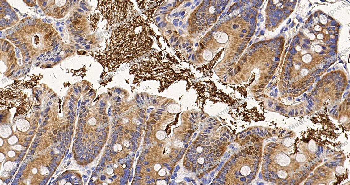 Immunohistochemical analysis of paraffin-embedded mouse colon tissue using P27 Kip1 Antibody. The section was pre-treated using heat mediated antigen retrieval with sodium citrate buffer (pH 6.0) for 8 minutes. The tissues were blocked in QuickBlock for 20 minutes at room temperature, washed with ddH2O and PBS, and then probed with the primary antibody at 1/100 dilution in 4℃ overnight. The detection was performed using an HRP conjugated compact polymer system. DAB was used as the chromogen. Tissues were counterstained with hematoxylin and mounted with DPX.
 -
Immunohistochemical analysis of paraffin-embedded mouse colon tissue using P27 Kip1 Antibody. The section was pre-treated using heat mediated antigen retrieval with sodium citrate buffer (pH 6.0) for 8 minutes. The tissues were blocked in QuickBlock for 20 minutes at room temperature, washed with ddH2O and PBS, and then probed with the primary antibody at 1/100 dilution in 4℃ overnight. The detection was performed using an HRP conjugated compact polymer system. DAB was used as the chromogen. Tissues were counterstained with hematoxylin and mounted with DPX.


Background
-
Function
p27 KIP 1 is important regulator of cell cycle progression. Inhibits the kinase activity of CDK2 bound to cyclin A, but has little inhibitory activity on CDK2 bound to SPDYA. Involved in G1 arrest. Potent inhibitor of cyclin E- and cyclin A-CDK2 complexes. Forms a complex with cyclin type D-CDK4 complexes and is involved in the assembly, stability, and modulation of CCND1-CDK4 complex activation. Acts either as an inhibitor or an activator of cyclin type D-CDK4 complexes depending on its phosphorylation state and/or stoichometry[1].
-
Subcellular Localization
Nucleus; Cytoplasm; Endosome
-
Expression
Tissue_specificity:It is expressed in the kidney (protein level) (PubMed: 15509543) . It is expressed in all tissues tested (PubMed: 8033212) . The highest content is found in skeletal muscle, and the lowest content is found in the liver and kidney (PubMed: 8033212) .
Induction:Maximal levels in quiescence cells and early G (1) . Levels decrease after mitogen stimulation as cells progress toward S-phase -
Subunit
Forms a ternary complex composed of CCNE1, CDK2 and CDKN1B. Interacts directly with CCNE1; the interaction is inhibited by CDK2-dependent phosphorylation on Thr-187.
-
SwissProt ID


Documentation

 Powered by Bioz
