
Anticancer agent 147
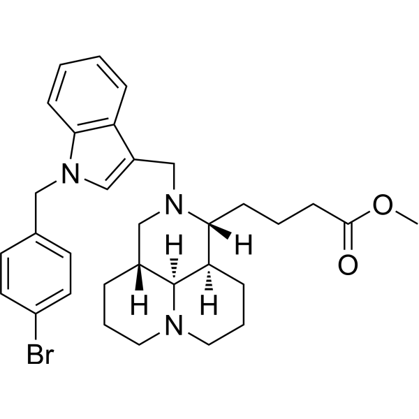
Anticancer agent 147 (compound 6j) is a derivative of sophoridine (HY-N1373) and is a ferroptosis inducer. Anticancer agent 147 can promote the accumulation of intracellular Fe2+, reactive oxygen species (ROS) and MDA, increase ER stress, and upregulate the expression of the activating transcription factor ATF3. Anticancer agent 147 has good anti-liver cancer effects in vitro and in vivo.
For research use only. We do not sell to patients.
- CAS No.: 2241283-11-2
- Formula: C32H40BrN3O2
- Molecular Weight:578.58
-
Storage:
Please store the product under the recommended conditions in the Certificate of Analysis.
Biological Activity
Description
Cellular Effect
|
Cell Line
|
Type | Value | Description | References |
|---|---|---|---|---|
| A549 | IC50 |
3.5 μM
Compound: 6j
|
Cytotoxicity against human A549 cells assessed as reduction in cell viability after 48 hrs by MTT assay
|
[PMID: 30025344] |
| CNE-2 | IC50 |
4.3 μM
Compound: 6j
|
Cytotoxicity against human CNE2 cells assessed as reduction in cell viability after 48 hrs by MTT assay
|
[PMID: 30025344] |
| HepG2 | IC50 |
4.8 μM
Compound: 6j
|
Cytotoxicity against human HepG2 cells assessed as reduction in cell viability after 48 hrs by MTT assay
|
[PMID: 30025344] |
Chemical Information
-
CAS No. 2241283-11-2
-
Molecular Weight 578.58
-
Formula C32H40BrN3O2
-
SMILES
COC(CCC[C@]1([H])[C@]2([H])[C@]3([H])[C@@](CCCN3CCC2)([H])CN1CC(C4=C5C=CC=C4)=CN5CC6=CC=C(Br)C=C6)=O
-
Shipping
Room temperature in continental US; may vary elsewhere.
-
Storage
Please store the product under the recommended conditions in the Certificate of Analysis.
Protocols
-
Nuclear Protein Extraction (High-Salt/Hypotonic Fractionation)
The high-salt/hypotonic fractionation method for nuclear protein extraction is based on the differential solubility of cellular components. Cytoplasmic proteins are extracted first using a hypotonic buffer that causes cell swelling and membrane rupture, followed by centrifugation to separate the cytoplasmic supernatant from the nuclear pellet. The nuclear pellet is then subjected to high-salt extraction (e. g. , 0. 4 M (NH4)2SO4 or 1 M NaCl) to solubilize tightly bound nuclear matrix proteins, including transcription factors, histones, and structural proteins associated with chromatin and the nuclear scaffold. This approach allows for the isolation of both soluble cytoplasmic proteins and salt-resistant nuclear proteins while minimizing cross-contamination.
-
RNA extraction experimental
By lysing cells, releasing RNA, and removing impurities such as proteins and DNA, high-purity RNA products are finally obtained. The commonly used traditional method is the guanidine isothiocyanate/phenol/chloroform method (Trizol), which is suitable for a variety of animal materials including animal tissues, microorganisms, cultured cells, etc., and most plant materials.
-
ROS/oxidative-stress fluorescent staining
ROS/oxidative-stress fluorescent staining uses cell-permeant fluorogenic probes that become fluorescent after oxidation inside cells or tissues; commonly used examples include DCFH-DA/DCFDA for broad cellular oxidant detection, DHE for superoxide-related signal detection, MitoSOX for mitochondrial superoxide-related signal detection, and CellROX probes for oxidative-stress-associated fluorescence readouts. The assay detects probe oxidation rather than a single ROS species unless the probe and analysis method have been chemically validated for that species. DCFH-DA enters cells, is deacetylated by intracellular esterases to DCFH, and produces fluorescent DCF after oxidation, so the readout is used as an operational measure of total cellular oxidative stress rather than a species-specific ROS measurement. DHE and MitoSOX can report superoxide-related oxidation, but red fluorescence alone can include non-specific ethidium-like oxidation products; HPLC or optimized spectral approaches are
-
Ferroptosis Solutions
Ferroptosis is an iron-dependent, non-apoptotic form of regulated cell death characterized by lethal lipid peroxidation and sensitivity to suppression by iron chelators or lipophilic radical-trapping antioxidants. The core pathway links cystine uptake through system Xc−, glutathione availability, GPX4-dependent detoxification of phospholipid hydroperoxides, iron-dependent oxidative reactions, and polyunsaturated-phospholipid metabolism into a cell-death program that is biochemically and morphologically distinct from apoptosis, necrosis, and autophagy. The ferroptosis pathway is experimentally linked to phenotype through chemical and genetic perturbation. Erastin induces ferroptosis by inhibiting cystine uptake through system Xc− and weakening antioxidant defenses, while GPX4 inhibition or depletion causes lipid peroxide accumulation and ferroptotic cancer-cell death. ACSL4 and oxidizable arachidonoyl- or adrenoyl-containing phosphatidylethanolamines shape ferroptosis sensitivity by con
-
Liver Cancer Modeling
Liver cancer can be classified into primary liver cancer and secondary liver cancer. Secondary liver cancer is the metastatic liver cancer. Primary liver cancer includes hepatocellular carcinoma (HCC), intrahepatic cholangiocarcinoma (ICC) and fibrolamellar HCC, of which HCC is the most common form, accounting for approximately 90% of primary liver cancers[1]. HCC mouse models include chemical agent-induced models, transplanted tumor models, and genetic engineered models.
Purity & Documentation
References
Calculators
Concentration (start) × Volume (start) = Concentration (final) × Volume (final)

