



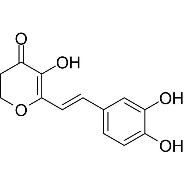
Antioxidant agent-8
Antioxidant agent-8 is an orally active inhibitor of Aβ1-42 deposition. Antioxidant agent-8 inhibits fibril aggregation (IC50=11.15 µM) and promotes fibril disaggregation (IC50=6.87 µM). Antioxidant agent-8 also inhibits Cu2+-induced Aβ1-42 fibril aggregation (IC50=3.69 µM) and promotes Cu2+-induced Aβ1-42 fibril disaggregation (IC50=3.35 µM). Antioxidant agent-8 has antioxidant activity, anti-inflammatory activity, biosafety, blood-brain barrier permeability and neuroprotective effect.
For research use only. We do not sell to patients.
- Formula: C13H12O5
- Molecular Weight:248.23
-
Storage:
Please store the product under the recommended conditions in the Certificate of Analysis.

To place orders, for customer services and technical support, please contact: MedChemExpress USA
Tel: 609-228-6898 E-mail: [email protected] [email protected]


-
 Biological Activity
-
Chemical Information
- Protocols
- Purity & Documentation
- References
-
 Help & FAQs




Biological Activity
Description
In Vitro
Antioxidant agent-8 (compound 30) (50 µM; 24 h) selectively chelates with Cu2+, Fe2+, Zn2+, Fe3+ and Al3+ metal ions, significantly inhibits self- and Cu2+-induced Aβ1-42 fibril aggregation and disaggregation[1].
Antioxidant agent-8 (2.5, 5 and 10 µM; 24 h) promotes BV-2 cells to clear Aβ1-42, reduces Aβ1-42 induced apoptosis and protects nerves with concentration-dependent manner[1].
MedChemExpress (MCE) has not independently confirmed the accuracy of these methods. They are for reference only. Further protocols information, click here.
-
Cell Line:Mouse microglia BV-2 cells.
-
Concentration:2.5, 5 and 10 µM.
-
Incubation Time:24 h.
-
Result:Reduced the expression level of Aβ1-42 in cells.
-
Cell Line:Mouse microglia BV-2 cells.
-
Concentration:2.5, 5 and 10 µM.
-
Incubation Time:24 h.
-
Result:Significantly reduced Aβ1-42 induced apoptosis (cell apoptosis rate were below 30%).
-
Cell Line:Mouse microglia BV-2 cells.
-
Concentration:2.5, 5 and 10 µM.
-
Incubation Time:24 h.
-
Result:Promoted cell viability and the cell survival was 75.50 % (10 µM).
In Vivo
Antioxidant agent-8 (2000 mg/kg; i.g.; single dose) exhibits biosafety[1].
Antioxidant agent-8 (20 mg/kg; p.o.; once daily for 25 d) significantly improves anxiety, memory impairment and cognitive impairment caused by Scopolamine (HY-N0296)[1].
MedChemExpress (MCE) has not independently confirmed the accuracy of these methods. They are for reference only.
-
Animal Model:Sprague-Dawley rats[1].
-
Dosage:15 mg/kg.
-
Administration:Intragastric administration; single dose.
-
Result:Appeared in plasma and hippocampus at 0.083, 0.167, 0.25, 0.5, 1, 2 and 4 hours after administration, and then gradually gathered in hippocampus.
-
Animal Model:Mice[1].
-
Dosage:2000 mg/kg.
-
Administration:Intragastric administration; single dose.
-
Result:Showed insignificant toxic and side effects on heart, liver, spleen and brain.
-
Animal Model:SCOP-induced cognitive impairment in ICR mice (25-28 g)[1].
-
Dosage:20 mg/kg.
-
Administration:Oral gavage; from day 7 to day 31, after 30 min of SCOP administration.
-
Result:Improved animal behavior, learning and memory.


Chemical Information
-
Molecular Weight 248.23
-
Formula C13H12O5
-
SMILES
O=C1CCOC(/C=C/C2=CC=C(C(O)=C2)O)=C1O
-
Shipping
Room temperature in continental US; may vary elsewhere.
-
Storage
Please store the product under the recommended conditions in the Certificate of Analysis.

Protocols
-
Cell Cytotoxicity Assay
Cytotoxicity assays are usually based on the assessment of cell membrane damage, which can also be indirectly detected by measuring cell viability. Detection methods include MTT assay, CKK-8 assay, LDH assay and ATP assay, etc.
-
Cotton Pellet Granuloma
Cotton pellet granuloma is a classical in vivo chronic inflammation model used to evaluate the anti-inflammatory potential of test substances by measuring their ability to inhibit granuloma tissue formation around an implanted foreign body (cotton pellet) in rodents. The method is based on the biological response to a sterile implanted material, which induces proliferative phase inflammation characterized by fibroblast proliferation and collagen-rich granuloma formation, and the final readout reflects the extent of chronic inflammatory tissue growth surrounding the pellet. In multiple preclinical pharmacological evaluations, inhibition of cotton pellet-induced granuloma formation has been used as an indicator of anti-inflammatory activity in both synthetic and natural product screening contexts.
-
Carrageenan-Induced Paw Edema
Carrageenan-induced paw edema is an acute inflammation model in which intraplantar injection of carrageenan induces localized inflammatory swelling characterized by vascular permeability, leukocyte infiltration, and production of inflammatory mediators such as prostaglandins and cytokines, making it widely used to evaluate anti-inflammatory agents in vivo. The resulting paw volume or thickness increase is quantified over time as a direct readout of inflammatory intensity and drug efficacy, typically reflecting cyclooxygenase-mediated prostaglandin-driven edema formation and immune cell recruitment in peripheral tissue[20].
-
Transepithelial/transendothelial electrical resistance assay
TEER measures electrical resistance across epithelial or endothelial monolayers cultured on permeable supports, and the readout reflects ionic conductance through the cell barrier, especially the paracellular pathway regulated by junctional integrity. TEER can be measured without destroying the monolayer and is commonly used before or during transport, permeability, barrier-disruption, and barrier-maturation experiments. TEER values are influenced by biological maturation and technical conditions; reported factors include temperature, medium formulation, passage number, electrode geometry, membrane properties, and junctional length during early monolayer maturation. Therefore, TEER should be interpreted with blank-insert subtraction, area normalization, repeated readings, and, when possible, orthogonal barrier readouts such as FITC-dextran flux or tight-junction staining.
-
Research Protocol for Inflammation-related Diseases
The NLRP3 inflammasome is a cytosolic innate immune signaling platform that integrates priming signals and danger-signal activation to promote caspase-1 activation, maturation of IL-1β and IL-18, and gasdermin D-mediated pyroptotic cell death. The core experimental logic is to determine whether inflammatory disease phenotypes are driven by increased NLRP3 expression, ASC-containing inflammasome assembly, caspase-1 cleavage, GSDMD cleavage, and extracellular release of IL-1β/IL-18 rather than by nonspecific cell injury alone. The pathway is strongly linked to inflammation-related disease phenotypes because monosodium urate crystals activate NALP3/NLRP3 inflammasome signaling in gout-like crystal inflammation, cholesterol crystals activate NLRP3 inflammasomes in atherogenesis models, and DSS-induced intestinal inflammation has been reported to involve NLRP3 inflammasome activity. However, experimental colitis studies also show context-dependent protective effects of NLRP3 inflammasome co


Purity & Documentation
References

Calculators
Concentration (start) × Volume (start) = Concentration (final) × Volume (final)


Antioxidant agent-8 Related Classifications
HY-152506 Related Classifications
 Powered by Bioz