



Crystal Violet-d6
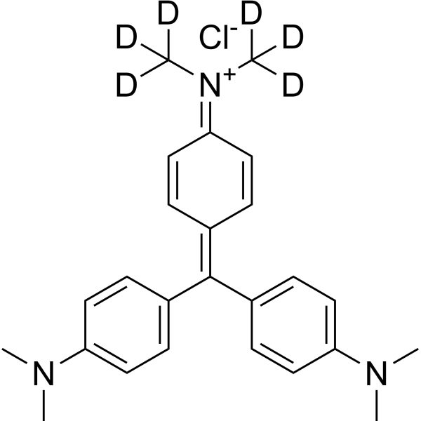
Crystal Violet-d6 is the deuterium labeled Crystal Violet (HY-B0324A). Crystal Violet, also known as Gentian violet, methyl violet 10B, is a triphenyl-methane, an alkaline dye that binds to DNA in the nucleus of a cell, staining it a deep purple. It is often used for Gram staining to classify bacteria, or for cell or histological staining.
Nur für Forschungszwecke. Wir verkaufen nicht an Patienten.
- CAS. Nr.: 1266676-01-0
- Formel: C25H24D6ClN3
- Molecular Weight:414.02
-
Speicherung:
Please store the product under the recommended conditions in the Certificate of Analysis.

To place orders, for customer services and technical support, please contact: MedChemExpress USA
Tel: 609-228-6898 E-mail: [email protected] [email protected]


-
 Biologische Aktivität
-
Chemical Information
- Protokoll
- Reinheit & Dokumentation
- Verweise
-
 Help & FAQs




Biologische Aktivität
Beschreibung
In Vitro
Stable heavy isotopes of hydrogen, carbon, and other elements have been incorporated into drug molecules, largely as tracers for quantitation during the drug development process. Deuteration has gained attention because of its potential to affect the pharmacokinetic and metabolic profiles of drugs[75].
MedChemExpress (MCE) has not independently confirmed the accuracy of these methods. They are for reference only.
Anwendung
1. This compound can be used as a tracer
2. This compound can be used as an internal standard for quantitative analysis by NMR, GC-MS, or LC-MS.


Chemical Information
-
CAS. Nr. 1266676-01-0
-
Unlabeled CAS 548-62-9
-
Molecular Weight 414.02
-
Formel C25H24D6ClN3
-
SMILES
CN(C)C(C=C1)=CC=C1/C(C2=CC=C(C=C2)N(C)C)=C3C=C/C(C=C/3)=[N+](C([2H])([2H])[2H])/C([2H])([2H])[2H].[Cl-]
-
Synonyms
Basic Violet 3-d6; Gentian Violet-d6; Methyl Violet 10B-d6
-
Versand
Room temperature in continental US; may vary elsewhere.
-
Speicherung
Please store the product under the recommended conditions in the Certificate of Analysis.


Reinheit & Dokumentation
Verweise

Calculators
Konzentration (Stammlösung) × Volumen (Stammlösung) = Konzentration (Ziellösung) × Volumen (Ziellösung)


Crystal Violet-d6 Related Classifications
HY-B0324AS Related Classifications
 Powered by Bioz