



Lipoxin A4
Based on 2 publication(s) in Google Scholar
Lipoxin A4 (LXA4), an endogenous lipoxygenase-derived eicosanoid mediator, has potent dual pro-resolving and anti-inflammatory properties. Lipoxin A4 inhibits proliferation and inflammatory cytokine/chemokine production of human epidermal keratinocytes (NHEKs) associated with the ERK1/2 and NF-kB pathways. Lipoxin A4 inhibits serum amyloid A (SAA)-mediated IL-8 release with an IC50 value of 25.74 nM.
For research use only. We do not sell to patients.
- Purity : 98.0%
- CAS No.: 89663-86-5
- Formula: C20H32O5
- Molecular Weight:352.47
-
Storage:
Solution, -20°C, 1 year

To place orders, for customer services and technical support, please contact: MedChemExpress USA
Tel: 609-228-6898 E-mail: [email protected] [email protected]


-
 Biological Activity
-
Chemical Information
- Protocols
- Purity & Documentation
- References
-
 Help & FAQs


-
HY-125527
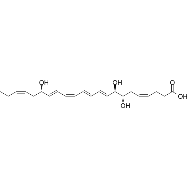
Resolvin D1Endogenous Inflammation Mediator -
HY-P1119
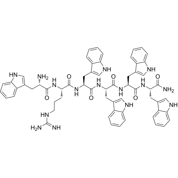
WRW4FPRL1 Antagonist -
HY-116953
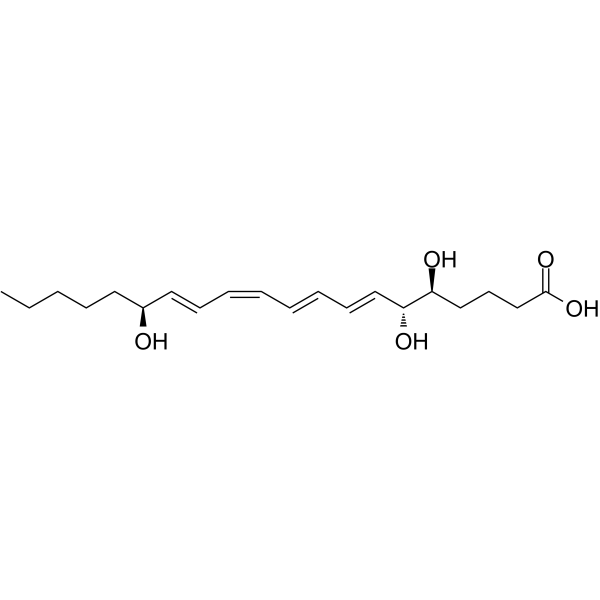
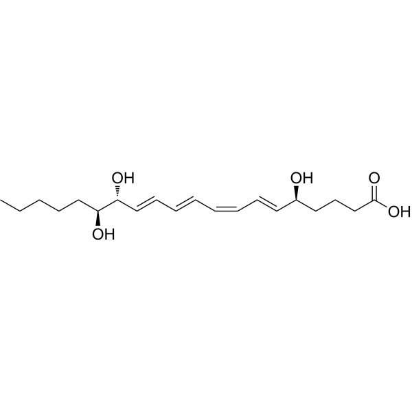
Lipoxin B4Anti-inflammatory Agent -
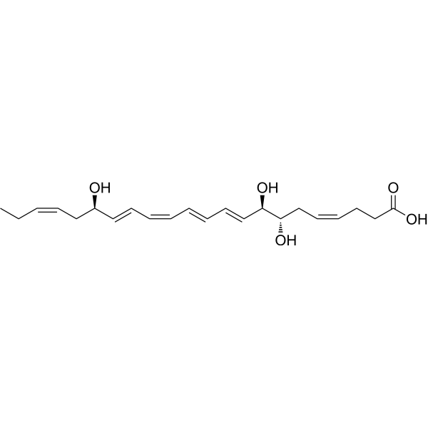
HY-125527A
17(R)-Resolvin D1TRPV3 Inhibitor -
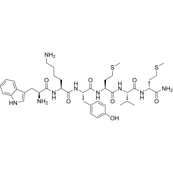
HY-P1120
WKYMVmN-Formyl Peptide Receptor Agonist -

HY-113509B
15(R)-Lipoxin A4Specialized Pro-resolving Mediator -

HY-P9917
TocilizumabIL-6R Neutralizing Antibody -
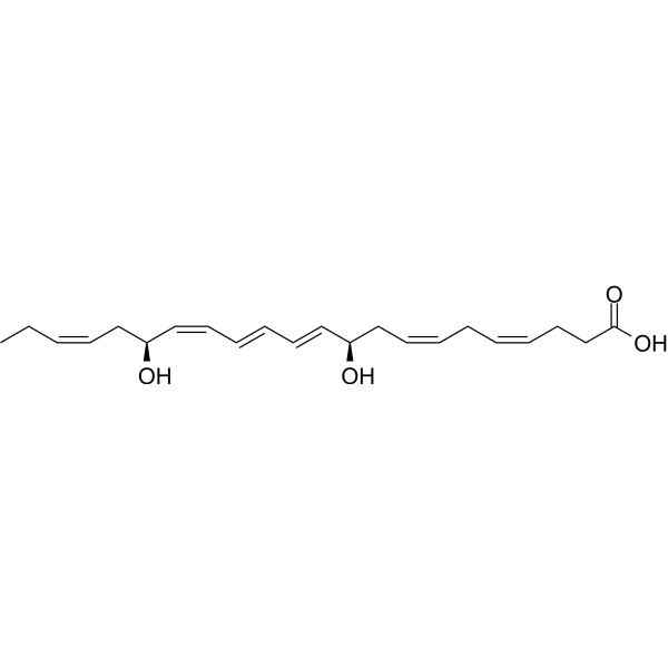
HY-130413
Protectin D1Neuroprotectin D1 -
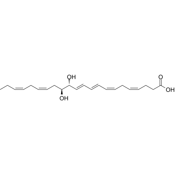
HY-119684
Maresin 2Anti-inflammatory And Pro-resolving Mediator -
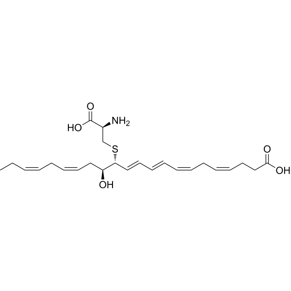
HY-125516
MCTR3Pro-resolving Mediator -
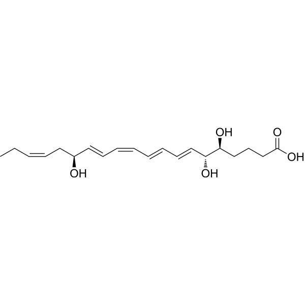
HY-115381
Lipoxin A5Endogenous Metabolite -
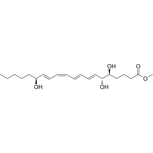
HY-126722
Lipoxin A4 methyl esterLipoxin Analog
Publications Citing Use of MedChemExpress (MCE) Lipoxin A4
MoreAll Endogenous Metabolite Isoforms
More

Biological Activity
Description
IC50 & Target
|
IL-6 |
IL-8 |
Human Endogenous Metabolite |
Cellular Effect
|
Cell Line

|
Type | Value | Description | References |
|---|---|---|---|---|
| THP-1 | EC50 |
5 nM
Compound: 1
|
Induction of phagocytosis in human THP-1-MF0 macrophages pretreated for 30 mins followed by fluorescently labeled Escherichia coli derived bio-particles addition and measured after 2 hrs
Induction of phagocytosis in human THP-1-MF0 macrophages pretreated for 30 mins followed by fluorescently labeled Escherichia coli derived bio-particles addition and measured after 2 hrs
|
[PMID: 34138563] |
| THP-1 | IC50 |
1 pM
Compound: 1
|
Anti-inflammatory activity against human THP-1 monocytes assessed as reduction in LPS-induced IFNgamma release pretreated for 30 mins followed LPS stimulation and measured after 24 hrs by multiplex chemiluminescence detection method
Anti-inflammatory activity against human THP-1 monocytes assessed as reduction in LPS-induced IFNgamma release pretreated for 30 mins followed LPS stimulation and measured after 24 hrs by multiplex chemiluminescence detection method
|
[PMID: 34138563] |
| THP-1 | IC50 |
1 pM
Compound: 1
|
Anti-inflammatory activity against human THP-1 monocytes assessed as reduction in LPS-induced IL-1beta release pretreated for 30 mins followed LPS stimulation and measured after 24 hrs by multiplex chemiluminescence detection method
Anti-inflammatory activity against human THP-1 monocytes assessed as reduction in LPS-induced IL-1beta release pretreated for 30 mins followed LPS stimulation and measured after 24 hrs by multiplex chemiluminescence detection method
|
[PMID: 34138563] |
| THP-1 | IC50 |
1 pM
Compound: 1
|
Anti-inflammatory activity against human THP-1 monocytes assessed as reduction in LPS-induced IL-6 release pretreated for 30 mins followed LPS stimulation and measured after 24 hrs by multiplex chemiluminescence detection method
Anti-inflammatory activity against human THP-1 monocytes assessed as reduction in LPS-induced IL-6 release pretreated for 30 mins followed LPS stimulation and measured after 24 hrs by multiplex chemiluminescence detection method
|
[PMID: 34138563] |
In Vitro
LXA4 pretreatment (100 nM for 30 minutes) downregulates the LPS-induced secretion and expression of HMGB1 in keratinocytes[1].
Lipoxin A4 (LXA4) inhibits the expression of IL-6 and IL-8 in NHEKs[2].
Lipoxin A4 downregulates the expression of cyclin D1[2].
Lipoxin A4 also suppresses the ERK1/2 phosphorylation and NF-kB-p65 nuclear translocation of NHEKs[2].
LXA4 (100 nM; preincubation for 30 minutes)inhibits the proliferation of NHEKs with or without stimulating by LPS (10 μg/mL)[2].
MedChemExpress (MCE) has not independently confirmed the accuracy of these methods. They are for reference only. Further protocols information, click here.
-
Cell Line:Normal human epidermal keratinocytes (NHEKs)
-
Concentration:100 nM
-
Incubation Time:30 minutes.
-
Result:HMGB1 protein levels in the cytoplasm of NHEKs were induced by LPS, which were decreased after preincubation with LXA4 but decreased in the nucleus after stimulation with LPS.
-
Cell Line:Normal human epidermal keratinocytes (NHEKs)
-
Concentration:100 nM
-
Incubation Time:30 minutes
-
Result:A significant increase in proliferation of NHEKs after 7 days of stimulation with LPS (10 μg/mL) was seen. However, there was a significant decrease in the proliferation of NHEKs when pretreated with LXA4 for 30 min.
In Vivo
MedChemExpress (MCE) has not independently confirmed the accuracy of these methods. They are for reference only.
-
Animal Model:Rat models of non-compressive lumbar disc herniation[4]
-
Dosage:10 and 100 ng
-
Administration:Intrathecal injection; 3 days
-
Result:Alleviated the development of neuropathic pain.
Inhibited the upregulation of pro-inflammatory cytokines (TNF-a and IL-1β).
Upregulated the expression of anti-inflammatory cytokines (TGF-β1 and IL-10) and attenuated the activation of NF-κB/p65, p-ERK, p-JNK, but not p-p38.


Chemical Information
-
CAS No. 89663-86-5
-
Appearance Liquid
-
Molecular Weight 352.47
-
Formula C20H32O5
-
Color Colorless to light yellow
-
SMILES
CCCCC[C@H](O)/C=C/C=C\C=C\C=C\[C@@H](O)[C@@H](O)CCCC(O)=O
-
Synonyms
LXA4
-
Structure Classification
-
Initial Source
-
Shipping
Shipping with dry ice.
-
Storage
Solution, -20°C, 1 year


Publications (2)
-
Journal Impact Factor
-
Most Recent
-
J Inflamm Res
Lipoxin A4 Attenuates E. coli-Induced ARDS-Like Lung Injury in Mice via ALX/FPR2-Dependent Macrophage Reprogramming. [Abstract]2026 Jul 16:19:597266. PMID: 42518610 -
Drug Alcohol Depend
The effect of the NLRP1 inflammasome on methamphetamine-induced cognitive impairment in rats. [Abstract]2022 Aug 1:237:109537. PMID: 35752024

Protocols
-
Cell Cytotoxicity Assay
Cytotoxicity assays are usually based on the assessment of cell membrane damage, which can also be indirectly detected by measuring cell viability. Detection methods include MTT assay, CKK-8 assay, LDH assay and ATP assay, etc.
-
Research Protocol for Inflammation-related Diseases
The NLRP3 inflammasome is a cytosolic innate immune signaling platform that integrates priming signals and danger-signal activation to promote caspase-1 activation, maturation of IL-1β and IL-18, and gasdermin D-mediated pyroptotic cell death. The core experimental logic is to determine whether inflammatory disease phenotypes are driven by increased NLRP3 expression, ASC-containing inflammasome assembly, caspase-1 cleavage, GSDMD cleavage, and extracellular release of IL-1β/IL-18 rather than by nonspecific cell injury alone. The pathway is strongly linked to inflammation-related disease phenotypes because monosodium urate crystals activate NALP3/NLRP3 inflammasome signaling in gout-like crystal inflammation, cholesterol crystals activate NLRP3 inflammasomes in atherogenesis models, and DSS-induced intestinal inflammation has been reported to involve NLRP3 inflammasome activity. However, experimental colitis studies also show context-dependent protective effects of NLRP3 inflammasome co
-
Amyloid: Congo Red Amyloid Staining
Congo red amyloid staining is a histochemical method used to detect extracellular amyloid deposits in tissue sections based on the affinity of Congo red dye for β-pleated sheet-rich protein aggregates. When bound to amyloid, Congo red produces characteristic apple-green birefringence under polarized light microscopy, which is widely regarded as a diagnostic feature of amyloid deposition in histopathology. The diagnostic principle relies on the combination of dye binding (congophilia) and optical anisotropy under polarized illumination, which distinguishes amyloid from most non-amyloid eosinophilic extracellular deposits in routine histological evaluation. Amyloid identification by Congo red staining remains a cornerstone in diagnostic pathology despite the availability of adjunct methods such as immunohistochemistry and mass spectrometry, particularly because of its ability to localize deposits directly within tissue architecture. The specificity of Congo red-positive deposits is incre
-
Cell Viability Determination by MTT Colorimetric Assay
The following protocol uses the MTT colorimetric assay as a classic literature-established method for assessing cell viability/metabolic activity in cultured mammalian cells. MTT[3-(4,5-dimethylthiazol-2-yl)-2,5-diphenyltetrazolium bromide] is reduced by metabolically active cells to a colored formazan product; the amount of formazan is quantified spectrophotometrically and provides an indirect measure of metabolically active viable cells. Importantly, MTT reduction reflects cellular oxidoreductase/metabolic activity rather than an absolute direct count of living cells, so changes in cellular metabolism can alter the signal independently of cell number.


Purity & Documentation
-
Data Sheet (270 KB)
-
SDS (544 KB)
- English - EN (544 KB)
- Français - FR (544 KB)
- Deutsch - DE (544 KB)
- Norwegian - NO (544 KB)
- Español - ES (544 KB)
- Swedish - SV (544 KB)
- Italian - IT (544 KB)
- Korean - KR (544 KB)
- Portuguese - PT (544 KB)
-
Handling Instructions (2659 KB)
References
[1]. Feng Hu, et al. Lipoxin A4 inhibits proliferation and inflammatory cytokine/chemokine production of human epidermal keratinocytes associated with the ERK1/2 and NF-κB pathways. J Dermatol Sci. 2015 Jun;78(3):181-8. [Content Brief]
[2]. Steven Bozinovski, et al. Serum amyloid A opposes lipoxin A₄ to mediate glucocorticoid refractory lung inflammation in chronic obstructive pulmonary disease. Proc Natl Acad Sci U S A. 2012 Jan 17;109(3):935-40. [Content Brief]
[3]. Xinxin Liu, et al. Lipoxin A4 and its analog suppress inflammation by modulating HMGB1 translocation and expression in psoriasis.Sci Rep. 2017 Aug 2;7(1):7100. [Content Brief]
[4]. Miao GS, et al. Lipoxin A4 attenuates radicular pain possibly by inhibiting spinal ERK, JNK and NF-κB/p65 and cytokine signals, but not p38, in a rat model of non-compressive lumbar disc herniation. Neuroscience. 2015 Aug 6;300:10-8. [Content Brief]

Calculators
Concentration (start) × Volume (start) = Concentration (final) × Volume (final)


Lipoxin A4 Related Classifications
HY-113509 Related Classifications
 Powered by Bioz