



β-Amyloid (1-9)
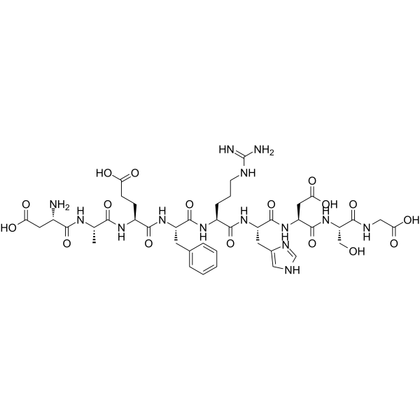
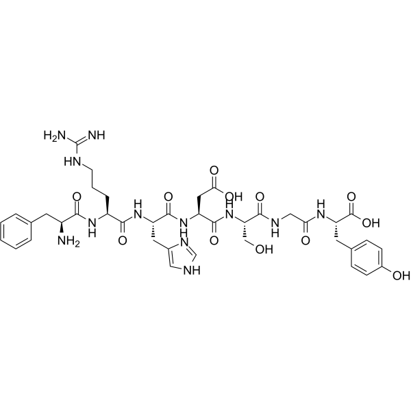
β-Amyloid (1-9), an N-terminal fragment of beta amyloid, consists of amino acid residues 1 to 9. β-Amyloid (1-9) contains a B cell epitope, but it does not include T cell epitopes. Omission of residues 1 to 9 from the full-length Alzheimer'sβ-Amyloid peptide 1 to 40 does not prevent the peptide from forming amyloid fibrils or eliminate fibril polymorphism .
For research use only. We do not sell to patients.
- CAS No.: 147529-30-4
- Formula: C42H60N14O17
- Molecular Weight:1033.01
-
Storage:
Please store the product under the recommended conditions in the Certificate of Analysis.

To place orders, for customer services and technical support, please contact: MedChemExpress USA
Tel: 609-228-6898 E-mail: [email protected] [email protected]


-
 Biological Activity
-
Chemical Information
- Protocols
- Purity & Documentation
- References
-
 Help & FAQs







Biological Activity
Description


Chemical Information
-
CAS No. 147529-30-4
-
Molecular Weight 1033.01
-
Formula C42H60N14O17
-
Sequence
Asp-Ala-Glu-Phe-Arg-His-Asp-Ser-Gly
-
Sequence Shortening
DAEFRHDSG
-
Shipping
Room temperature in continental US; may vary elsewhere.
-
Storage
Please store the product under the recommended conditions in the Certificate of Analysis.

Protocols
-
Amyloid: Congo Red Amyloid Staining
Congo red amyloid staining is a histochemical method used to detect extracellular amyloid deposits in tissue sections based on the affinity of Congo red dye for β-pleated sheet-rich protein aggregates. When bound to amyloid, Congo red produces characteristic apple-green birefringence under polarized light microscopy, which is widely regarded as a diagnostic feature of amyloid deposition in histopathology. The diagnostic principle relies on the combination of dye binding (congophilia) and optical anisotropy under polarized illumination, which distinguishes amyloid from most non-amyloid eosinophilic extracellular deposits in routine histological evaluation. Amyloid identification by Congo red staining remains a cornerstone in diagnostic pathology despite the availability of adjunct methods such as immunohistochemistry and mass spectrometry, particularly because of its ability to localize deposits directly within tissue architecture. The specificity of Congo red-positive deposits is incre
-
Research Protocol for Cancer Immunology
Cancer immunology studies how the immune system recognizes, suppresses, edits, or fails to eliminate malignant cells through tumor antigen release, antigen presentation, T-cell priming, immune trafficking, tumor-cell killing, and feedback inhibition in the tumor microenvironment. The cancer-immunity cycle links tumor antigenicity, dendritic-cell priming, CD8+ T-cell infiltration, cytotoxic function, and immune-checkpoint regulation to tumor rejection or immune escape. Immune-checkpoint pathways such as PD-1/PD-L1 and CTLA-4 suppress antitumor T-cell activity and can be therapeutically blocked, but many tumors remain resistant because of poor antigen presentation, weak T-cell infiltration, suppressive myeloid cells, regulatory T cells, and tumor-intrinsic immune-exclusion programs. Unresolved questions include which immune-cell states predict response, how tumor-intrinsic pathways exclude immune cells, how myeloid suppression limits checkpoint blockade, and which combination strategies


Purity & Documentation
References
[1]. Zamora E, et al. Papillomavirus-like particles are an effective platform for amyloid-beta immunization in rabbitsand transgenic mice. J Immunol. 2006 Aug 15;177(4):2662-2670. [Content Brief]
[2]. Paravastu AK, et al. Polymorphic fibril formation by residues 10-40 of the Alzheimer's beta-amyloid peptide. Biophys J. 2006 Jun 15;90(12):4618-29. [Content Brief]

Calculators
Concentration (start) × Volume (start) = Concentration (final) × Volume (final)


β-Amyloid (1-9) Related Classifications
HY-P1854 Related Classifications
 Powered by Bioz