



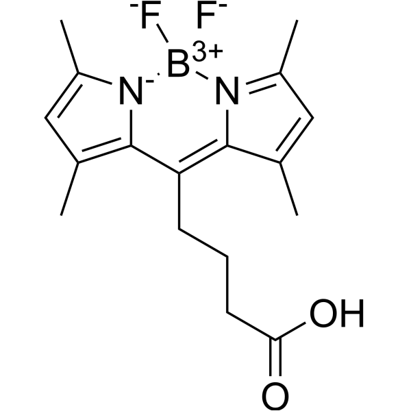
BODIPY 505/515-8-C3-COOH

Based on 1 Customer Validation
BODIPY 505/515-8-C3-COOH is a green fluorescing derivative, as a fluorescent dye for imaging lipid droplets in nannochloropsis. BODIPY 505/515-8-C3-COOH can be used for the research of flow cytometric high-throughput screening and cell sorting.
For research use only. We do not sell to patients.
- Purity : 99.92%
- CAS No.: 878674-84-1
- Formula: C17H21BF2N2O2
- Molecular Weight:334.17
-
Storage:
4°C, protect from light
* In solvent : -80°C, 6 months; -20°C, 1 month (protect from light)

To place orders, for customer services and technical support, please contact: MedChemExpress USA
Tel: 609-228-6898 E-mail: [email protected] [email protected]


-
 Biological Activity
-
Chemical Information
- Protocols
- Purity & Documentation
- References
-
 Help & FAQs




Biological Activity
Description
In Vitro
Guide (Following is our recommended protocol. This protocol only provides a guideline, and should be modified according to your specific needs).
Labeling of Cells
1. Dissolve BODIPY 505/515-8-C3-COOH in DMSO at a concentration of 4 mg/mL, then dilute to different working stock concentrations with DMSO.
2. Add an appropriate amount of BODIPY 505/515-8-C3-COOH working stock solution to the cell suspension to achieve a final DMSO volume fraction of 2-10% (v/v) and a final BODIPY 505/515-8-C3-COOH concentration of 0.8-4 μg/mL.
3. After mixing the cells and the dye thoroughly, incubate in the dark for 15 min, then detect using a flow cytometer.
MedChemExpress (MCE) has not independently confirmed the accuracy of these methods. They are for reference only. Further protocols information, click here.
Emission (Em)
515
Excitation (Ex)
505


Chemical Information
-
CAS No. 878674-84-1
-
Appearance Solid
-
Molecular Weight 334.17
-
Formula C17H21BF2N2O2
-
Color Orange to red
-
SMILES
O=C(CCCC1=C2C(C)=CC(C)=[N]2[B+3]([F-])([N-]3C1=C(C)C=C3C)[F-])O
-
Shipping
Room temperature in continental US; may vary elsewhere.
-
Storage
4°C, protect from light
* In solvent : -80°C, 6 months; -20°C, 1 month (protect from light)

Protocols
-
Research Protocol for Drug Screening technologies
Drug screening technologies are experimental and computational strategies used to identify small molecules or chemical probes that modulate a defined molecular target, signaling pathway, cellular phenotype, disease model, or patient-derived response profile. High-throughput screening tests many compounds in miniaturized assay formats, while quantitative high-throughput screening tests compounds across concentration ranges so that potency and efficacy can be inferred from concentration-response behavior rather than from a single-point signal. The core biological function of a drug-screening strategy is to connect compound exposure with measurable pathway activity, target modulation, cell-state change, viability, cytotoxicity, morphology, or disease-relevant phenotype. Assay performance must be evaluated before screening because hit identification depends on the separation between positive and negative controls, control variability, plate effects, outliers, and the statistical framework
-
Flow Cytometry
Flow cytometry (FC) is a technique for high-speed, step-by-step quantitative analysis and sorting of single cells or other biological particles in a suspension by detecting labeled fluorescent signals.
-
Fluorescence activated cell sorting
Fluorescence activated cell sorting (FACS) is a special technology for identifying and separating rare cell populations with high purity.
-
Fluorescence-Activated Cell Sorting (FACS)-Based Cell Sorting
Fluorescence-Activated Cell Sorting (FACS) separates cells in suspension after flow-cytometric measurement of light scatter and fluorescence; classic droplet-based instruments interrogate cells with a laser, convert optical signals into electronic signals, charge droplets containing target cells, and electrostatically deflect them into collection vessels. FACS detects phenotypes defined by fluorescent antibodies, fluorescent proteins, viability dyes, or intracellular markers, and the readout is the sorted fraction, purity, recovery, and post-sort viability of the gated population.
-
Lipid Droplets: Oil Red O/Sudan Dye Lipid Staining
Lipid droplets are intracellular organelles with a neutral-lipid core that stores triacylglycerols and sterol esters, and Oil Red O or Sudan dyes detect these hydrophobic lipid deposits by partitioning into retained lipids in fresh or frozen specimens. Oil Red O stains neutral triglycerides and lipids in frozen tissue sections or air-dried cytologic preparations, while Sudan Black B has also been used as a histochemical fat stain for lipid-rich tissue structures.


Purity & Documentation
-
Data Sheet (273 KB)
-
SDS (252 KB)
- English - EN (252 KB)
- Français - FR (252 KB)
- Deutsch - DE (252 KB)
- Norwegian - NO (252 KB)
- Español - ES (252 KB)
- Swedish - SV (252 KB)
- Italian - IT (252 KB)
- Korean - KR (252 KB)
- Portuguese - PT (252 KB)
-
Handling Instructions (2659 KB)
References

Calculators
Concentration (start) × Volume (start) = Concentration (final) × Volume (final)


BODIPY 505/515-8-C3-COOH Related Classifications
HY-D1581 Related Classifications
 Powered by Bioz