



BPDA2

Based on 1 Customer Validation
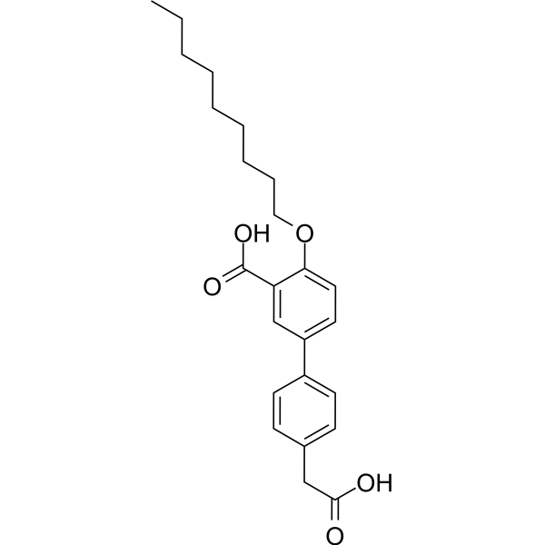
BPDA2 is a highly selective and competitive active site SHP2 inhibitor with IC50s of 92.0 nM, 33.39 μM, 40.71 μM for SHP2, SHP1, SHP1B, respectively. DBDA2 downregulates mitogenic and cell survival signaling and RTK expression. BPDA2 suppresses SHP2 mediated signaling and breast cancer cell phenotypes.
For research use only. We do not sell to patients.
- Purity : 99.30%
- CAS No.: 2907659-86-1
- Formula: C24H30O5
- Molecular Weight:398.49
-
Storage:Powder -20°C, 3 years , 4°C, 2 years ; In solvent -80°C, 6 months , -20°C, 1 month

To place orders, for customer services and technical support, please contact: MedChemExpress USA
Tel: 609-228-6898 E-mail: [email protected] [email protected]


-
 Biological Activity
-
Chemical Information
- Protocols
- Purity & Documentation
- References
-
 Help & FAQs




Biological Activity
Description
In Vitro
BPDA2 (0.2-3.2 μM) inhibits basal activation of Akt and ERK1/2 in a concentration dependent manner[1].
BPDA2 (0.25-4.0 μM; for 10 days) suppresses the anchorage independent growth and cancer stem cell properties of breast cancer cells in a concentration dependent manner[1].
MedChemExpress (MCE) has not independently confirmed the accuracy of these methods. They are for reference only.
-
Cell Line:JIMT-1, MDA-MB468 cell
-
Concentration:0.2, 0.4, 0.8, 1.6, 3.2 μM
-
Incubation Time:
-
Result:Inhibited basal activation of Akt and ERK1/2 in a concentration dependent manner.


Chemical Information
-
CAS No. 2907659-86-1
-
Appearance Solid
-
Molecular Weight 398.49
-
Formula C24H30O5
-
Color White to off-white
-
SMILES
CCCCCCCCCOC1=CC=C(C=C1C(O)=O)C2=CC=C(C=C2)CC(O)=O
-
Shipping
Room temperature in continental US; may vary elsewhere.
-
Storage
Powder -20°C 3 years 4°C 2 years In solvent -80°C 6 months -20°C 1 month


Purity & Documentation
-
Data Sheet (264 KB)
-
SDS (251 KB)
- English - EN (251 KB)
- Français - FR (251 KB)
- Deutsch - DE (251 KB)
- Norwegian - NO (251 KB)
- Español - ES (251 KB)
- Swedish - SV (251 KB)
- Italian - IT (251 KB)
- Korean - KR (251 KB)
- Portuguese - PT (251 KB)
-
Handling Instructions (2659 KB)
References

Calculators
Concentration (start) × Volume (start) = Concentration (final) × Volume (final)


BPDA2 Related Classifications
HY-152208 Related Classifications
 Powered by Bioz