



Buprofezin
Based on 2 publication(s) in Google Scholar
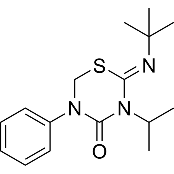
Buprofezin is a broad-spectrum insecticide and chitin synthesis inhibitor that targets developmental stage coleopteran pests.Buprofezin promotes the conversion of energy metabolism from the aerobic tricarboxylic acid (TCA) cycle and oxidative phosphorylation to anaerobic glycolysis. Buprofezin also promotes the production of reactive oxygen species (ROS) by inhibiting cytochrome c oxidase.
For research use only. We do not sell to patients.
- Purity : 99.55%
- CAS No.: 69327-76-0
- Formula: C16H23N3OS
- Molecular Weight:305.44
-
Storage:Powder -20°C, 3 years , 4°C, 2 years ; In solvent -80°C, 6 months , -20°C, 1 month

To place orders, for customer services and technical support, please contact: MedChemExpress USA
Tel: 609-228-6898 E-mail: [email protected] [email protected]


-
 Biological Activity
-
Chemical Information
-
Solvent & Solubility
- Protocols
- Purity & Documentation
- References
-
 Help & FAQs


-
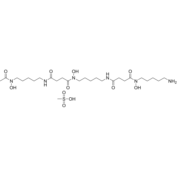
HY-B0988
Deferoxamine mesylateIron Chelator -
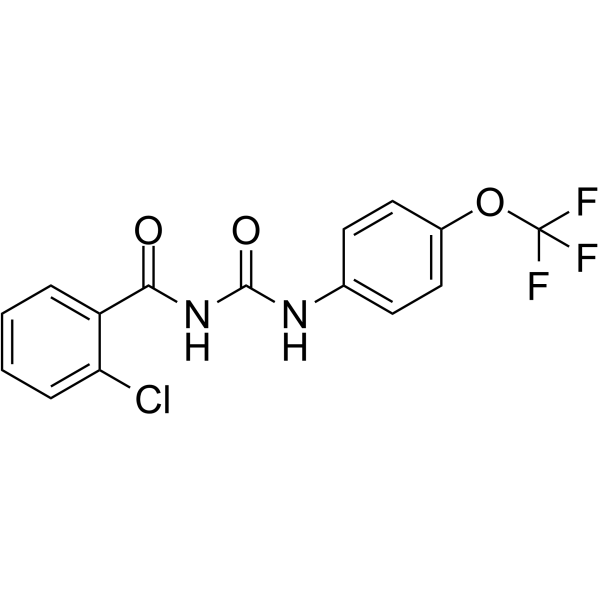
HY-B2049
TriflumuronChitin Synthesis Inhibitor -
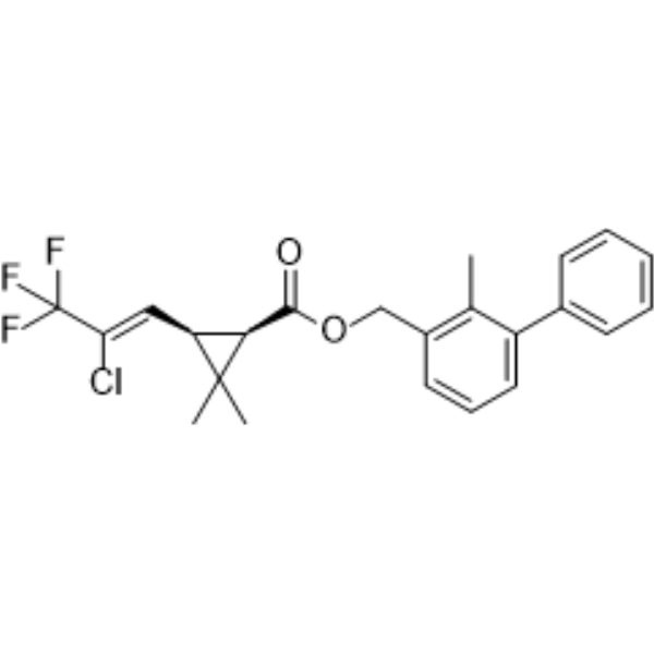
HY-B0824
BifenthrinSodium Channel Activator -
HY-119649
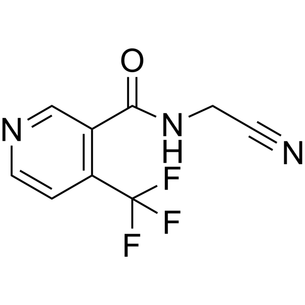
FlonicamidInsecticide -
HY-B1111
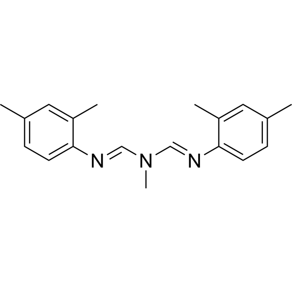
AmitrazParasite Inhibitor -

HY-B0831R
Buprofezin (Standard)Chitin Synthesis Inhibitor -
HY-B0840
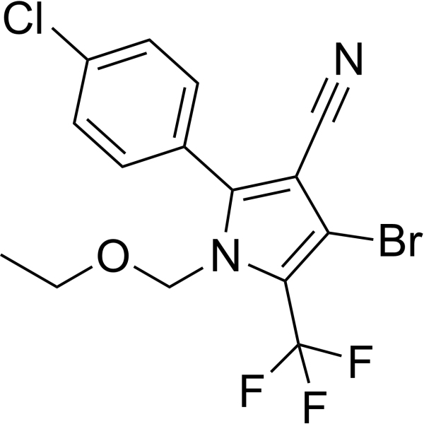
ChlorfenapyrPyrrole Insecticide -

HY-B0840R
Chlorfenapyr (Standard)Pyrrole Insecticide -
HY-B2050
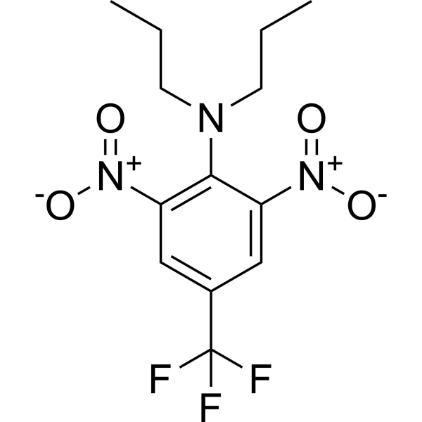
TrifluralinPre-Emergence Herbicide -
HY-119687
BifenazateGABA Receptor Modulator -
HY-B2046
SimazineHerbicide -

HY-B1111R
Amitraz (Standard)Parasite Inhibitor
Publications Citing Use of MedChemExpress (MCE) Buprofezin
More

Biological Activity
Description
In Vitro
Buprofezin (100, 300, 1000 μM) significantly reduces viability of HepG2 cells[1].
Buprofezin (3, 10, 30 μM; 24 h) promotes the conversion of energy metabolism from the aerobic tricarboxylic acid (TCA) cycle and oxidative phosphorylation to anaerobic glycolysis in HepG2 cells[1].
Buprofezin (3, 10, 30 μM; 24 h) inhibits the activity of Complex IV in HepG2 cells[1].
MedChemExpress (MCE) has not independently confirmed the accuracy of these methods. They are for reference only. Further protocols information, click here.
-
Cell Line:HepG2 cells
-
Concentration:3, 10, 30 µM
-
Incubation Time:24 h
-
Result:Significantly increased lactate dehydrogenase B (LDHB) levels when at 30 µM, and slightly increased 6-phosphofructo-2-kinase/fructose-2,6-biphosphatase 3 (PFKFB3).
Decreased ATP levels in a concentration-dependent manner to 91.3, 87.9 and 67.2% of the levels in the vehicle control under treatment with 3, 10 and 30 µM buprofezin, respectively.
Significantly increased the lactate levels.
-
Cell Line:HepG2 cells
-
Concentration:3, 10, 30 µM
-
Incubation Time:24 h
-
Result:Significantly inhibited the activity of Complex IV to 82.2, 69.2 and 63.4% of the vehicle control levels following buprofezin treatment at 3, 10 and 30 µM, respectively.
-
Cell Line:HepG2 cells
-
Concentration:3, 10, 30 µM
-
Incubation Time:24 h
-
Result:Significantly increased the intracellular ROS levels in a concentrate-independent manner, and decreased mtDNA contents.
In Vivo
Buprofezin (46.3, 139, 417 mg/kg; p.o.; single) elevates the MDA level in all organs (especially in the liver and brain) in mice[1].
MedChemExpress (MCE) has not independently confirmed the accuracy of these methods. They are for reference only.
-
Animal Model:Male C57BL/6 mice (6 to 8-week-old)[1].
-
Dosage:46.3, 139, 417 mg/kg
-
Administration:Oral administration; single
-
Result:Tended to elevate the MDA level in all organs, and the most significant concentration-dependent increases were observed in the liver and brain.
Exhibited the highest concentrations in the liver (35.84%) followed by the brain (23.58%), stomach (21.94%) and kidney (18.64%), while the levels in the mouse spleen and heart were below the limit of detection.


Chemical Information
-
CAS No. 69327-76-0
-
Appearance Solid
-
Molecular Weight 305.44
-
Formula C16H23N3OS
-
Color White to off-white
-
SMILES
O=C1N(C(C)C)/C(SCN1C2=CC=CC=C2)=N/C(C)(C)C
-
Shipping
Room temperature in continental US; may vary elsewhere.
-
Storage
Powder -20°C 3 years 4°C 2 years In solvent -80°C 6 months -20°C 1 month


Publications (2)
-
Journal Impact Factor
-
Most Recent
-
ACS Environ Au
Machine Learning-Assisted Recognition of Environmental Sulfur-Containing Chemicals in Nontargeted Mass Spectrometry Analysis of Inadequate Mass Resolution. [Abstract]2025 Aug 5;5(6):573-582. PMID: 41277996 -
Anal Chem
Exposome-Scale Investigation of Cl-/Br-Containing Chemicals Using High-Resolution Mass Spectrometry, Multistage Machine Learning, and Cloud Computing. [Abstract]2025 Jun 3;97(21):11099-11109. PMID: 40401576


Solvent & Solubility
In Vitro:
DMSO : 100 mg/mL (327.40 mM; Need ultrasonic; Hygroscopic DMSO has a significant impact on the solubility of product, please use newly opened DMSO)
Please refer to the solubility information to select the appropriate solvent. Once prepared, please aliquot and store the solution to prevent product inactivation from repeated freeze-thaw cycles.
Storage method and period of stock solution: -80°C, 6 months; -20°C, 1 month. When stored at -80°C, please use it within 6 months. When stored at -20°C, please use it within 1 month.
Please refer to the solubility information to select the appropriate solvent. Once prepared, please aliquot and store the solution to prevent product inactivation from repeated freeze-thaw cycles.
Storage method and period of stock solution: -80°C, 6 months; -20°C, 1 month. When stored at -80°C, please use it within 6 months. When stored at -20°C, please use it within 1 month.
Concentration (start) × Volume (start) = Concentration (final) × Volume (final)
In Vivo:
Select the appropriate dissolution method based on your experimental animal and administration route.
- For the following dissolution methods, please ensure to first prepare a clear stock solution using an In Vitro approach and then sequentially add co-solvents:
- To ensure reliable experimental results, the clarified stock solution can be appropriately stored based on storage conditions. As for the working solution for In Vivo experiments, it is recommended to prepare freshly and use it on the same day.
- The percentages shown for the solvents indicate their volumetric ratio in the final prepared solution. If precipitation or phase separation occurs during preparation, heat and/or sonication can be used to aid dissolution.
Add each solvent one by one: 10% DMSO 40% PEG300 5% Tween-80 45% Saline
Solubility: ≥ 2.5 mg/mL (8.18 mM); Clear solution
This protocol yields a clear solution of ≥ 2.5 mg/mL (saturation unknown).
Taking 1 mL working solution as an example, add 100 μL DMSO stock solution (25.0 mg/mL) to 400 μL PEG300, and mix evenly; then add 50 μL Tween-80 and mix evenly; then add 450 μL Saline to adjust the volume to 1 mL.
Preparation of Saline: Dissolve 0.9 g sodium chloride in ddH₂O and dilute to 100 mL to obtain a clear Saline solution.
Add each solvent one by one: 10% DMSO 90% (20% SBE-β-CD in Saline)
Solubility: ≥ 2.5 mg/mL (8.18 mM); Clear solution
This protocol yields a clear solution of ≥ 2.5 mg/mL (saturation unknown).
Taking 1 mL working solution as an example, add 100 μL DMSO stock solution (25.0 mg/mL) to 900 μL 20% SBE-β-CD in Saline, and mix evenly.
Preparation of 20% SBE-β-CD in Saline (4°C, storage for one week): 2 g SBE-β-CD powder is dissolved in 10 mL Saline, completely dissolve until clear.
In Vivo Dissolution Calculator
Please enter the basic information of animal experiments:
-
-
-
-
Recommended: Prepare an additional quantity of animals to account for potential losses during experiments.
Please enter your animal formula composition:
-
%DMSO +
Recommended: Keep the proportion of DMSO in working solution below 2% if your animal is weak.
-
%+
-
+%Tween-80 + +
-
%Saline +
The co-solvents required include: DMSO, . All of co-solvents are available by MedChemExpress (MCE). , Tween 80. All of co-solvents are available by MedChemExpress (MCE).
Working solution concentration: 0.22 mg/mL
Method for preparing stock solution: mg drug dissolved in μL DMSO. Stock solution concentration: mg/mL.
1. Take μL DMSO stock solution;
2. Add μL .
μL , mix evenly;
3. Then add μL Tween 80, mix evenly;
4. Then add μL
Please ensure that the stock solution in the first step is dissolved to a clear state, and add co-solvents in sequence. You can use ultrasonic heating (ultrasonic cleaner, recommended frequency 20-40 kHz), vortexing, etc. to assist dissolution.

Protocols
-
Kinase activity and phosphorylation assays
Kinase activity assays measure the ability of kinases to transfer phosphate groups from ATP to specific substrates, while phosphorylation assays detect the presence and levels of phosphorylated proteins. Common methods include radiolabeled ATP incorporation (e. g. ,), ADP release detection via bioluminescence (e. g. ,[3]), enzyme-linked immunosorbent assays (ELISA) for phospho-specific epitopes (e. g. ,[6]), and microtiter-based formats for high-throughput screening (e. g. ,[8]). The ADP-Glo assay quantifies kinase activity by measuring ADP produced during phosphorylation using a luciferase-based system. Radiometric assays involve autoradiography or scintillation counting after incorporation of 32P-labeled ATP into substrate proteins. ELISA-based approaches rely on phospho-specific antibodies to detect activated kinases in cell lysates or purified samples.
-
Western Blot
Western blotting (WB) is a commonly used experimental method in molecular biology, biochemistry, and immunogenetics for identifying and quantifying target proteins. It combines gel electrophoresis with immunoassay, enabling researchers to analyze protein expression, post-translational modifications, and molecular weight.
-
ROS/oxidative-stress fluorescent staining
ROS/oxidative-stress fluorescent staining uses cell-permeant fluorogenic probes that become fluorescent after oxidation inside cells or tissues; commonly used examples include DCFH-DA/DCFDA for broad cellular oxidant detection, DHE for superoxide-related signal detection, MitoSOX for mitochondrial superoxide-related signal detection, and CellROX probes for oxidative-stress-associated fluorescence readouts. The assay detects probe oxidation rather than a single ROS species unless the probe and analysis method have been chemically validated for that species. DCFH-DA enters cells, is deacetylated by intracellular esterases to DCFH, and produces fluorescent DCF after oxidation, so the readout is used as an operational measure of total cellular oxidative stress rather than a species-specific ROS measurement. DHE and MitoSOX can report superoxide-related oxidation, but red fluorescence alone can include non-specific ethidium-like oxidation products; HPLC or optimized spectral approaches are
-
Protocol for Kinase activity and phosphorylation assays
Kinase activity assays measure transfer of phosphate from ATP to a protein or peptide substrate, generating phosphorylated substrate, ADP, or incorporated radiolabeled phosphate as the readout; phosphorylation assays measure site-specific phosphorylation in cells or tissues as a proxy for kinase-pathway activation, inhibition, or substrate regulation. Phosphorylation can be detected by phospho-specific Western blot, immunoprecipitation kinase assay, phospho-immunofluorescence, phospho-flow cytometry, luminescent ADP detection, radiolabeled ATP incorporation, or reporter-based pathway assays, and these readouts can be applied to cancer cells, primary neurons, mouse tumors, organoids, inflammatory macrophages, ferroptosis studies, and mitophagy studies when the kinase target is biologically relevant.


Purity & Documentation
-
Data Sheet (281 KB)
-
SDS (563 KB)
- English - EN (563 KB)
- Français - FR (563 KB)
- Deutsch - DE (563 KB)
- Norwegian - NO (563 KB)
- Español - ES (563 KB)
- Swedish - SV (563 KB)
- Italian - IT (563 KB)
- Korean - KR (563 KB)
- Portuguese - PT (563 KB)
-
Handling Instructions (2659 KB)
References


Complete Stock Solution Preparation Table
Please refer to the solubility information to select the appropriate solvent. Once prepared, please aliquot and store the solution to prevent product inactivation from repeated freeze-thaw cycles.
Storage method and period of stock solution: -80°C, 6 months; -20°C, 1 month. When stored at -80°C, please use it within 6 months. When stored at -20°C, please use it within 1 month.
| Optional Solvent | Concentration Solvent Mass | 1 mg | 5 mg | 10 mg | 25 mg |
|---|---|---|---|---|---|
| DMSO | 1 mM | 3.2740 mL | 16.3698 mL | 32.7397 mL | 81.8491 mL |
| 5 mM | 0.6548 mL | 3.2740 mL | 6.5479 mL | 16.3698 mL | |
| 10 mM | 0.3274 mL | 1.6370 mL | 3.2740 mL | 8.1849 mL | |
| 15 mM | 0.2183 mL | 1.0913 mL | 2.1826 mL | 5.4566 mL | |
| 20 mM | 0.1637 mL | 0.8185 mL | 1.6370 mL | 4.0925 mL | |
| 25 mM | 0.1310 mL | 0.6548 mL | 1.3096 mL | 3.2740 mL | |
| 30 mM | 0.1091 mL | 0.5457 mL | 1.0913 mL | 2.7283 mL | |
| 40 mM | 0.0818 mL | 0.4092 mL | 0.8185 mL | 2.0462 mL | |
| 50 mM | 0.0655 mL | 0.3274 mL | 0.6548 mL | 1.6370 mL | |
| 60 mM | 0.0546 mL | 0.2728 mL | 0.5457 mL | 1.3642 mL | |
| 80 mM | 0.0409 mL | 0.2046 mL | 0.4092 mL | 1.0231 mL | |
| 100 mM | 0.0327 mL | 0.1637 mL | 0.3274 mL | 0.8185 mL |


Buprofezin Related Classifications
HY-B0831 Related Classifications
 Powered by Bioz