



Fibronectin Adhesion-promoting Peptide TFA
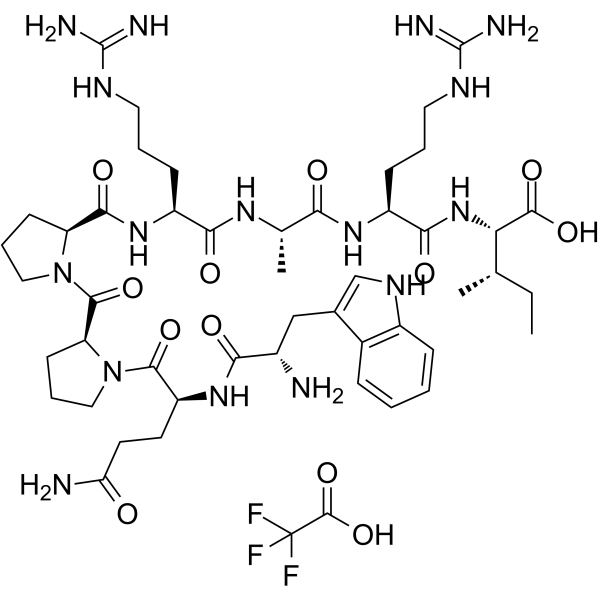
Fibronectin Adhesion-promoting Peptide (Heparin Binding Peptide) is one of the heparin-binding amino acid sequences found in the carboxy-terminal heparin-binding domain of fibronectin. It promotes assembly of mesenchymal stem cell (MSC) spheroids into larger aggregates. Fibronectin Adhesion-promoting Peptide directly promotes the adhesion, spreading, and migration of endothelial cells by reacting with heparin binding domains of cells.
Nos produits utilisent uniquement pour la recherche. Nous ne vendons pas aux patients.
- Formule: C49H75F3N16O12
- Masse moléculaire:1137.22
-
Stockage:
Please store the product under the recommended conditions in the Certificate of Analysis.

To place orders, for customer services and technical support, please contact: MedChemExpress USA
Tel: 609-228-6898 E-mail: [email protected] [email protected]


-
 Activité biologique
-
Chemical Information
- Protocole
- Pureté et documentation
- Références
-
 Help & FAQs




Activité biologique
Description
In Vitro
GRGDS and Fibronectin Adhesion-promoting Peptide TFA (Heparin Binding Peptide TFA: WQPPRARI) micropatterns increase the endothelialisation of prosthetic materials in vitro. Fibronectin Adhesion-promoting Peptide also enhances haptotactic cell migration. The anti-apoptotic effects of fibronectin require the presence of the WQPPRARI sequence, which may be due to synergistic survival signals mediated by this sequence and by the RGD motif[1].
RDGS and Fibronectin Adhesion-promoting Peptide TFA promote cell adhesion through different activation pathways that induce different forms of adhesion which can be highlighted through the analysis of focal and fibrillar contacts[2].
MedChemExpress (MCE) has not independently confirmed the accuracy of these methods. They are for reference only. Further protocols information, click here.


Chemical Information
-
Masse moléculaire 1137.22
-
Formule C49H75F3N16O12
-
Synonyms
Heparin Binding Peptide TFA
-
Sequence
Trp-Gln-Pro-Pro-Arg-Ala-Arg-Ile
-
Sequence Shortening
WQPPRARI
-
Livraison
Room temperature in continental US; may vary elsewhere.
-
Stockage
Please store the product under the recommended conditions in the Certificate of Analysis.

Protocole
-
Cell migration
Cell migration is a method that plays an important role in wound healing, cell differentiation, embryonic development, etc.
-
Endothelial Tube Formation Assay
Endothelial tube formation assay evaluates the ability of endothelial cells to attach, migrate, align, and organize into capillary-like networks when cultured on gelled basement membrane extract or Matrigel; the readout is the morphology and quantity of tube-like networks, which reflects an in vitro endothelial morphogenesis step related to angiogenesis. Basement membrane extract/Matrigel provides laminin-rich extracellular matrix cues that support endothelial differentiation into capillary-like structures, but it can contain biologically active growth factors, so growth-factor-reduced matrix is preferred when testing defined angiogenic stimulators or inhibitors.


Pureté et documentation
Références
[1]. Hoesli CA, et al. A fluorophore-tagged RGD peptide to control endothelial cell adhesion to micropatterned surfaces. Biomaterials. 2014 Jan;35(3):879-90. [Content Brief]
[2]. Chollet C, et al. Impact of peptide micropatterning on endothelial cell actin remodeling for cell alignment under shear stress. Macromol Biosci. 2012 Dec;12(12):1648-59. [Content Brief]
[3]. Lei J, Murphy WL, et al. Combination of Heparin Binding Peptide and Heparin Cell Surface Coatings for Mesenchymal Stem Cell Spheroid Assembly. Bioconjug Chem. 2018 Apr 18;29(4):878-884. [Content Brief]

Calculators
Concentration (start) × Volume (start) = Concentration (final) × Volume (final)


Fibronectin Adhesion-promoting Peptide TFA Related Classifications
HY-P0306A Related Classifications
 Powered by Bioz