



Calcium Green 1AM

Based on 1 Customer Validation
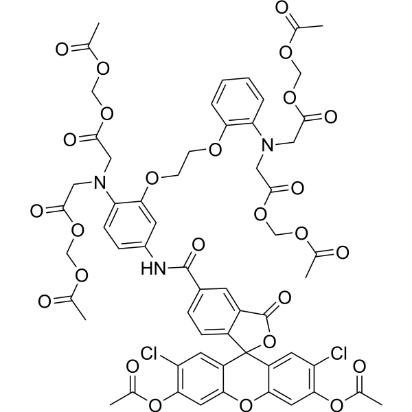
Calcium Green 1AM is a cell-permeable fluorescent calcium indicator dye. Calcium Green 1AM detects intracellular calcium signals and reports calcium transients in spontaneously rhythmic tissues as relative fluorescence changes. Calcium Green 1AM is excited by an argon laser, captures emission light at 488 nm, and is detected using an epifluorescence illumination system on an upright microscope equipped with a fluorescein filter block.
For research use only. We do not sell to patients.
- Purity : 95.0%
- CAS No.: 186501-28-0
- Formula: C59H53Cl2N3O26
- Molecular Weight:1290.96
-
Storage:
-20°C, protect from light
* In solvent : -80°C, 6 months; -20°C, 1 month (protect from light)

To place orders, for customer services and technical support, please contact: MedChemExpress USA
Tel: 609-228-6898 E-mail: [email protected] [email protected]


-
 Biological Activity
-
Chemical Information
-
Solvent & Solubility
- Protocols
- Purity & Documentation
- References
-
 Help & FAQs


-
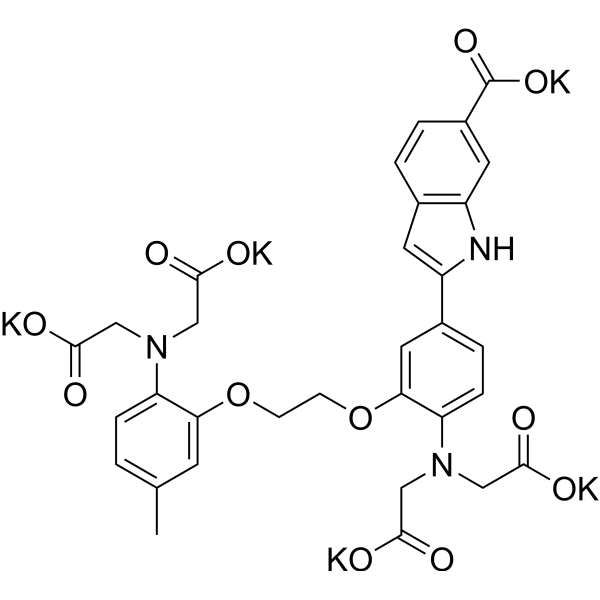
HY-D0121A
INDO 1 pentapotassiumCa2+ Indicator -
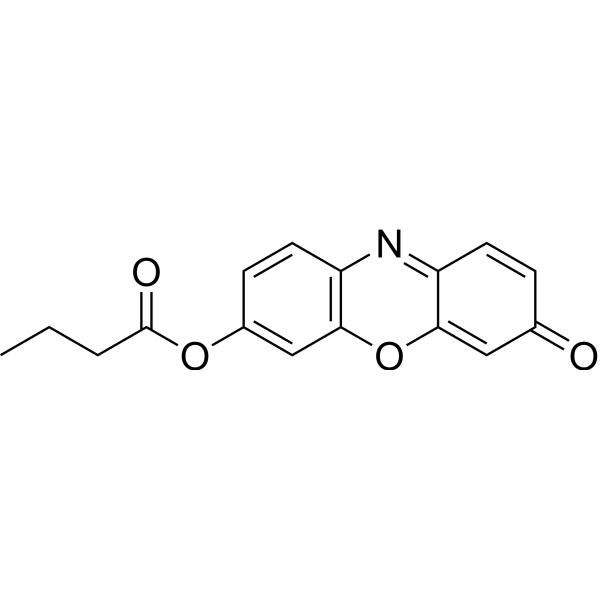
HY-116583
Resorufin butyrateFluorogenic Substrate -
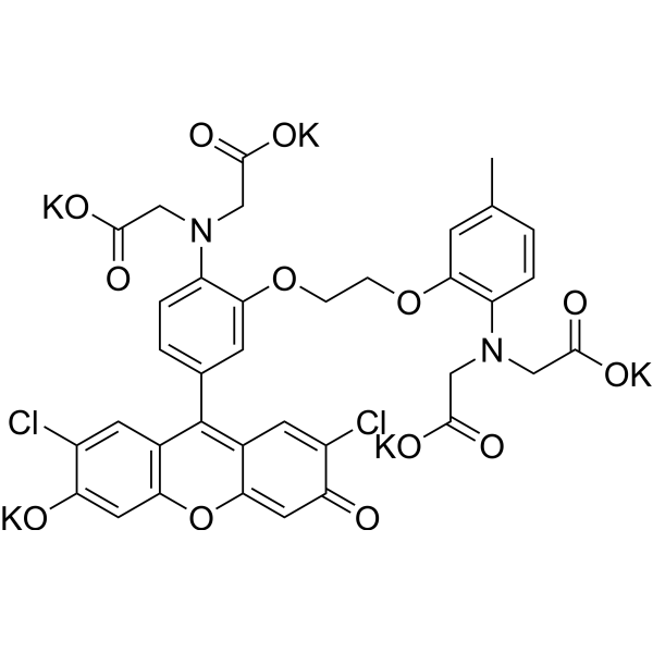
HY-129547
Fluo-3 pentapotassiumFluorochrome -
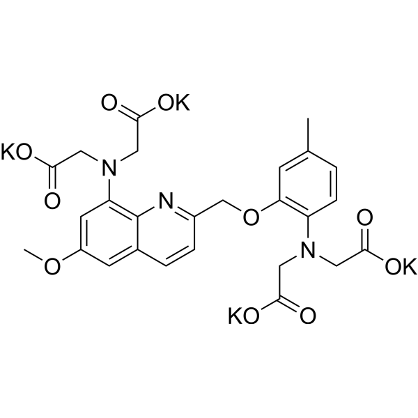
HY-135269
Quin II potassiumBiochemical Assay Reagent -
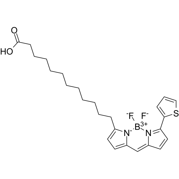
HY-138226
BODIPY 558/568 C12Lipid Droplets Probe -
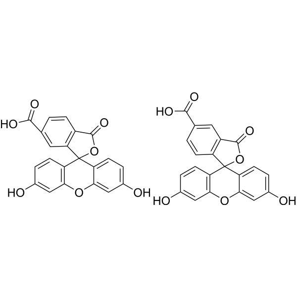
HY-15940
5(6)-CarboxyfluoresceinFluorescent Probe -
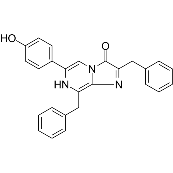
HY-D1024
Coelenterazine hRLuc8 Luminescent Substrate -
HY-130412
FlAsH-EDT2Protein Labeling Reagent -

HY-158779
Sulfane sulfur probe 4Fluorescent Indicator -

HY-DY1061
BODIPY 558/568 C12 (solution)Lipid Droplets Probe -
HY-138159
Boc-Lys(Ac)-AMCFluorometric HDAC Substrate -
HY-126831
SBFI-AMFluorescent Indicator


Biological Activity
Description
In Vitro
Protocol (The following is our recommended protocol. This protocol is for reference only and should be modified according to your specific needs).
1. Stock Solution Preparation
Solvent: DMSO
2. Working Solution Preparation
2.1 Working concentration selection:
- Calcium Green 1AM 4 mM.
- Calcium Green 1AM 10 mM, dissolved in oxygenated artificial cerebrospinal fluid (a-CSF).
2.2 a-CSF composition for 10 mM working solution: 120 mM NaCl, 8 mM KCl, 1.26 mM CaCl2, 1.5 mM MgCl2, 21 mM NaHCO3, 0.5 mM Na2HPO4, 30 mM glucose, pH 7.4.
2.3 Note: Adjust the working solution concentration as needed; prepare fresh before use.
3. Staining Procedure
3.1 For primary hypothalamic astrocytes passaged onto poly-D-lysine-coated glass coverslips[1]:
- Load cells with 4 mM Calcium Green 1AM and incubate at 37°C for 30 min.
- Place the coverslip in a temperature-controlled perfusion chamber on the stage of an inverted microscope.
- Perfuse with oxygenated Kreb's solution at a rate of 1.0 mL/min.
3.2 For isolated embryonic mouse hindbrain preparations[2]:
- Use isolated brainstem for embryonic parafacial oscillator (e-pF) experiments and transverse medullary slices for preBötzinger complex (preBötC) experiments.
- For e-pF imaging, place the isolated brainstem ventral side (pial side) up.
- For preBötC imaging, place the transverse slice rostral side up.
- Incubate the preparation in oxygenated a-CSF containing 10 mM Calcium Green 1AM for 40 min.
- After loading, transfer the preparation to the recording chamber and continuously perfuse with oxygenated a-CSF at 30°C.
- Place the preparation in the recording chamber and recover in oxygenated a-CSF for 30 min to wash out excess dye.
- Allow spontaneous firing activity to appear during this recovery period.
4. Controls
4.1 Negative control: Cells on control coverslips perfused with Kreb's solution alone show no change in calcium signal.
4.2 Positive control: ATP (50 mM) applied for 30 s reliably evokes calcium oscillations and, at the end of the study, induces a strong and sustained increase in calcium response on each coverslip.
4.3 Specificity control: No calcium influx is observed in cells perfused with heat-inactivated leptin or chemical inhibitors of mitogen-activated protein kinases (MAPK), indicating specificity.
5. Detection and Analysis
5.1 Instrument type:
- Inverted microscope.
- Epifluorescence microscope equipped with a cooled CCD camera.
5.2 Wavelength settings:
- Calcium Green is excited by an argon laser, and emission is captured at 488 nm.
- Use a fluorescein filter block to excite the dye and capture emission.
5.3 Imaging parameters:
- Perform real-time imaging using a 40× objective at a frequency of 2 frames/s, capturing 600 frames in total.
- Capture images in overlapping mode with 100 ms exposure during the 30 to 120 sec time period.
5.4 Result analysis:
- Leptin (6.2 nM) induces a strong increase in intracellular Calcium Green fluorescence in nearly 100% of astrocytes; the response appears immediately after the start of 2 min perfusion, rapidly returns to baseline, with an average increase of 22.7 ± 3.3% (n=20).
- Repeated application of leptin on the same coverslip after 5 min induces a much smaller response amplitude (4.5 ± 1.1%), indicating desensitization.
- Calculate the percentage of responsive cells and the magnitude of increase relative to baseline fluorescence intensity for each coverslip.
- All results are repeated at least four times.
- Calculate the average intensity within the region of interest for each frame.
- Measure the average baseline fluorescence between two spontaneous events, normalize the DF signal, and display as ΔF/F.
- Analyze cells that take up the dye and can be detected using the imaging parameters used as active cells.
- Expected results: Rhythmic organized calcium transients are detected in active cells, manifested as fluorescence changes.
MedChemExpress (MCE) has not independently confirmed the accuracy of these methods. They are for reference only. Further protocols information, click here.
Emission (Em)
512


Chemical Information
-
CAS No. 186501-28-0
-
Appearance Solid
-
Molecular Weight 1290.96
-
Formula C59H53Cl2N3O26
-
Color Off-white to light yellow
-
SMILES
O=C1OC2(C3=CC(Cl)=C(C=C3OC4=CC(OC(C)=O)=C(C=C42)Cl)OC(C)=O)C5=CC=C(C(NC6=CC=C(N(CC(OCOC(C)=O)=O)CC(OCOC(C)=O)=O)C(OCCOC7=C(N(CC(OCOC(C)=O)=O)CC(OCOC(C)=O)=O)C=CC=C7)=C6)=O)C=C15
-
Shipping
Room temperature in continental US; may vary elsewhere.
-
Storage
-20°C, protect from light
* In solvent : -80°C, 6 months; -20°C, 1 month (protect from light)


Solvent & Solubility
In Vitro:
DMSO : 6.45 mg/mL (5.00 mM; Need ultrasonic and warming; Hygroscopic DMSO has a significant impact on the solubility of product, please use newly opened DMSO)
Please refer to the solubility information to select the appropriate solvent. Once prepared, please aliquot and store the solution to prevent product inactivation from repeated freeze-thaw cycles.
Storage method and period of stock solution: -80°C, 6 months; -20°C, 1 month (protect from light). When stored at -80°C, please use it within 6 months. When stored at -20°C, please use it within 1 month.
Please refer to the solubility information to select the appropriate solvent. Once prepared, please aliquot and store the solution to prevent product inactivation from repeated freeze-thaw cycles.
Storage method and period of stock solution: -80°C, 6 months; -20°C, 1 month (protect from light). When stored at -80°C, please use it within 6 months. When stored at -20°C, please use it within 1 month.
Concentration (start) × Volume (start) = Concentration (final) × Volume (final)

Protocols
-
Calcium Spark Assay
Calcium sparks are localized, transient increases in intracellular calcium concentration ([Ca2+]i) that occur in cardiac myocytes and represent elementary events underlying excitation-contraction coupling. These events are generated by the coordinated opening of clusters of ryanodine receptors (RyRs) on the sarcoplasmic reticulum membrane, leading to a brief release of Ca2+ into the cytosol. The detection and analysis of calcium sparks provide insights into the mechanisms of calcium handling and signaling in cardiac cells. Imaging techniques using fluorescent calcium indicators such as Fluo-3 are employed to visualize these subcellular calcium transients with high spatial and temporal resolution. The protocol is based on established methodologies described in primary literature for both experimental measurement and automated analysis of calcium sparks.
-
Ca2+ Staining Technique
Ca2+ staining is an experimental technique that utilizes specific fluorescent probes (such as Fluo-4 AM, Fura-2, etc.) to qualitatively or quantitatively detect dynamic changes in intracellular Ca2+ concentrations; this is achieved by monitoring the changes in fluorescent signals generated when these probes bind to free intracellular calcium ions. The underlying principle relies primarily on the presence of chelating groups within the probe's molecular structure that possess high affinity for calcium ions.


Purity & Documentation
-
Data Sheet (292 KB)
-
SDS (252 KB)
- English - EN (252 KB)
- Français - FR (252 KB)
- Deutsch - DE (252 KB)
- Norwegian - NO (252 KB)
- Español - ES (252 KB)
- Swedish - SV (252 KB)
- Italian - IT (252 KB)
- Korean - KR (252 KB)
- Portuguese - PT (252 KB)
-
Handling Instructions (2659 KB)
References


Complete Stock Solution Preparation Table
Please refer to the solubility information to select the appropriate solvent. Once prepared, please aliquot and store the solution to prevent product inactivation from repeated freeze-thaw cycles.
Storage method and period of stock solution: -80°C, 6 months; -20°C, 1 month (protect from light). When stored at -80°C, please use it within 6 months. When stored at -20°C, please use it within 1 month.
| Optional Solvent | Concentration Solvent Mass | 1 mg | 5 mg | 10 mg | 25 mg |
|---|---|---|---|---|---|
| DMSO | 1 mM | 0.7746 mL | 3.8731 mL | 7.7462 mL | 19.3654 mL |


Calcium Green 1AM Related Classifications
HY-D1176 Related Classifications
 Powered by Bioz
Keywords
- Calcium Green 1AM
- 186501-28-0
- Fluorescent Dye
- astrocytic calcium signaling
- calcium imaging
- calcium transients
- embryonic hindbrain preparations
- embryonic parafacial oscillator
- fluorescent calcium indicator
- intracellular calcium signals
- neuronal activity
- preB?tzinger complex
- primary mouse hypothalamic astrocytes
- Inhibitor
- inhibitor
- inhibit