



ABBV-467

Based on 1 Customer Validation
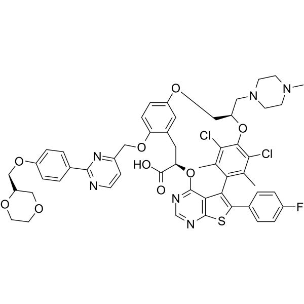
ABBV-467 is a selective MCL-1 inhibitor (Ki: <0.01 nM). ABBV-467 induces apoptosis. ABBV-467 induces cancer cell death and inhibits tumor growth in models of hematological malignancies, such as multiple myeloma.
Nur für Forschungszwecke. Wir verkaufen nicht an Patienten.
- Reinheit : 99.78%
- CAS. Nr.: 2287186-66-5
- Formel: C53H51Cl2FN6O9S
- Molecular Weight:1037.98
-
Speicherung:
-20°C, protect from light, stored under nitrogen
* In solvent : -80°C, 6 months; -20°C, 1 month (protect from light, stored under nitrogen)

To place orders, for customer services and technical support, please contact: MedChemExpress USA
Tel: 609-228-6898 E-mail: [email protected] [email protected]


-
 Biologische Aktivität
-
Chemical Information
-
Lösungsmittel & Löslichkeit
- Protokoll
- Reinheit & Dokumentation
- Verweise
-
 Help & FAQs


-
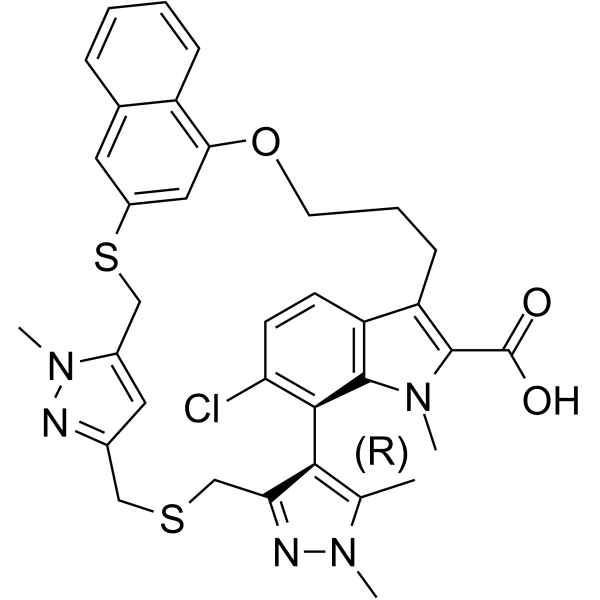
HY-101533
AZD-5991Mcl-1 Inhibitor -
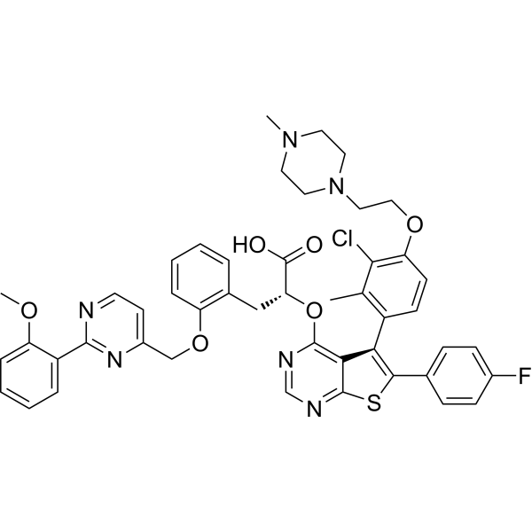
HY-112218
MIK665MCL1 Inhibitor -
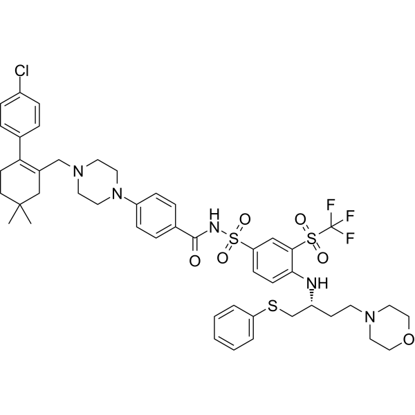
HY-10087
NavitoclaxBcl-2 Inhibitor -
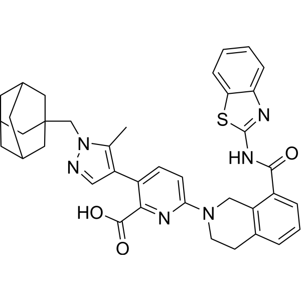
HY-19741
A-1331852Bcl-xL Inhibitor -
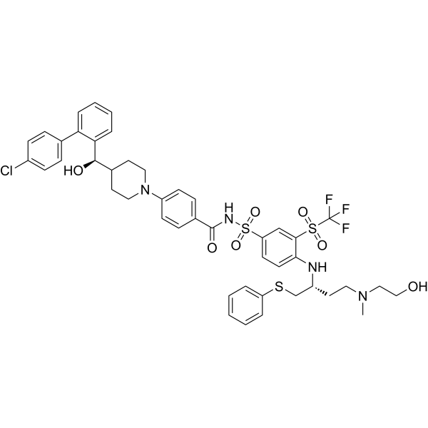
HY-112416
AZD4320BCL2/BCLxL Inhibitor -
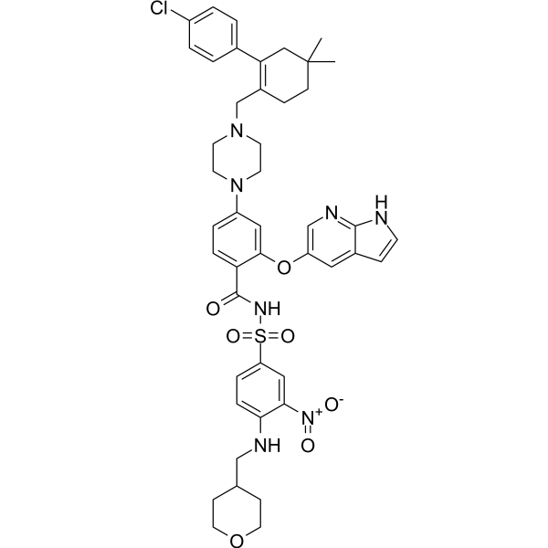
HY-15531
VenetoclaxBCL-2 Inhibitor -
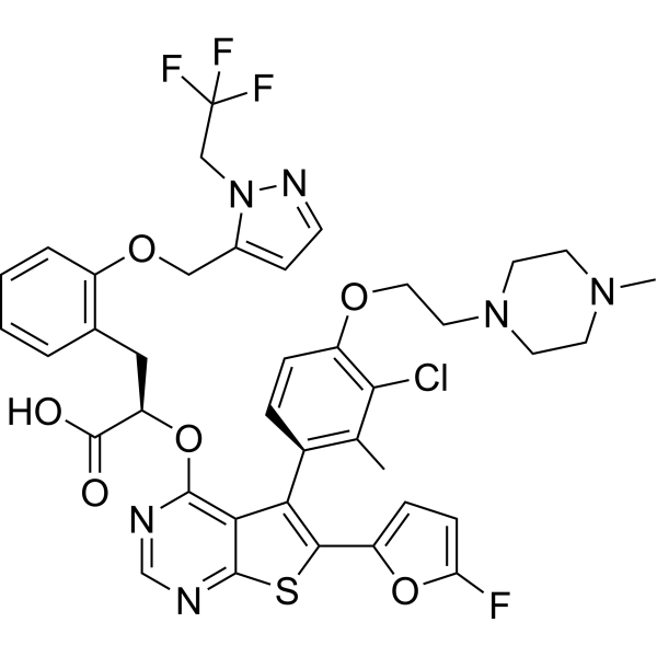
HY-100741
S63845MCL1 Inhibitor -

HY-18739
Phorbol 12-myristate 13-acetatePKC Activator -
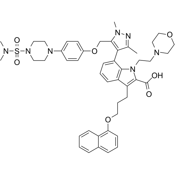
HY-12468
A-1210477Mcl-1 Inhibitor -
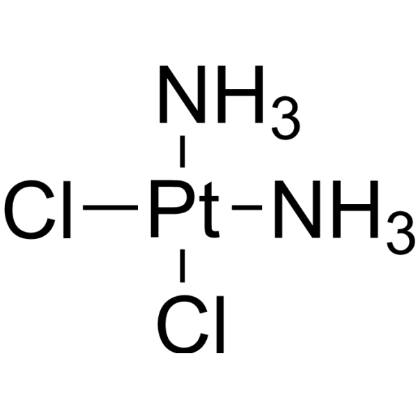
HY-17394
CisplatinDNA Alkylator -
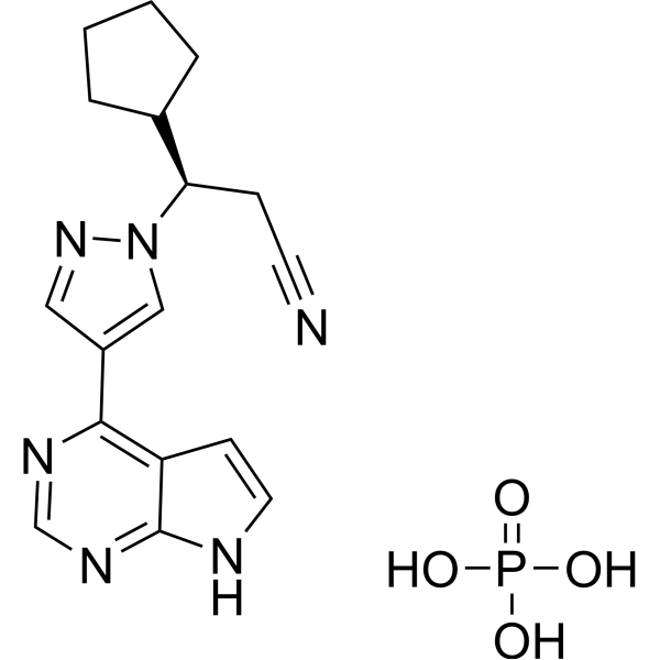
HY-50858
Ruxolitinib phosphateJAK1/2 Inhibitor -
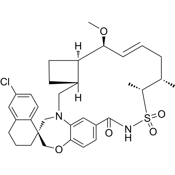
HY-101565
TapotoclaxMCL-1 Inhibitor


Biologische Aktivität
Beschreibung
IC50 & Target
MCL-1[1].
Cellular Effect
|
Cell Line

|
Type | Value | Description | References |
|---|---|---|---|---|
| AMO1 | EC50 |
0.16 nM
Compound: ABBV-467
|
Antiproliferative activity against human AMO1 cells assessed as reduction in cell viability
Antiproliferative activity against human AMO1 cells assessed as reduction in cell viability
|
[PMID: 38597264] |
| MV4-11 | EC50 |
3.91 nM
Compound: ABBV-467
|
Antiproliferative activity against human MV4-11 cells assessed as reduction in cell viability
Antiproliferative activity against human MV4-11 cells assessed as reduction in cell viability
|
[PMID: 38597264] |
| NCI-H929 | EC50 |
0.47 nM
Compound: ABBV-467
|
Antiproliferative activity against human NCI-H929 cells assessed as reduction in cell viability
Antiproliferative activity against human NCI-H929 cells assessed as reduction in cell viability
|
[PMID: 38597264] |
In Vitro
ABBV-467 (1 nM - 10 μM, 24 h) inhibits BAK protein expression in BAK-deficient SKBR3 cells[1]. ABBV-467 (0.01–10 nM, 24 hours) induces apoptosis in AMO-1 cell line[1].
MedChemExpress (MCE) has not independently confirmed the accuracy of these methods. They are for reference only.
-
Cell Line:BAK-deficient SKBR3 cells (breast cancer)
-
Concentration:1 nM to 10 μM
-
Incubation Time:24 h
-
Result:Induced dose-dependent apoptosis after treatment.
-
Cell Line:AMO-1 cell line (multiple myeloma)
-
Concentration:1 nM to 10 μM
-
Incubation Time:24 h
-
Result:Resulted in a significant reduction in BAK protein expression.
In Vivo
MedChemExpress (MCE) has not independently confirmed the accuracy of these methods. They are for reference only.
-
Animal Model:t(4;14) (chromosomal translocation between chromosome 4 and chromosome 14)-translocated high-risk multiple myeloma NOD-SCID mice[1]
-
Dosage:6.25 mg/kg and 12.5 mg/kg
-
Administration:Intravenous injection (i.v.), once per week for 3 weeks
-
Result:Resulted in a maximal tumor delay of 82% at 6.25 mg/kg and complete tumor regression at 12.5 mg/kg.
- Detection of Apoptosis
- TUNEL staining for apoptotic DNA fragmentation
- Cell Cytotoxicity Assay
- Mammalian live/dead viability and cytotoxicity staining
- Annexin V plus membrane-impermeant dye apoptosis staining
- Patient-Derived Xenograft (PDX)
- Apoptosis Solutions
- Patient-Derived Orthotopic Xenograft (PDOX)
- Subcutaneous Cell-Line-Derived Xenograft
- Orthotopic Cell-Line Xenograft
Clinical Trial
| NCT Number | Sponsor | Condition | Start Date |
Phase
|
|---|---|---|---|---|
| NCT01329991 | 2011-05 | PHASE1 |


Chemical Information
-
CAS. Nr. 2287186-66-5
-
Appearance Solid
-
Molecular Weight 1037.98
-
Formel C53H51Cl2FN6O9S
-
Color White to light yellow
-
SMILES
O=C(O)[C@@H](CC1=CC2=CC=C1OCC3=NC(C4=CC=C(OC[C@H]5OCCOC5)C=C4)=NC=C3)OC6=NC=NC7=C6C(C8=C(C)C(Cl)=C(O[C@@H](CO2)CN9CCN(C)CC9)C(Cl)=C8C)=C(C%10=CC=C(F)C=C%10)S7
-
Versand
Room temperature in continental US; may vary elsewhere.
-
Speicherung
-20°C, protect from light, stored under nitrogen
* In solvent : -80°C, 6 months; -20°C, 1 month (protect from light, stored under nitrogen)


Lösungsmittel & Löslichkeit
In Vitro:
DMSO : 100 mg/mL (96.34 mM; Need ultrasonic; Hygroscopic DMSO has a significant impact on the solubility of product, please use newly opened DMSO)
Please refer to the solubility information to select the appropriate solvent. Once prepared, please aliquot and store the solution to prevent product inactivation from repeated freeze-thaw cycles.
Storage method and period of stock solution: -80°C, 6 months; -20°C, 1 month (protect from light, stored under nitrogen). When stored at -80°C, please use it within 6 months. When stored at -20°C, please use it within 1 month.
Please refer to the solubility information to select the appropriate solvent. Once prepared, please aliquot and store the solution to prevent product inactivation from repeated freeze-thaw cycles.
Storage method and period of stock solution: -80°C, 6 months; -20°C, 1 month (protect from light, stored under nitrogen). When stored at -80°C, please use it within 6 months. When stored at -20°C, please use it within 1 month.
Konzentration (Stammlösung) × Volumen (Stammlösung) = Konzentration (Ziellösung) × Volumen (Ziellösung)


Reinheit & Dokumentation
-
Data Sheet (279 KB)
-
SDS (252 KB)
- English - EN (252 KB)
- Français - FR (252 KB)
- Deutsch - DE (252 KB)
- Norwegian - NO (252 KB)
- Español - ES (252 KB)
- Swedish - SV (252 KB)
- Italian - IT (252 KB)
- Korean - KR (252 KB)
- Portuguese - PT (252 KB)
-
Handling Instructions (2659 KB)
Verweise


Complete Stock Solution Preparation Table
Please refer to the solubility information to select the appropriate solvent. Once prepared, please aliquot and store the solution to prevent product inactivation from repeated freeze-thaw cycles.
Storage method and period of stock solution: -80°C, 6 months; -20°C, 1 month (protect from light, stored under nitrogen). When stored at -80°C, please use it within 6 months. When stored at -20°C, please use it within 1 month.
| Optional Solvent | Concentration Solvent Mass | 1 mg | 5 mg | 10 mg | 25 mg |
|---|---|---|---|---|---|
| DMSO | 1 mM | 0.9634 mL | 4.8170 mL | 9.6341 mL | 24.0852 mL |
| 5 mM | 0.1927 mL | 0.9634 mL | 1.9268 mL | 4.8170 mL | |
| 10 mM | 0.0963 mL | 0.4817 mL | 0.9634 mL | 2.4085 mL | |
| 15 mM | 0.0642 mL | 0.3211 mL | 0.6423 mL | 1.6057 mL | |
| 20 mM | 0.0482 mL | 0.2409 mL | 0.4817 mL | 1.2043 mL | |
| 25 mM | 0.0385 mL | 0.1927 mL | 0.3854 mL | 0.9634 mL | |
| 30 mM | 0.0321 mL | 0.1606 mL | 0.3211 mL | 0.8028 mL | |
| 40 mM | 0.0241 mL | 0.1204 mL | 0.2409 mL | 0.6021 mL | |
| 50 mM | 0.0193 mL | 0.0963 mL | 0.1927 mL | 0.4817 mL | |
| 60 mM | 0.0161 mL | 0.0803 mL | 0.1606 mL | 0.4014 mL | |
| 80 mM | 0.0120 mL | 0.0602 mL | 0.1204 mL | 0.3011 mL |


ABBV-467 Related Classifications
HY-149672 Related Classifications
 Powered by Bioz