



CFDA-SE (solution)

Based on 1 Customer Validation
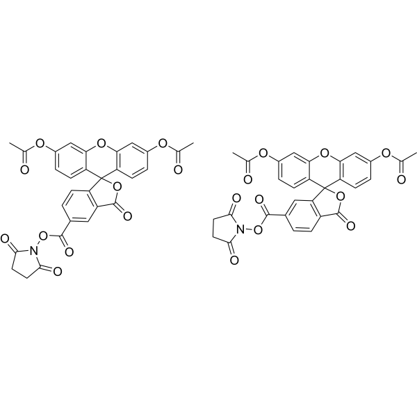
CFDA-SE (solution) is a fluorescent dye that can penetrate the cell membrane. It can react with the free amine group in the cytoskeleton protein inside the cell, and finally form a protein complex with fluorescence. After entering the cell, CFDA-SE locates in the cell membrane, cytoplasm and nucleus, and the fluorescence staining is strongest in the nucleus. CFDA-SE dye can be uniformly inherited by the cells with cell division and proliferation, and its attenuation is proportional to the number of cell divisions. This phenomenon can be detected and analyzed by flow cytometry under the excitation light of 488 nm, and can be used to detect the proliferation of cells.
Solvent and concentration: DMSO: 5 mM
Nur für Forschungszwecke. Wir verkaufen nicht an Patienten.
- CAS. Nr.: 150347-59-4
- Formel: C58H38N2O22
- Molecular Weight:1114.93
-
Speicherung:
Please store the product under the recommended conditions in the Certificate of Analysis.


To place orders, for customer services and technical support, please contact: MedChemExpress USA
Tel: 609-228-6898 E-mail: [email protected] [email protected]


-
 Biologische Aktivität
-
Chemical Information
- Protokoll
- Reinheit & Dokumentation
- Verweise
-
 Help & FAQs


-
HY-D0815
Propidium IodideFluorochrome -
HY-D0938
CFDA-SECell Proliferation Fluorescent Probe -
HY-66019
FITCFluorochrome -
HY-D0711
Indocyanine greenFluorescent Agent -
HY-D0819
CY5-SEReactive Dye -
HY-116215
2-NBDGFluorescent Indicator -
HY-D0040
CalceinFluorescent Dye -
HY-12591
D-Luciferin sodiumSubstrate for Luciferase -

HY-DY1063
CY5-SE (solution)Reactive Dye -

HY-107910
Hyaluronidase, Bovine testesHyaluronic Acid Degrader -
HY-15924
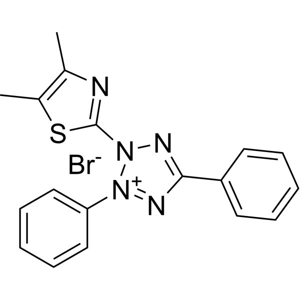
Thiazolyl BlueCytotoxicity Detection Reagent -
HY-15534
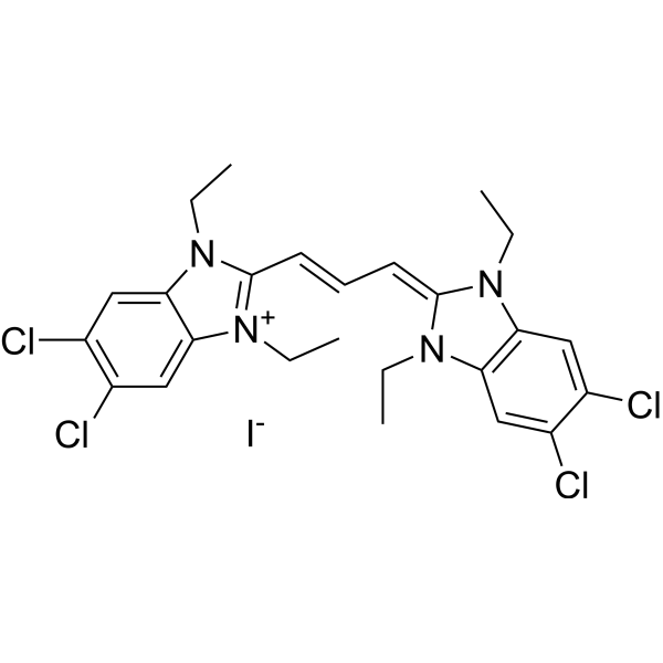
JC-1Mitochondrial Membrane Potential Probe


Biologische Aktivität
Beschreibung
Solvent and concentration: DMSO: 5 mM
In Vitro
Guide (The following is the experimental plan we recommend. This plan serves only as a reference guide. The specific operations should be adjusted according to your actual needs.)
1. Preparation of CFDA-SE working solution
Dilute the original solution in serum-free cell culture medium or PBS to obtain a 5-10 μM CFDA-SE working solution.
Note: Please adjust the concentration of CFDA-SE working solution according to the actual situation.
2. Cell Staining
2.1 Cell Preparation
Suspended cells: Centrifuge at 1000 g for 3-5 minutes at 4°C, then discard the supernatant. Wash twice with PBS, each time for 5 minutes.
Monolayer cells: Remove the cell culture medium, add trypsin to dissociate the cells and prepare a single-cell suspension. Centrifuge at 1000 g for 3-5 minutes at 4°C, then discard the supernatant. Wash twice with PBS, each time for 5 minutes.
2.2 Add 1 mL of CFDA-SE working solution and incubate at room temperature for 30 minutes.
2.3 Centrifuge at 4°C for 3-4 minutes with 400 g
2.4 Wash twice with PBS, 5 minutes each time.
2.5 Resuspend the cells in serum-free cell culture medium or PBS, and then detect them using a fluorescence microscope or flow cytometer.
MedChemExpress (MCE) has not independently confirmed the accuracy of these methods. They are for reference only.
- Flow Cytometry
- Flow cytometric DNA-content cell-cycle staining
- Immunoprecipitation
- Protocol for Phospho-flow cytometry
- CFSE Dye Dilution Proliferation Assay
- Dye-dilution cell tracking and proliferation staining
- Phalloidin F-actin cytoskeleton staining
- Co-Immunoprecipitation
- Cell Viability Determination by MTT Colorimetric Assay


Chemical Information
-
CAS. Nr. 150347-59-4
-
Appearance Liquid
-
Molecular Weight 1114.93
-
Formel C58H38N2O22
-
Color Colorless to light yellow
-
SMILES
CC(OC1=CC=C(C2(C(C=CC(C(ON3C(CCC3=O)=O)=O)=C4)=C4C(O2)=O)C(C=CC(OC(C)=O)=C5)=C5O6)C6=C1)=O.CC(OC7=CC=C(C8(C(C=C(C(ON9C(CCC9=O)=O)=O)C=C%10)=C%10C(O8)=O)C(C=CC(OC(C)=O)=C%11)=C%11O%12)C%12=C7)=O
-
Versand
Room temperature in continental US; may vary elsewhere.
-
Speicherung
Please store the product under the recommended conditions in the Certificate of Analysis.


Reinheit & Dokumentation
-
Data Sheet (273 KB)
-
SDS (251 KB)
- English - EN (251 KB)
- Français - FR (251 KB)
- Deutsch - DE (251 KB)
- Norwegian - NO (251 KB)
- Español - ES (251 KB)
- Swedish - SV (251 KB)
- Italian - IT (251 KB)
- Korean - KR (251 KB)
- Portuguese - PT (251 KB)
-
Handling Instructions (2659 KB)
Verweise
[1]. Banks HT, et al. Quantifying CFSE Label Decay in Flow Cytometry Data. Appl Math Lett. 2013 May 1;26(5):571-577. [Content Brief]
[2]. Cui J, et al. Prostaglandin E3 attenuates macrophage-associated inflammation and prostate tumour growth by modulating polarization. J Cell Mol Med. 2021 Jun;25(12):5586-5601. [Content Brief]
[3]. A Bruce Lyons, et al. Flow cytometric analysis of cell division by dilution of CFSE and related dyes. Curr Protoc Cytom. 2013;Chapter 9:Unit9.11. [Content Brief]
[4]. Xiuyun Jiang et al. Long-lived pancreatic ductal adenocarcinoma slice cultures enable precise study of the immune microenvironment. Oncoimmunology. 2017; 6(7): e1333210. [Content Brief]

Calculators
Konzentration (Stammlösung) × Volumen (Stammlösung) = Konzentration (Ziellösung) × Volumen (Ziellösung)


CFDA-SE (solution) Related Classifications
HY-DY1009 Related Classifications
 Powered by Bioz