



FGL peptide

Based on 1 Customer Validation
FGL peptide is a fibroblast growth factor receptor (FGFR) modulator and blood-brain barrier-penetrant. FGL peptide activates NCAM-FGFR and FGFR1 signaling pathways. FGL peptide alters expression of apoptosis, signal transduction and metabolism regulator genes in traumatic brain injury contexts. FGL peptide can be used for the research of traumatic brain injury.
Nur für Forschungszwecke. Wir verkaufen nicht an Patienten.
- Reinheit : 99.69%
- CAS. Nr.: 499993-62-3
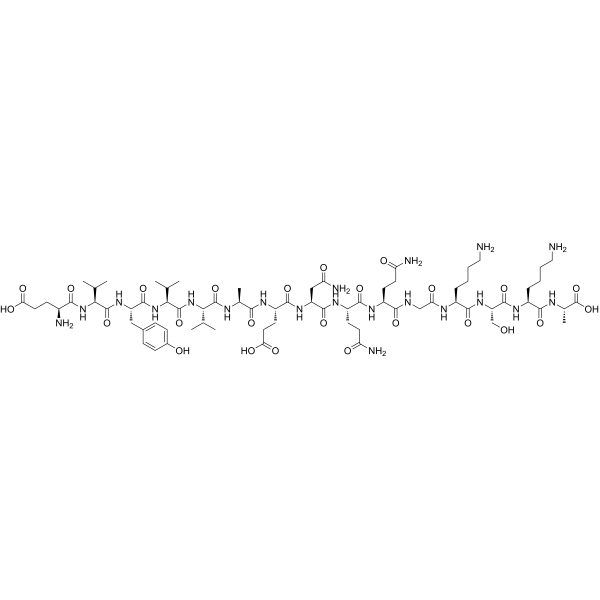
- Formel: C71H116N20O25
- Molecular Weight:1649.80
-
Speicherung:
Sealed storage, away from moisture.
Powder -80°C, 2 years , -20°C, 1 year* In solvent : -80°C, 6 months; -20°C, 1 month (sealed storage, away from moisture)

To place orders, for customer services and technical support, please contact: MedChemExpress USA
Tel: 609-228-6898 E-mail: [email protected] [email protected]


-
 Biologische Aktivität
-
Chemical Information
-
Lösungsmittel & Löslichkeit
- Protokoll
- Reinheit & Dokumentation
- Verweise
-
 Help & FAQs




Biologische Aktivität
Beschreibung
In Vivo
FGL peptide (2.6-26.0 µg; i.n.; on PND1, 2, and 3) accelerates maturation of vestibular-dependent sensorimotor coordination skills in newborn Wistar rats, specifically reducing surface righting reflex latency and increasing successful negative geotaxis responses on PND4-6, without affecting general motor activity, muscle strength, or body weight[2].
MedChemExpress (MCE) has not independently confirmed the accuracy of these methods. They are for reference only.
-
Animal Model:Wistar rats (male, adolescent, 100 g, traumatic brain injury model via dry ice application to skull)[1]
-
Dosage:10 mg/kg
-
Administration:s.c.; every third day
-
Result:Altered the expression of 82 genes via interaction with TBI in injured animals. Upregulated a gene cluster including Fas apoptotic inhibitory molecule, regulator of G-protein signaling 14, and solute carrier family 7 member 1 at 4 days post-injury. Modulated expression of genes involved in metabolism, transcription, and cytoskeleton maintenance, with some genes showing upregulation at 6 hours and 4 days but downregulation at 1 day post-injury. Upregulated genes encoding neurofilament, adenylate cyclase activating polypeptide 1, and RNA binding protein p45AUF1 in non-injured animals. Downregulated genes encoding RNA helicase II, mitochondrial ribosomal protein 9, and metaxin 2 in non-injured animals.


Chemical Information
-
CAS. Nr. 499993-62-3
-
Appearance Solid
-
Molecular Weight 1649.80
-
Formel C71H116N20O25
-
Color White to off-white
-
Sequence
Glu-Val-Tyr-Val-Val-Ala-Glu-Asn-Gln-Gln-Gly-Lys-Ser-Lys-Ala
-
Sequence Shortening
EVYVVAENQQGKSKA
-
Versand
Room temperature in continental US; may vary elsewhere.
-
Speicherung
Sealed storage, away from moisture
Powder -80°C 2 years -20°C 1 year * In solvent : -80°C, 6 months; -20°C, 1 month (sealed storage, away from moisture)


Lösungsmittel & Löslichkeit
In Vitro:
H2O : ≥ 100 mg/mL (60.61 mM)
* "≥" means soluble, but saturation unknown.
Please refer to the solubility information to select the appropriate solvent. Once prepared, please aliquot and store the solution to prevent product inactivation from repeated freeze-thaw cycles.
Storage method and period of stock solution: -80°C, 6 months; -20°C, 1 month (sealed storage, away from moisture). When stored at -80°C, please use it within 6 months. When stored at -20°C, please use it within 1 month.
* Note: If you choose water as the stock solution, please dilute it to the working solution, then filter and sterilize it with a 0.22 μm filter before use.
Please refer to the solubility information to select the appropriate solvent. Once prepared, please aliquot and store the solution to prevent product inactivation from repeated freeze-thaw cycles.
Storage method and period of stock solution: -80°C, 6 months; -20°C, 1 month (sealed storage, away from moisture). When stored at -80°C, please use it within 6 months. When stored at -20°C, please use it within 1 month.
* Note: If you choose water as the stock solution, please dilute it to the working solution, then filter and sterilize it with a 0.22 μm filter before use.
Konzentration (Stammlösung) × Volumen (Stammlösung) = Konzentration (Ziellösung) × Volumen (Ziellösung)


Reinheit & Dokumentation
-
Data Sheet (273 KB)
-
SDS (252 KB)
- English - EN (252 KB)
- Français - FR (252 KB)
- Deutsch - DE (252 KB)
- Norwegian - NO (252 KB)
- Español - ES (252 KB)
- Swedish - SV (252 KB)
- Italian - IT (252 KB)
- Korean - KR (252 KB)
- Portuguese - PT (252 KB)
-
Handling Instructions (2659 KB)
Verweise
[1]. Pedersen MV, et al. The synthetic NCAM-derived peptide, FGL, modulates the transcriptional response to traumatic brain injury. Neurosci Lett. 2008;437(2):148-153. [Content Brief]
[2]. Secher T, et al. A neural cell adhesion molecule-derived fibroblast growth factor receptor agonist, the FGL-peptide, promotes early postnatal sensorimotor development and enhances social memory retention. Neuroscience. 2006;141(3):1289-1299. [Content Brief]


Complete Stock Solution Preparation Table
Please refer to the solubility information to select the appropriate solvent. Once prepared, please aliquot and store the solution to prevent product inactivation from repeated freeze-thaw cycles.
Storage method and period of stock solution: -80°C, 6 months; -20°C, 1 month (sealed storage, away from moisture). When stored at -80°C, please use it within 6 months. When stored at -20°C, please use it within 1 month.
| Optional Solvent | Concentration Solvent Mass | 1 mg | 5 mg | 10 mg | 25 mg |
|---|---|---|---|---|---|
| H2O | 1 mM | 0.6061 mL | 3.0307 mL | 6.0613 mL | 15.1534 mL |
| 5 mM | 0.1212 mL | 0.6061 mL | 1.2123 mL | 3.0307 mL | |
| 10 mM | 0.0606 mL | 0.3031 mL | 0.6061 mL | 1.5153 mL | |
| 15 mM | 0.0404 mL | 0.2020 mL | 0.4041 mL | 1.0102 mL | |
| 20 mM | 0.0303 mL | 0.1515 mL | 0.3031 mL | 0.7577 mL | |
| 25 mM | 0.0242 mL | 0.1212 mL | 0.2425 mL | 0.6061 mL | |
| 30 mM | 0.0202 mL | 0.1010 mL | 0.2020 mL | 0.5051 mL | |
| 40 mM | 0.0152 mL | 0.0758 mL | 0.1515 mL | 0.3788 mL | |
| 50 mM | 0.0121 mL | 0.0606 mL | 0.1212 mL | 0.3031 mL | |
| 60 mM | 0.0101 mL | 0.0505 mL | 0.1010 mL | 0.2526 mL |
* Note: If you choose water as the stock solution, please dilute it to the working solution, then filter and sterilize it with a 0.22 μm filter before use.


FGL peptide Related Classifications
HY-P3281 Related Classifications
 Powered by Bioz