



Oloctinebart

Based on 1 Customer Validation
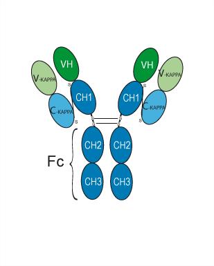
Oloctinebart is a humanized immunoglobulin G4-kappa, anti-LGALS3 monoclonal antibody. Oloctinebart is used for reduction of amyloid beta oligomers formation.
Nur für Forschungszwecke. Wir verkaufen nicht an Patienten.
- Reinheit : 98.00%
- CAS. Nr.: 2669063-84-5
- Molecular Weight:144.876 kDa
-
Speicherung:
Please store the product under the recommended conditions in the Certificate of Analysis.

To place orders, for customer services and technical support, please contact: MedChemExpress USA
Tel: 609-228-6898 E-mail: [email protected] [email protected]


-
 Biologische Aktivität
-
Chemical Information
- Protokoll
- Reinheit & Dokumentation
- Verweise
-
 Help & FAQs




Biologische Aktivität
Beschreibung
Isotype
Human IgG4 kappa
Recommend Isotype Controls
Species Reactivity
Human
IC50 & Target
Galectin-3/LGALS3
Clinical Trial
| NCT Number | Sponsor | Condition | Start Date |
Phase

|
|---|---|---|---|---|
| NCT01329991 | Plexxikon| | 2011-05 | PHASE1 |
Gene ID
Accession
AAH53667.1
Conjugated
Unconjugated
Reconsititution
The product can be reconstituted/diluted with sterile PBS or saline.
Format
-
Human IgG4 kappa
Anwendung
ELISA, FACS, Functional assay
Verified Bioactivity
-
 Loaded Oloctinebart on AHC2 biosensor, can bind Galectin-3/LGALS3 Protein, Human (HEK293, His, HY-P70171) with an affinity constant of 5.984E-08 M as determined in BLI assay.
 -
 Flow Cytometry analysis of Hela cells labelling LGALS3 (red) with Oloctinebart (anti-LGALS3) (HY-P990081). Goat Anti-Human IgG (Alexa Fluor 488) (HY-P83776) at a dilution of 1/1000 was used as the secondary antibody. Blue-Human IgG4 (S228P) kappa (HY-P99003). Black-Unlabelled control, cells without incubation with primary antibody.


Chemical Information
-
CAS. Nr. 2669063-84-5
-
Appearance Liquid
-
Molecular Weight 144.876 kDa
-
Color Colorless to light yellow
-
SMILES
[Oloctinebart]
-
Synonyms
TB-006
-
Versand
Shipping with dry ice.
-
Formulation
Please refer to the lot-specific COA for specific buffer information.
-
Speicherung
Please store the product under the recommended conditions in the Certificate of Analysis.


Reinheit & Dokumentation
-
Data Sheet (255 KB)
-
SDS (251 KB)
- English - EN (251 KB)
- Français - FR (251 KB)
- Deutsch - DE (251 KB)
- Norwegian - NO (251 KB)
- Español - ES (251 KB)
- Swedish - SV (251 KB)
- Italian - IT (251 KB)
- Korean - KR (251 KB)
- Portuguese - PT (251 KB)
-
Inhibitory Antibodies User Guide (603 KB)
Verweise

Calculators
Konzentration (Stammlösung) × Volumen (Stammlösung) = Konzentration (Ziellösung) × Volumen (Ziellösung)


Oloctinebart Related Classifications
HY-P990081 Related Classifications
 Powered by Bioz