



Praliciguat
Based on 2 publication(s) in Google Scholar
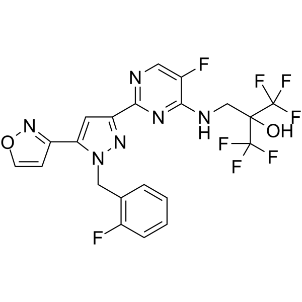
Praliciguat (IW-1973) is a potent and orally active soluble guanylate cyclase (sGC) activator. Praliciguat can increases cGMP via the nitric oxide (NO)-sGC pathway. Praliciguat can inhibit the expression of proinflammatory cytokines and inhibit apoptosis. Praliciguat can promote vasodilation. Praliciguat can be used for the researches of metabolic disease and cardiovascular disease, such as hypertension, diabetes and heart failure.
Nur für Forschungszwecke. Wir verkaufen nicht an Patienten.
- Reinheit : 99.35%
- CAS. Nr.: 1628730-49-3
- Formel: C21H14F8N6O2
- Molecular Weight:534.36
-
Speicherung:Powder -20°C, 3 years , 4°C, 2 years ; In solvent -80°C, 2 years , -20°C, 1 year

To place orders, for customer services and technical support, please contact: MedChemExpress USA
Tel: 609-228-6898 E-mail: [email protected] [email protected]


-
 Biologische Aktivität
-
Chemical Information
-
Lösungsmittel & Löslichkeit
- Protokoll
- Reinheit & Dokumentation
- Verweise
-
 Help & FAQs


-
HY-109136
RuncaciguatsGC Activator -
HY-14181
CinaciguatGuanylate Cyclase Activator -
HY-16774
VericiguatsCG Stimulator -
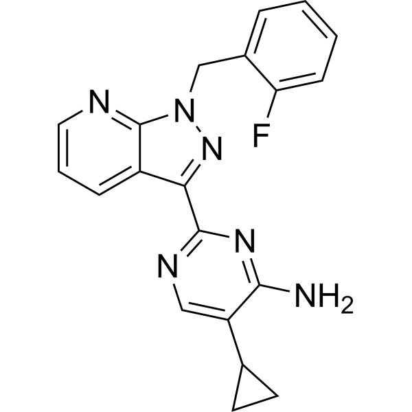
HY-12376
BAY 41-2272Guanylate Cyclase Activator -
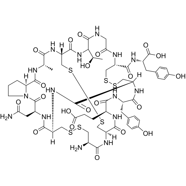
HY-17584
LinaclotideGuanylate Cyclase Agonist -
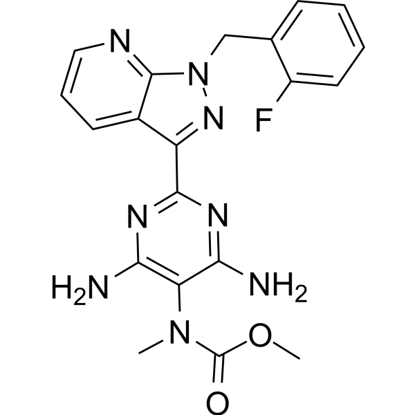
HY-14779
Riociguat -
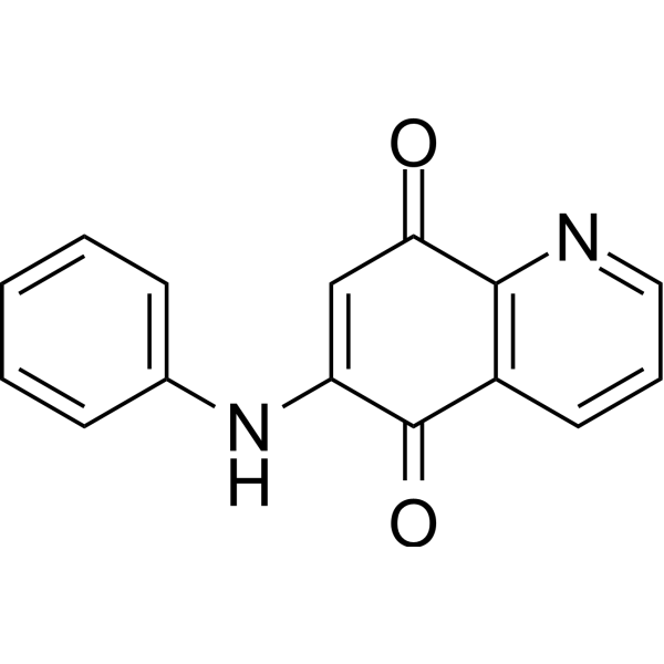
HY-W013386
LY83583sGC Inhibitor -
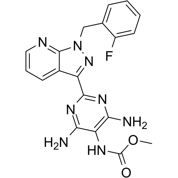
HY-78237
NelociguatGuanylate Cyclase Activator -
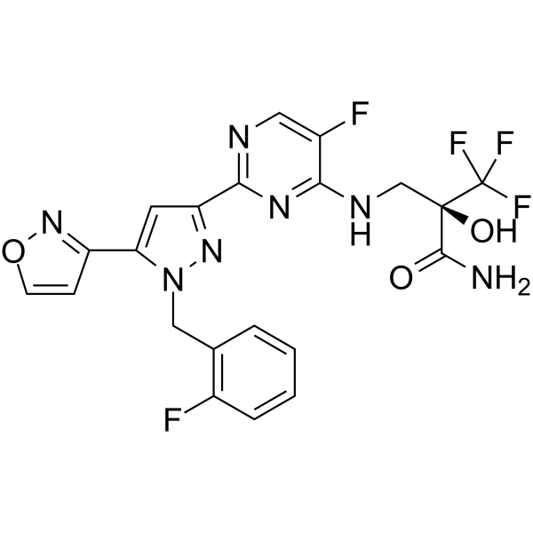
HY-109066
Olinciguat -

HY-16774R
Vericiguat (Standard)sCG Stimulator -
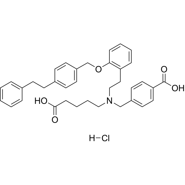
HY-14181A
Cinaciguat hydrochlorideGuanylate Cyclase Activator -
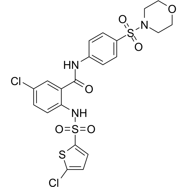
HY-17500
AtaciguatsGC Activator
Publications Citing Use of MedChemExpress (MCE) Praliciguat
More

Biologische Aktivität
Beschreibung
IC50 & Target
EC50: 197 nM (sGC)[1]
In Vitro
Praliciguat stimulates soluble guanylate cyclase in HEK-293 cells with an EC50 of 197 nM[1].
Praliciguat demonstrates concentration-dependent relaxation of pre-contracted subcutaneous resistance arteries (EC50 = 34.7 nM)[1].
Praliciguat (10 μM, 48 h) inhibits the expression of proinflammatory cytokines and secretion of monocyte chemoattractant protein 1 in TNF-α-challenged proximal tubular epithelial cells (RPTC)[2].
Praliciguat (1-10 μM, 48 h) inhibits TGF-β-mediated apoptosis in RPTC[2].
MedChemExpress (MCE) has not independently confirmed the accuracy of these methods. They are for reference only. Further protocols information, click here.
In Vivo
Praliciguat (1-10 mg/kg, p.o.) attenuates proteinuria in obese ZSF1 rats model of diabetic nephropathy[2].
Praliciguat (1-10 mg/kg, p.o., daily for 2-7 weeks) attenuates inflammation, fibrosis, and end-organ damage in the Dahl rats model of cardiorenal failure[3].
MedChemExpress (MCE) has not independently confirmed the accuracy of these methods. They are for reference only.
-
Animal Model:Obese ZSF1 rat model of diabetic nephropathy [2]
-
Dosage:1, 3 and 10 mg/kg
-
Administration:Orally administration
-
Result:Attenuated proteinuria.
Reduced fasting glucose and cholesterol.
-
Animal Model:Dahl rats model of cardiorenal failure[3]
-
Dosage:1, 3 and 10 mg/kg
-
Administration:Orally administration
-
Result:Reduced blood pressure.
Improved cardiorenal damage, daily for 2-7 weeks.
Attenuated the increase in circulating markers of inflammation and fibrosis.
Clinical Trial
| NCT Number | Sponsor | Condition | Start Date |
Phase

|
|---|---|---|---|---|
| NCT01329991 | Plexxikon| | 2011-05 | PHASE1 |


Chemical Information
-
CAS. Nr. 1628730-49-3
-
Appearance Solid
-
Molecular Weight 534.36
-
Formel C21H14F8N6O2
-
Color White to off-white
-
SMILES
FC1=C(C=CC=C1)CN2C(C3=NOC=C3)=CC(C4=NC=C(F)C(NCC(C(F)(F)F)(O)C(F)(F)F)=N4)=N2
-
Synonyms
IW-1973
-
Versand
Room temperature in continental US; may vary elsewhere.
-
Speicherung
Powder -20°C 3 years 4°C 2 years In solvent -80°C 2 years -20°C 1 year


Publications (2)
-
Journal Impact Factor
-
Most Recent
-
-
Biomed Pharmacother
Revisiting soluble guanylate cyclase pharmacology: Additive potential of stimulators and activators. [Abstract]2025 Dec:193:118762. PMID: 41252787


Lösungsmittel & Löslichkeit
In Vitro:
DMSO : 250 mg/mL (467.85 mM; Need ultrasonic; Hygroscopic DMSO has a significant impact on the solubility of product, please use newly opened DMSO)
Please refer to the solubility information to select the appropriate solvent. Once prepared, please aliquot and store the solution to prevent product inactivation from repeated freeze-thaw cycles.
Storage method and period of stock solution: -80°C, 2 years; -20°C, 1 year. When stored at -80°C, please use it within 2 years. When stored at -20°C, please use it within 1 year.
Please refer to the solubility information to select the appropriate solvent. Once prepared, please aliquot and store the solution to prevent product inactivation from repeated freeze-thaw cycles.
Storage method and period of stock solution: -80°C, 2 years; -20°C, 1 year. When stored at -80°C, please use it within 2 years. When stored at -20°C, please use it within 1 year.
Konzentration (Stammlösung) × Volumen (Stammlösung) = Konzentration (Ziellösung) × Volumen (Ziellösung)
In Vivo:
Select the appropriate dissolution method based on your experimental animal and administration route.
- For the following dissolution methods, please ensure to first prepare a clear stock solution using an In Vitro approach and then sequentially add co-solvents:
- To ensure reliable experimental results, the clarified stock solution can be appropriately stored based on storage conditions. As for the working solution for In Vivo experiments, it is recommended to prepare freshly and use it on the same day.
- The percentages shown for the solvents indicate their volumetric ratio in the final prepared solution. If precipitation or phase separation occurs during preparation, heat and/or sonication can be used to aid dissolution.
Add each solvent one by one: 10% DMSO 40% PEG300 5% Tween-80 45% Saline
Solubility: ≥ 2.08 mg/mL (3.89 mM); Clear solution
This protocol yields a clear solution of ≥ 2.08 mg/mL (saturation unknown).
Taking 1 mL working solution as an example, add 100 μL DMSO stock solution (20.8 mg/mL) to 400 μL PEG300, and mix evenly; then add 50 μL Tween-80 and mix evenly; then add 450 μL Saline to adjust the volume to 1 mL.
Preparation of Saline: Dissolve 0.9 g sodium chloride in ddH₂O and dilute to 100 mL to obtain a clear Saline solution.
In Vivo Dissolution Calculator
Please enter the basic information of animal experiments:
-
-
-
-
Recommended: Prepare an additional quantity of animals to account for potential losses during experiments.
Please enter your animal formula composition:
-
%DMSO +
Recommended: Keep the proportion of DMSO in working solution below 2% if your animal is weak.
-
%+
-
+%Tween-80 + +
-
%Saline +
The co-solvents required include: DMSO, . All of co-solvents are available by MedChemExpress (MCE). , Tween 80. All of co-solvents are available by MedChemExpress (MCE).
Working solution concentration: 0.22 mg/mL
Method for preparing stock solution: mg drug dissolved in μL DMSO. Stock solution concentration: mg/mL.
1. Take μL DMSO stock solution;
2. Add μL .
μL , mix evenly;
3. Then add μL Tween 80, mix evenly;
4. Then add μL
Please ensure that the stock solution in the first step is dissolved to a clear state, and add co-solvents in sequence. You can use ultrasonic heating (ultrasonic cleaner, recommended frequency 20-40 kHz), vortexing, etc. to assist dissolution.

Protokoll
-
RNA extraction experimental
By lysing cells, releasing RNA, and removing impurities such as proteins and DNA, high-purity RNA products are finally obtained. The commonly used traditional method is the guanidine isothiocyanate/phenol/chloroform method (Trizol), which is suitable for a variety of animal materials including animal tissues, microorganisms, cultured cells, etc., and most plant materials.
-
Apoptosis
Apoptosis, also called programmed cell death, is generally characterized by distinct morphological characteristics.
-
TUNEL staining for apoptotic DNA fragmentation
TUNEL staining detects DNA strand breaks by using terminal deoxynucleotidyl transferase to add labeled nucleotides to exposed 3′-OH DNA termini, generating either microscopic staining in fixed cells or tissue sections, or fluorescence/cytometric signal in cell suspensions. TUNEL positivity reflects DNA fragmentation but should not be interpreted alone as definitive apoptosis, because TUNEL can also label necrotic, autolytic, mechanically damaged, or DNA-repair-associated DNA breaks.
-
Annexin V plus membrane-impermeant dye apoptosis staining
Annexin V-based apoptosis assays rely on the detection of phosphatidylserine (PS) externalization from the inner leaflet of the plasma membrane to the outer leaflet, an early biochemical hallmark of apoptosis. Fluorescently labeled Annexin V binds PS in a calcium-dependent manner, enabling identification of early apoptotic cells by flow cytometry or fluorescence microscopy. When combined with a membrane-impermeant DNA-binding dye (e. g. , propidium iodide), this approach allows discrimination between viable (Annexin V−/dye−), early apoptotic (Annexin V+/dye−), and late apoptotic or necrotic (Annexin V+/dye+) cell populations by assessing membrane integrity and PS exposure.
-
Apoptosis Solutions
Apoptosis is a regulated, generally non-lytic cell-death pathway that removes unwanted, damaged, infected, or abnormal cells through coordinated morphological changes, caspase activation, DNA fragmentation, and membrane remodeling. The intrinsic apoptosis pathway is controlled mainly by mitochondrial outer membrane permeabilization, BCL-2 family proteins, cytochrome c release, apoptosome formation, caspase-9 activation, and downstream executioner caspase-3/7 activation. The extrinsic apoptosis pathway is initiated by death receptors such as Fas, TNFR, and TRAIL receptors, which recruit adaptor proteins and activate caspase-8 before engaging executioner caspases or mitochondrial amplification through BID cleavage. Apoptosis is linked to many phenotypes, including cancer cell killing, tissue homeostasis, immune regulation, neurodegeneration, infection response, and treatment-induced cytotoxicity; unresolved questions include how apoptosis interacts with necroptosis, pyroptosis, ferroptos
-
Research Protocol for Cardiovascular Diseases
Cardiovascular disease can be modeled as maladaptive cardiac remodeling, where ischemic injury or pressure overload activates inflammatory signaling, fibroblast activation, extracellular-matrix deposition, cardiomyocyte hypertrophy, vascular remodeling, and progressive ventricular dysfunction. The TGF-β/SMAD axis is a central profibrotic pathway after myocardial injury and pressure overload, while innate immune and cytokine pathways regulate leukocyte recruitment, scar formation, and adverse remodeling. Key unresolved questions include which inflammatory signals are reparative versus harmful, when fibrosis is protective versus maladaptive, and whether pathway inhibition improves function without weakening necessary infarct healing or compensatory remodeling.
-
Research Protocol for Metabolic Diseases
AMP-activated protein kinase, AMPK, is a conserved cellular energy sensor that responds to reduced cellular energy status and coordinates metabolism by increasing ATP-generating catabolic pathways while suppressing ATP-consuming anabolic processes. In metabolic disease research, the AMPK pathway is experimentally relevant because it regulates hepatic lipid synthesis, fatty acid oxidation, glucose production, skeletal-muscle glucose disposal, mTORC1-linked biosynthesis, autophagy, mitochondrial homeostasis, and whole-body energy balance. The central pathway logic is that energy stress, metformin, exercise-like stimulation, or direct AMPK activators increase AMPKα Thr172 phosphorylation and downstream substrate phosphorylation, including ACC and RAPTOR. Phosphorylation of ACC suppresses lipogenesis and supports fatty acid oxidation, whereas phosphorylation of RAPTOR suppresses mTORC1 signaling and links cellular energy status to growth and protein synthesis control. The pathway is linked


Reinheit & Dokumentation
-
Data Sheet (277 KB)
-
SDS (252 KB)
- English - EN (252 KB)
- Français - FR (252 KB)
- Deutsch - DE (252 KB)
- Norwegian - NO (252 KB)
- Español - ES (252 KB)
- Swedish - SV (252 KB)
- Italian - IT (252 KB)
- Korean - KR (252 KB)
- Portuguese - PT (252 KB)
-
Handling Instructions (2659 KB)
Verweise
[2]. Liu G, et al. Praliciguat inhibits progression of diabetic nephropathy in ZSF1 rats and suppresses inflammation and apoptosis in human renal proximal tubular cells. Am J Physiol Renal Physiol. 2020 Oct 1;319(4):F697-F711. [Content Brief]
[3]. Shea CM, et al. Soluble guanylate cyclase stimulator praliciguat attenuates inflammation, fibrosis, and end-organ damage in the Dahl model of cardiorenal failure. Am J Physiol Renal Physiol. 2020 Jan 1;318(1):F148-F159. [Content Brief]


Complete Stock Solution Preparation Table
Please refer to the solubility information to select the appropriate solvent. Once prepared, please aliquot and store the solution to prevent product inactivation from repeated freeze-thaw cycles.
Storage method and period of stock solution: -80°C, 2 years; -20°C, 1 year. When stored at -80°C, please use it within 2 years. When stored at -20°C, please use it within 1 year.
| Optional Solvent | Concentration Solvent Mass | 1 mg | 5 mg | 10 mg | 25 mg |
|---|---|---|---|---|---|
| DMSO | 1 mM | 1.8714 mL | 9.3570 mL | 18.7140 mL | 46.7849 mL |
| 5 mM | 0.3743 mL | 1.8714 mL | 3.7428 mL | 9.3570 mL | |
| 10 mM | 0.1871 mL | 0.9357 mL | 1.8714 mL | 4.6785 mL | |
| 15 mM | 0.1248 mL | 0.6238 mL | 1.2476 mL | 3.1190 mL | |
| 20 mM | 0.0936 mL | 0.4678 mL | 0.9357 mL | 2.3392 mL | |
| 25 mM | 0.0749 mL | 0.3743 mL | 0.7486 mL | 1.8714 mL | |
| 30 mM | 0.0624 mL | 0.3119 mL | 0.6238 mL | 1.5595 mL | |
| 40 mM | 0.0468 mL | 0.2339 mL | 0.4678 mL | 1.1696 mL | |
| 50 mM | 0.0374 mL | 0.1871 mL | 0.3743 mL | 0.9357 mL | |
| 60 mM | 0.0312 mL | 0.1559 mL | 0.3119 mL | 0.7797 mL | |
| 80 mM | 0.0234 mL | 0.1170 mL | 0.2339 mL | 0.5848 mL | |
| 100 mM | 0.0187 mL | 0.0936 mL | 0.1871 mL | 0.4678 mL |


Praliciguat Related Classifications
HY-109039 Related Classifications
 Powered by Bioz