



Ferroheme
Based on 1 publication(s) in Google Scholar
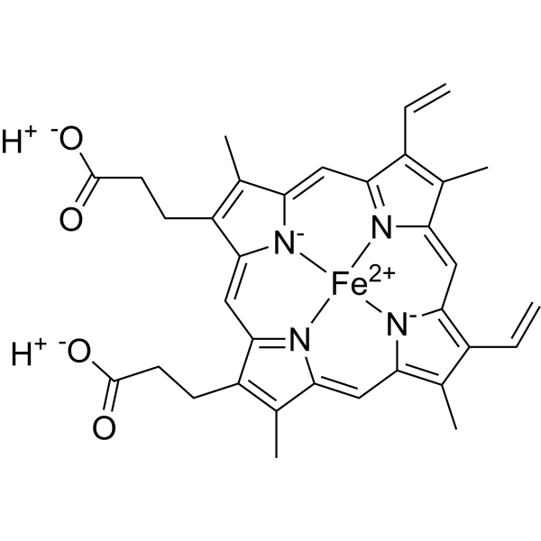
Ferroheme is the ferrous form of heme in hemoglobin, reversibly binding oxygen as an oxygen carrier. Its free form induces oxidative stress and ferroptosis by releasing iron ions, which catalyze reactive oxygen species generation via Fenton reactions, leading to lipid peroxidation and cell death. This mechanism is critical in pathological contexts like intracerebral hemorrhage and neurodegenerative diseases, making it a target for studying iron-overload disorders and ferroptosis-related pathologies[1][2][3].
Nos produits utilisent uniquement pour la recherche. Nous ne vendons pas aux patients.
- Pureté : 99.70%
- CAS No.: 14875-96-8
- Formule: C34H32FeN4O4
- Masse moléculaire:616.49
-
Stockage:
-20°C, protect from light, stored under nitrogen
* In solvent : -80°C, 6 months; -20°C, 1 month (protect from light, stored under nitrogen)

To place orders, for customer services and technical support, please contact: MedChemExpress USA
Tel: 609-228-6898 E-mail: [email protected] [email protected]


-
 Activité biologique
-
Chemical Information
-
Solvant et solubilité
- Protocole
- Pureté et documentation
- Références
-
 Help & FAQs


-
HY-19424
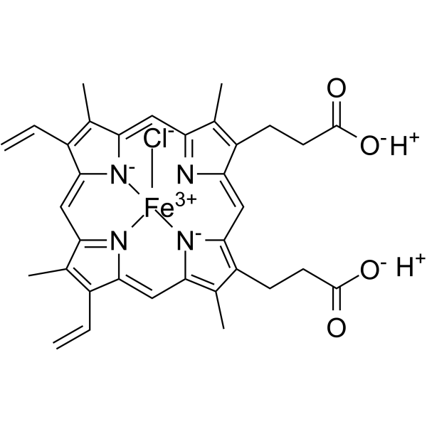
HeminHeme Oxygenase-1 Inducer -
HY-137808
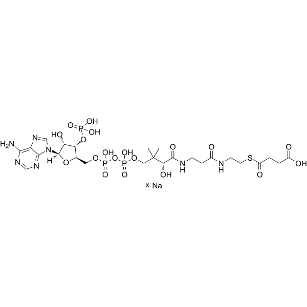
Succinyl-Coenzyme A sodiumCoenzyme A Derivative -
HY-B0511
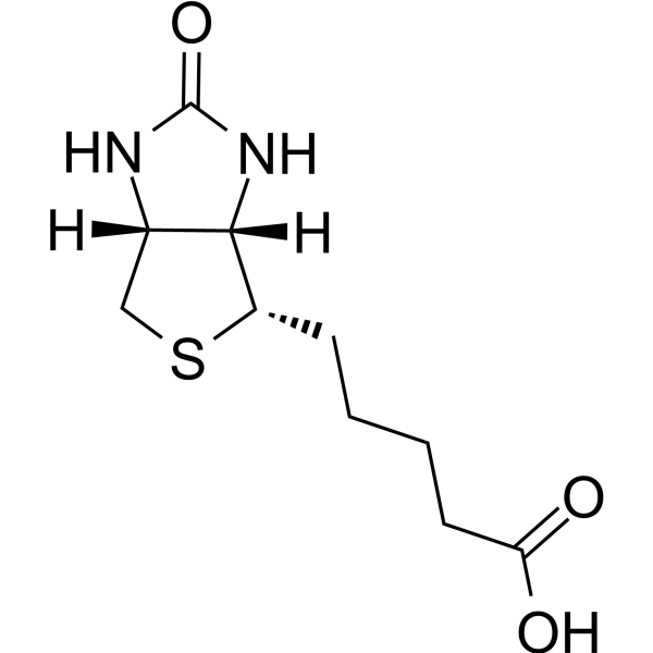
BiotinVitamin B7 -
HY-W355129
MeIQxDietary Aromatic Amine -
HY-B0399
L-CarnitineCo-factor for β-oxidation -
HY-N0323
BilirubinAntioxidant -

HY-B1342
Vitamin A -
HY-B1247
Protoporphyrin IXEndogenous Metabolite,Cancer Inhibitor -
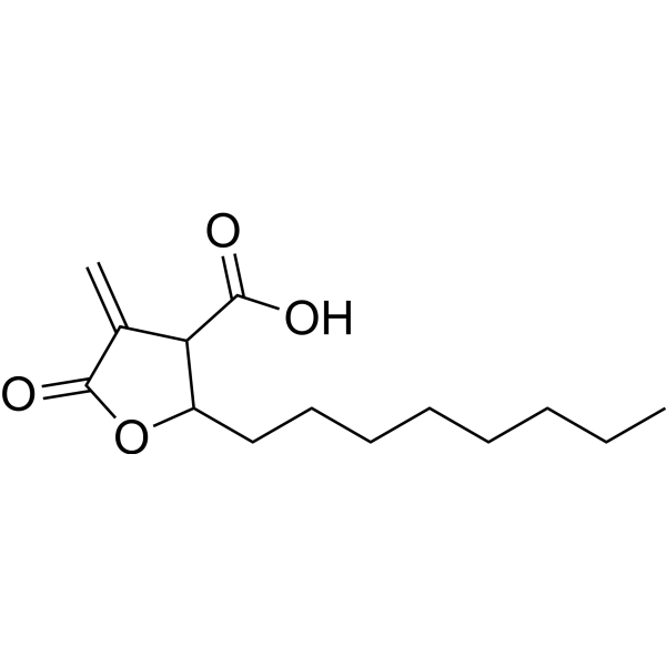
HY-12364
C75Fatty Acid Synthase Inhibitor -
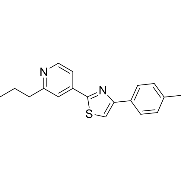
HY-14452
FatostatinSREBP Activation Inhibitor -
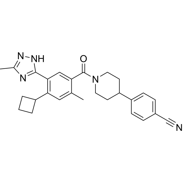
HY-112829
DenifanstatFASN Inhibitor -
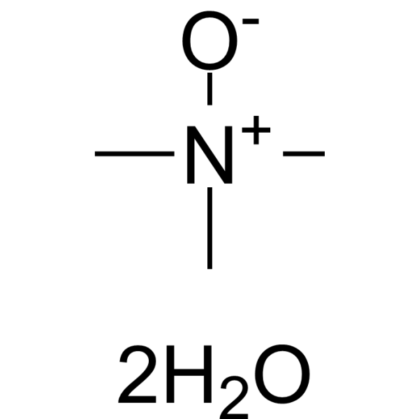
HY-108915
Trimethylamine N-oxide dihydrateROS/NLRP3 Inflammasome Activator
Publications Citing Use of MedChemExpress (MCE) Ferroheme
More

Activité biologique
Description
In Vitro
Ferroheme (5 μM; 24 h) significantly reduces neuronal viability and induces ferroptosis-related lipid peroxidation[1]. Ferroheme (10 μM; 48 h) promotes glioma cell ferroptosis via upregulation of ACSL4 and LPCAT3, accompanied by decreased GPX4 expression[2].
MedChemExpress (MCE) has not independently confirmed the accuracy of these methods. They are for reference only.
In Vivo
MedChemExpress (MCE) has not independently confirmed the accuracy of these methods. They are for reference only.


Chemical Information
-
CAS No. 14875-96-8
-
Appearance Solid
-
Masse moléculaire 616.49
-
Formule C34H32FeN4O4
-
Color Brown to black
-
SMILES
[O-]C(CCC(C1=CC(C(CCC([O-])=O)=C2C)=[N](C2=C3)[Fe+2]4([N-]5C3=C6C)[N-]1C7=CC(C(C=C)=C8C)=[N]4C8=CC5=C6C=C)=C7C)=O.[H+].[H+]
-
Structure Classification
-
Initial Source
-
Livraison
Room temperature in continental US; may vary elsewhere.
-
Stockage
-20°C, protect from light, stored under nitrogen
* In solvent : -80°C, 6 months; -20°C, 1 month (protect from light, stored under nitrogen)


Publications (1)
-
Journal Impact Factor
-
Most Recent
-
J Immunol
TLR4al senses heme as a key damage/danger-associated molecular pattern to activate immune responses in lower vertebrates. [Abstract]2025 Aug 19:vkaf192. PMID: 40829171


Solvant et solubilité
In Vitro:
1 M NaOH : 50 mg/mL (81.10 mM; Need ultrasonic)
H2O : 10 mg/mL (16.22 mM; ultrasonic and adjust pH to 10 with 1M NaOH)
Please refer to the solubility information to select the appropriate solvent. Once prepared, please aliquot and store the solution to prevent product inactivation from repeated freeze-thaw cycles.
Storage method and period of stock solution: -80°C, 6 months; -20°C, 1 month (protect from light, stored under nitrogen). When stored at -80°C, please use it within 6 months. When stored at -20°C, please use it within 1 month.
* Note: If you choose water as the stock solution, please dilute it to the working solution, then filter and sterilize it with a 0.22 μm filter before use.
Please refer to the solubility information to select the appropriate solvent. Once prepared, please aliquot and store the solution to prevent product inactivation from repeated freeze-thaw cycles.
Storage method and period of stock solution: -80°C, 6 months; -20°C, 1 month (protect from light, stored under nitrogen). When stored at -80°C, please use it within 6 months. When stored at -20°C, please use it within 1 month.
* Note: If you choose water as the stock solution, please dilute it to the working solution, then filter and sterilize it with a 0.22 μm filter before use.
Concentration (start) × Volume (start) = Concentration (final) × Volume (final)


Pureté et documentation
-
Fiche technique (281 KB)
-
SDS (393 KB)
- English - EN (393 KB)
- Français - FR (393 KB)
- Deutsch - DE (393 KB)
- Norwegian - NO (393 KB)
- Español - ES (393 KB)
- Swedish - SV (393 KB)
- Italian - IT (393 KB)
- Korean - KR (393 KB)
- Portuguese - PT (393 KB)
-
Instruction de manipulation (2659 KB)
Références
[1]. J M Stewart, et al. Ferriheme and ferroheme are isosteric inhibitors of fatty acid binding to rat liver fatty acid binding protein. Biochem Cell Biol. 1996;74(2):249-55. [Content Brief]
[2]. L Pauling, et al. The Magnetic Properties and Structure of Hemoglobin, Oxyhemoglobin and Carbonmonoxyhemoglobin. Proc Natl Acad Sci U S A. 1936 Apr;22(4):210-6. [Content Brief]
[3]. Derry P J, et al. The chemical basis of intracerebral hemorrhage and cell toxicity with contributions from eryptosis and ferroptosis. Frontiers in Cellular Neuroscience, 2020, 14: 603043. [Content Brief]


Complete Stock Solution Preparation Table
Please refer to the solubility information to select the appropriate solvent. Once prepared, please aliquot and store the solution to prevent product inactivation from repeated freeze-thaw cycles.
Storage method and period of stock solution: -80°C, 6 months; -20°C, 1 month (protect from light, stored under nitrogen). When stored at -80°C, please use it within 6 months. When stored at -20°C, please use it within 1 month.
| Optional Solvent | Concentration Solvent Mass | 1 mg | 5 mg | 10 mg | 25 mg |
|---|---|---|---|---|---|
| H2O / 1 M NaOH | 1 mM | 1.6221 mL | 8.1104 mL | 16.2209 mL | 40.5522 mL |
| 5 mM | 0.3244 mL | 1.6221 mL | 3.2442 mL | 8.1104 mL | |
| 10 mM | 0.1622 mL | 0.8110 mL | 1.6221 mL | 4.0552 mL | |
| 15 mM | 0.1081 mL | 0.5407 mL | 1.0814 mL | 2.7035 mL | |
| 1 M NaOH | 20 mM | 0.0811 mL | 0.4055 mL | 0.8110 mL | 2.0276 mL |
| 25 mM | 0.0649 mL | 0.3244 mL | 0.6488 mL | 1.6221 mL | |
| 30 mM | 0.0541 mL | 0.2703 mL | 0.5407 mL | 1.3517 mL | |
| 40 mM | 0.0406 mL | 0.2028 mL | 0.4055 mL | 1.0138 mL | |
| 50 mM | 0.0324 mL | 0.1622 mL | 0.3244 mL | 0.8110 mL | |
| 60 mM | 0.0270 mL | 0.1352 mL | 0.2703 mL | 0.6759 mL | |
| 80 mM | 0.0203 mL | 0.1014 mL | 0.2028 mL | 0.5069 mL |
* Note: If you choose water as the stock solution, please dilute it to the working solution, then filter and sterilize it with a 0.22 μm filter before use.


Ferroheme Related Classifications
HY-111914A Related Classifications
 Powered by Bioz