



FITC (solution)

Based on 1 Customer Validation
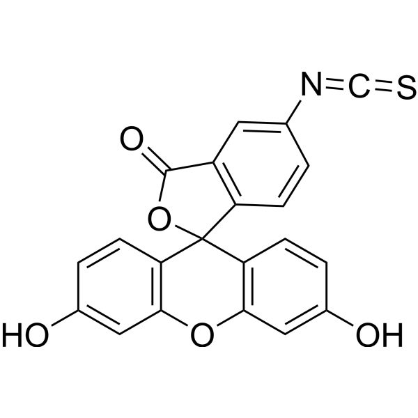
FITC (Fluorescein Isothiocyanate) (solution) , is one of the green fluorescein derivatives widely used in biology. FITC has the characteristics of high absorptivity and excellent fluorescence quantum yield. The isothiocyanate group of FITC can be combined with amino, sulfhydryl, imidazole, tyrosyl, carbonyl and other groups on the protein, so as to achieve protein labeling including antibodies and lectins. In addition to its use as a protein marker, FITC can also be used as a fluorescent protein tracer to rapidly identify pathogens by labeling antibodies, or for microsequencing of proteins and peptides (HPLC). The maximum excitation wavelength of FITC is 494 nm. Once excited, it fluoresces yellow-green at a maximum emission wavelength of 520 nm. In addition, FITC is also a hapten that can induce contact hypersensitivity (CHS) and induce an atopic dermatitis model.
Solvent and concentration: DMSO: 20 mM
The 1 mL volume is defined as the base specification. All larger sizes correspond to incremental volumes of this base.
Nos produits utilisent uniquement pour la recherche. Nous ne vendons pas aux patients.
- CAS No.: 3326-32-7
- Formule: C21H11NO5S
- Masse moléculaire:389.38
-
Stockage:
Please store the product under the recommended conditions in the Certificate of Analysis.


To place orders, for customer services and technical support, please contact: MedChemExpress USA
Tel: 609-228-6898 E-mail: [email protected] [email protected]


-
 Activité biologique
-
Chemical Information
- Protocole
- Pureté et documentation
- Références
-
 Help & FAQs


-
HY-66019
FITCFluorochrome -
HY-D0718
Nile RedHydrophobic Fluorescent Stain -
HY-D0083
DiIFluorescent Dye -
HY-D0938
CFDA-SECell Proliferation Fluorescent Probe -
HY-15938
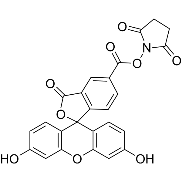
5-FAM SEFluorescent Labeling Reagent -
HY-15534
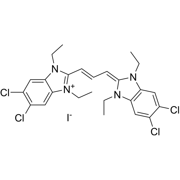
JC-1Mitochondrial Membrane Potential Probe -

HY-DY1008
Nile Red (solution)Hydrophobic Fluorescent Stain -
HY-B0178A
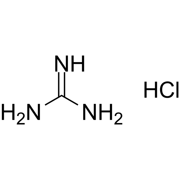
Guanidine hydrochlorideProtein Denaturant -
HY-D0914
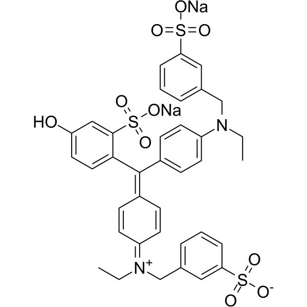
Fast Green FCFSea Green Triarylmethane Food Dye -
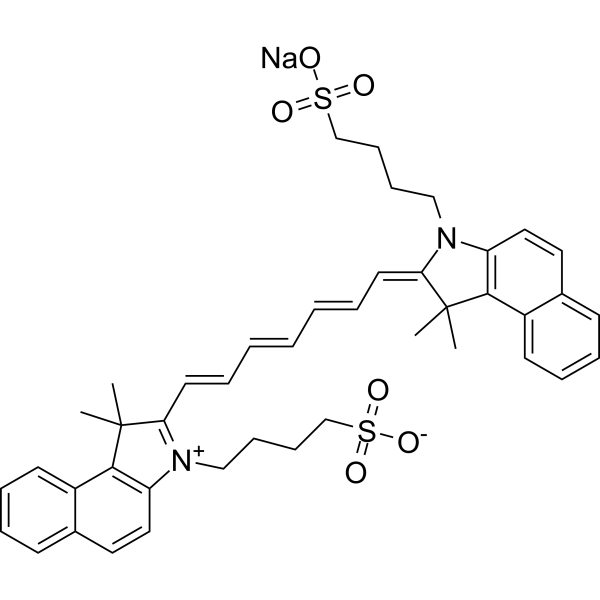
HY-D0711
Indocyanine greenFluorescent Agent -
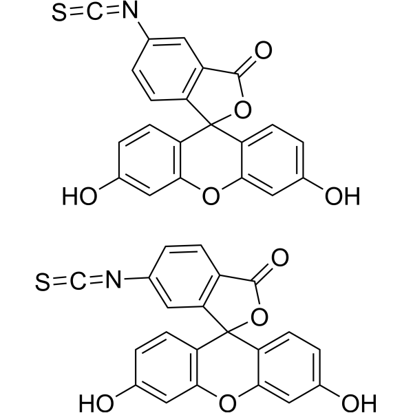
HY-15941
5(6)-FITCFluorescent Dye -
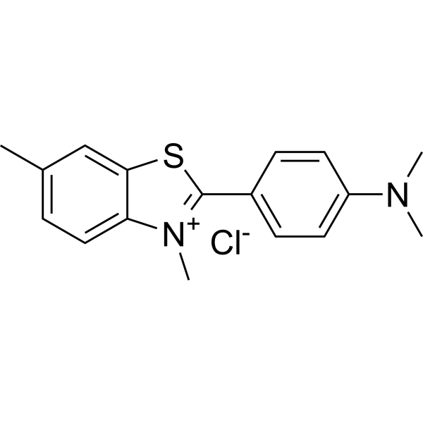
HY-D0218
Thioflavin Tβ-amyloid Dye


Activité biologique
Description
Solvent and concentration: DMSO: 20 mM
The 1 mL volume is defined as the base specification. All larger sizes correspond to incremental volumes of this base.
Cellular Effect
|
Cell Line

|
Type | Value | Description | References |
|---|---|---|---|---|
| L1210 | IC50 |
700 μM
Compound: FITC
|
Cytotoxicity against mouse L1210 cells after 24 hrs by WST8 assay
Cytotoxicity against mouse L1210 cells after 24 hrs by WST8 assay
|
[PMID: 18951028] |
In Vitro
Guide (The following is our recommended protocol. This protocol is only a guide and should be modified according to your specific needs).
1. Protein Preparation
To obtain the best labeling effect, please prepare the protein (antibody) concentration to 1 mg/mL.
1.1 The pH value of the protein solution should be 8.5 ± 0.5. If the pH is lower than 8.0, adjust it with 1 M sodium bicarbonate.
1.2 If the protein concentration is lower than 1 mg/mL, the labeling efficiency will be greatly reduced. To obtain the best labeling efficiency, it is recommended that the final protein concentration range be 1-10 mg/mL.
2.Calculation of dye dosage
The amount of FITC required for reaction depends on the amount of protein to be labeled, and the optimal molar ratio of FITC to protein is about 10.
Example: assuming the required marker protein is 1 mL 2 mg/mL IgG (MW=150,000), use 1 mL DMSO dissolve 1 mg FITC, the required FITC volume is 40 μL.
3. Running the Coupling Reaction
1) Slowly add the calculated volume of freshly prepared 10 mg/mL dye to 0.5 mL of protein sample solution, gently shake to mix, and then briefly centrifuge to collect the sample at the bottom of the reaction tube. Avoid vigorous mixing to prevent denaturation and inactivation of the protein sample.
2) Place the reaction tube in a dark place and incubate gently with shaking for 60 minutes at room temperature. Every 10-15 minutes, gently invert the reaction tube several times to thoroughly mix the reactants and improve labeling efficiency.
4. Purification of Conjugates
The following protocol is an example of purifying dye-protein conjugates using a Labelling Kits Centrifugation-Based Rapid Desalting Column (5KD) (HY-D3014).
4.1 Prepare the desalting column according to the instructions.
4.2 Load the reaction mixture onto the top of the desalting column.
4.3 Once the sample has run below the resin surface, immediately add PBS (pH 7.2-7.4).
4.4 Add more PBS (pH 7.2-7.4) to the target sample to complete column purification. Collect the fraction containing the desired dye-protein conjugate.
MedChemExpress (MCE) has not independently confirmed the accuracy of these methods. They are for reference only.


Chemical Information
-
CAS No. 3326-32-7
-
Appearance Liquid (Density: 1.548±0.14 g/cm3)
-
Masse moléculaire 389.38
-
Formule C21H11NO5S
-
Color Light yellow to yellow
-
SMILES
O=C1OC2(C3=C(OC4=C2C=CC(O)=C4)C=C(O)C=C3)C5=C1C=C(N=C=S)C=C5
-
Livraison
Room temperature in continental US; may vary elsewhere.
-
Stockage
Please store the product under the recommended conditions in the Certificate of Analysis.


Pureté et documentation
-
Fiche technique (292 KB)
-
SDS (251 KB)
- English - EN (251 KB)
- Français - FR (251 KB)
- Deutsch - DE (251 KB)
- Norwegian - NO (251 KB)
- Español - ES (251 KB)
- Swedish - SV (251 KB)
- Italian - IT (251 KB)
- Korean - KR (251 KB)
- Portuguese - PT (251 KB)
-
Instruction de manipulation (2659 KB)
Références
[1]. Gao SG, et al. Phosphorylation of osteopontin has proapoptotic and proinflammatory effects on human knee osteoarthritis chondrocytes. Exp Ther Med. 2016 Nov;12(5):3488-3494. Epub 2016 Oct 5. [Content Brief]
[2]. Zhu X, et al. Ratiometric, visual, dual-signal fluorescent sensing and imaging of pH/copper ions in real samples based on carbon dots-fluorescein isothiocyanate composites. Talanta. 2017 Jan 1;162:65-71 [Content Brief]
[3]. Shigeno T, et al. Phthalate ester‐induced thymic stromal lymphopoietin mediates allergic dermatitis in mice[J]. Immunology, 2009, 128(1pt2): e849-e857. [Content Brief]

Calculators
Concentration (start) × Volume (start) = Concentration (final) × Volume (final)


FITC (solution) Related Classifications
HY-DY1007 Related Classifications
 Powered by Bioz