



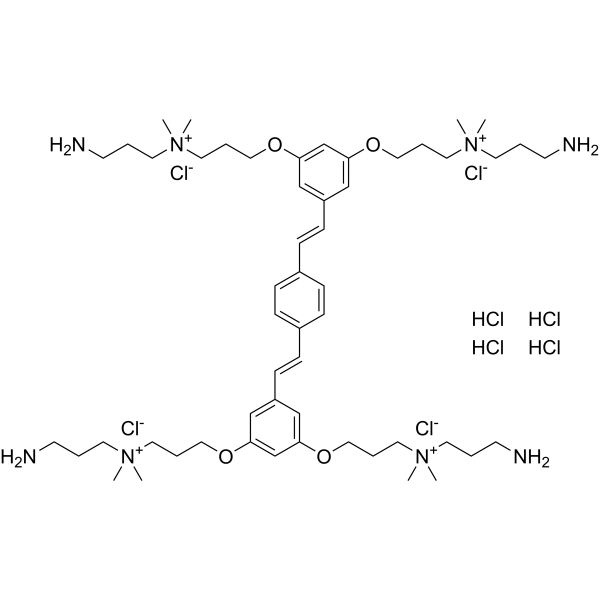
COE-PNH2
COE-PNH2 exhibits antibacterial activity against Mycobacterium abscessus (Mab) with MIC90 of 26 μM. COE-PNH2 affects the integrity of the bacterial envelope and mycomembrane. COE-PNH2 reveals intracelluar penetration without mitochondrial toxicity.
For research use only. We do not sell to patients.
- Formula: C54H98Cl8N8O4
- Molecular Weight:1207.03
-
Storage:
Please store the product under the recommended conditions in the Certificate of Analysis.

To place orders, for customer services and technical support, please contact: MedChemExpress USA
Tel: 609-228-6898 E-mail: [email protected] [email protected]


-
 Biological Activity
-
Chemical Information
- Protocols
- Purity & Documentation
- References
-
 Help & FAQs




Biological Activity
Description
In Vitro
COE-PNH2 (0-128 μg/mL) exhibits a stable antibacterial activity and extreme antibiotic tolerance against intracellular, replicating and nutrient-starved, non-replicating Mab[1].
COE-PNH2 exhibits a low propensity for resistance development with consistent MIC values in 14 passages of Mab[1].
COE-PNH2 (0-64 μg/mL) induces a depletion of ATP, accumulation of intracellular lipid inclusions (ILIs) and bacterial elongation in Mab[1].
MedChemExpress (MCE) has not independently confirmed the accuracy of these methods. They are for reference only.
In Vivo
MedChemExpress (MCE) has not independently confirmed the accuracy of these methods. They are for reference only.
-
Animal Model:Acute lung infection in C3HeB/FeJ mice[1]
-
Dosage:2.5-5 mg/kg
-
Administration:Intratracheal administration every other day for 12 days
-
Result:Reduced the colony-forming units (CFU) and bacilli in lung without weight loss.


Chemical Information
-
Molecular Weight 1207.03
-
Formula C54H98Cl8N8O4
-
SMILES
C[N+](C)(CCCN)CCCOC1=CC(/C=C/C2=CC=C(/C=C/C3=CC(OCCC[N+](C)(CCCN)C)=CC(OCCC[N+](C)(CCCN)C)=C3)C=C2)=CC(OCCC[N+](C)(CCCN)C)=C1.[Cl-].[Cl-].[Cl-].[Cl-].Cl.Cl.Cl.Cl
-
Shipping
Room temperature in continental US; may vary elsewhere.
-
Storage
Please store the product under the recommended conditions in the Certificate of Analysis.


Purity & Documentation
References

Calculators
Concentration (start) × Volume (start) = Concentration (final) × Volume (final)


COE-PNH2 Related Classifications
HY-158003 Related Classifications
 Powered by Bioz