



Cremophor EL-10
Cremophor EL-10 is a nonionic polyoxyethylene castor oil surfactant with multiple functions including organic solubilizer, mixed zinc anode corrosion inhibitor and dendrite growth inhibitor. By forming a physical barrier to block electrolyte contact and constructing oriented hydration channels to reduce the desolvation energy barrier of Zn2+ , Cremophor EL-10 achieves a corrosion inhibition efficiency of over 99%. Cremophor EL-10 exerts no significant effects on the mitochondrial activity and cell viability of epithelial cells, making it suitable for in vitro drug delivery and biopharmaceutical research at concentrations ≤10% v/v. However, Cremophor EL-10 may induce sustained non-endothelium-dependent contraction in rat aortic rings and exert concentration-dependent inhibitory effects on acetylcholine-induced endothelium-dependent relaxation responses.
For research use only. We do not sell to patients.
- CAS No.: 61791-12-6
-
Storage:
Please store the product under the recommended conditions in the Certificate of Analysis.


To place orders, for customer services and technical support, please contact: MedChemExpress USA
Tel: 609-228-6898 E-mail: [email protected] [email protected]


-
 Biological Activity
-
Chemical Information
- Protocols
- Purity & Documentation
- References
-
 Help & FAQs


-
HY-Y1890
Cremophor EL-40Surfactant -
HY-401424
Cremophor EL-36Surfactan -

HY-Y1890C
Cremophor EL-20Surfactant -

HY-Y1890D
Cremophor EL-60Cosolvent -

HY-Y1890E
Cremophor EL-80Cosolvent -

HY-Y1890H
Cremophor EL-90Cosolvent -

HY-Y1891
Tween 80 -
HY-141415
Tween 20Nonionic Surfactant -
HY-Y1883
Polyethylene glycol mono(4-tert-octylphenyl) ether surfactantNonionic Surfactant -
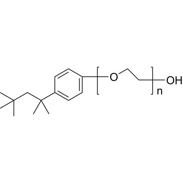
HY-Y1883A
Polyethylene glycol mono(4-tert-octylphenyl) etherNonionic Surfactant -
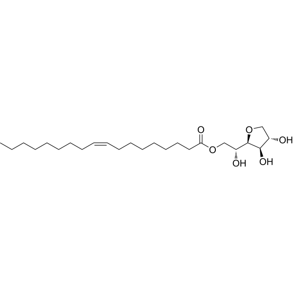
HY-N1446C
Sorbitan monooleateEmulsifier -
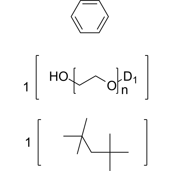
HY-W134422
(1,1,3,3-Tetramethylbutyl)phenyl-polyethylene glycol X-114Surfactant


Biological Activity
Description
In Vitro
Cremophor EL-10 (100-500 mg/L; 6 h) potently inhibits static corrosion of pure zinc plates in 6 M KOH solution, achieving >90% inhibition efficiency at 100 mg/L and reaching a near-maximum inhibition efficiency near 98% at concentrations ≥400 mg/L after 6 h immersion[3].
Cremophor EL-10 (400 mg/L; 6 h) adsorbs onto pure zinc plates in 6 M K+OH? solution at 400 mg/L after 6 h immersion, forming a surface film composed of its molecular components as confirmed by characteristic ATR-FTIR peaks[3].
MedChemExpress (MCE) has not independently confirmed the accuracy of these methods. They are for reference only. Further protocols information, click here.


Chemical Information
-
CAS No. 61791-12-6
-
SMILES
[Cremophor EL-10]
-
Shipping
Room temperature in continental US; may vary elsewhere.
-
Storage
Please store the product under the recommended conditions in the Certificate of Analysis.

Protocols
-
Cell Cytotoxicity Assay
Cytotoxicity assays are usually based on the assessment of cell membrane damage, which can also be indirectly detected by measuring cell viability. Detection methods include MTT assay, CKK-8 assay, LDH assay and ATP assay, etc.
-
Mammalian live/dead viability and cytotoxicity staining
Live/dead viability and cytotoxicity staining assays are based on the simultaneous detection of intracellular esterase activity in metabolically active (viable) cells and membrane integrity loss in non-viable cells. In commonly used dual-staining approaches, membrane-permeant fluorogenic substrates are converted by intracellular esterases into fluorescent products in live cells, while impermeant DNA-binding dyes selectively enter cells with compromised plasma membranes and label nucleic acids in dead or dying cells, enabling discrimination between viable and non-viable populations by fluorescence microscopy or flow cytometry.
-
CCK-8/WST-8 Cell Proliferation Assay
The CCK-8/WST-8 assay is based on the reduction of the water-soluble tetrazolium salt WST-8 to a water-soluble formazan product by cellular dehydrogenases in metabolically active cells, where the generated formazan amount is proportional to the number of living cells and is quantified by measuring absorbance in the visible range, providing a colorimetric readout for cell viability and proliferation assessment. This class of tetrazolium-based assays improves upon earlier MTT-based systems by producing a water-soluble formazan, eliminating the need for organic solubilization steps and enabling direct spectrophotometric measurement in culture medium.
-
MTT Cell Proliferation Assay
The MTT assay is a colorimetric endpoint assay for estimating viable cell number, cell growth, cytotoxicity, or cell activation in cultured mammalian cells. Living cells reduce the yellow tetrazolium salt MTT into purple/blue formazan, while dead cells do not generate the same signal; the resulting color can be quantified with a multiwell spectrophotometer. MTT reduction is commonly interpreted as a readout of metabolic activity that often correlates with viable cell number, but it should not be treated as a direct cell-counting method unless the assay is optimized for the cell type and experimental condition. Studies show that MTT reduction can involve mitochondrial and non-mitochondrial reducing systems, and formazan may accumulate in intracellular lipid droplets rather than simply marking mitochondria.
-
Cell Viability Determination by MTT Colorimetric Assay
The following protocol uses the MTT colorimetric assay as a classic literature-established method for assessing cell viability/metabolic activity in cultured mammalian cells. MTT[3-(4,5-dimethylthiazol-2-yl)-2,5-diphenyltetrazolium bromide] is reduced by metabolically active cells to a colored formazan product; the amount of formazan is quantified spectrophotometrically and provides an indirect measure of metabolically active viable cells. Importantly, MTT reduction reflects cellular oxidoreductase/metabolic activity rather than an absolute direct count of living cells, so changes in cellular metabolism can alter the signal independently of cell number.


Purity & Documentation
References
[1]. MacDonald C, et al. Water-soluble organic solubilizers for in vitro drug delivery studies with respiratory epithelial cells: selection based on various toxicity indicators. Drug Deliv. 2010;17(6):434-442. [Content Brief]
[2]. Zengil H, et al. Effects of polysorbates and Cremophor EL on vascular responses in rat aorta. Experientia. 1995;51(11):1055-1059. [Content Brief]
[3]. Cen H, et al. Nonlinear Correlation of Polyoxyethylene Castor Oil between Dynamic-Static Corrosion Inhibition and Cyclic Stability as Zinc-Air Battery Additives. Langmuir. 2026;42(1):1627-1643. [Content Brief]

Calculators
Concentration (start) × Volume (start) = Concentration (final) × Volume (final)


Cremophor EL-10 Related Classifications
HY-Y1890B Related Classifications
 Powered by Bioz