



Cy3 methyltetrazine

Based on 1 Customer Validation
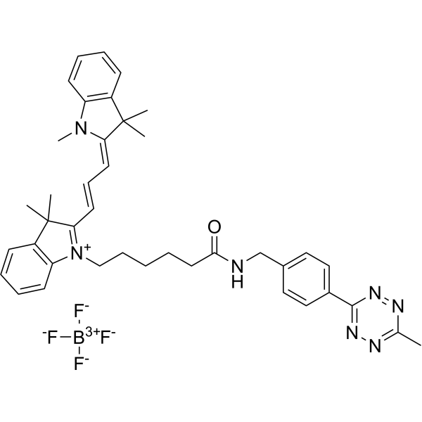
Cy3 methyltetrazine (TZ-Cy3) is a click chemistry reagent with methyltetrazine building blocks that is highly reactive towards cyclooctene. Cy3 methyltetrazine is also a tetrazine-modified fluorescent probe that can be used to analyze protein phosphorylation in solution and living cells.
For research use only. We do not sell to patients.
- Purity : 97.36%
- CAS No.: 2183473-57-4
- Formula: C40H46BF4N7O
- Molecular Weight:727.64
-
Storage:
-20°C, sealed storage, away from moisture and light
* In solvent : -80°C, 6 months; -20°C, 1 month (sealed storage, away from moisture and light)

To place orders, for customer services and technical support, please contact: MedChemExpress USA
Tel: 609-228-6898 E-mail: [email protected] [email protected]


-
 Biological Activity
-
Chemical Information
-
Solvent & Solubility
- Protocols
- Purity & Documentation
- References
-
 Help & FAQs




Biological Activity
Description
In Vitro
Guide (Following is our recommended protocol. This protocol only provides a guideline, and should be modified according to your specific needs).
Confocal imaging of phosphorylation in cells:(take H1299 cells as an example)
1. Culture H1299 cells in a coverglass-bottom imaging dish for 24 h.
2. Incubate H1299 cells with different concentrations of ATP-NB (a ATP analogue functionalized by norbornene, which can penetrate cells and efficiently phosphorylate proteins in living cells) for 1 h.
3. Add Cy3 methyltetrazine to label the phosphorylated protein with fluorescence.
4. Incubate with fresh medium for 1 h.
5. Wash the cells twice with PBS to release the free Cy3 methyltetrazine probe.
6.Incubate the cells with 4% paraformaldehyde for 15 min and with 0.1% Triton X-100 solution at room temperature for 10 min.
7. Wash cells 4 times with PBS.
8. Use a confocal microscopy imaging system to observe (Ex = 488 nm; Em = 561 nm).
MedChemExpress (MCE) has not independently confirmed the accuracy of these methods. They are for reference only.
Emission (Em)
570
Excitation (Ex)
550


Chemical Information
-
CAS No. 2183473-57-4
-
Appearance Solid
-
Molecular Weight 727.64
-
Formula C40H46BF4N7O
-
Color Brown to reddish brown
-
SMILES
O=C(CCCCC[N+]1=C(/C=C/C=C2N(C)C3=C(C\2(C)C)C=CC=C3)C(C)(C)C4=C1C=CC=C4)NCC5=CC=C(C6=NN=C(C)N=N6)C=C5.[F-][B+3]([F-])([F-])[F-]
-
Synonyms
TZ-Cy3
-
Shipping
Room temperature in continental US; may vary elsewhere.
-
Storage
-20°C, sealed storage, away from moisture and light
* In solvent : -80°C, 6 months; -20°C, 1 month (sealed storage, away from moisture and light)


Solvent & Solubility
In Vitro:
DMSO : 62.5 mg/mL (85.89 mM; Need ultrasonic; Hygroscopic DMSO has a significant impact on the solubility of product, please use newly opened DMSO)
Please refer to the solubility information to select the appropriate solvent. Once prepared, please aliquot and store the solution to prevent product inactivation from repeated freeze-thaw cycles.
Storage method and period of stock solution: -80°C, 6 months; -20°C, 1 month (sealed storage, away from moisture and light). When stored at -80°C, please use it within 6 months. When stored at -20°C, please use it within 1 month.
Please refer to the solubility information to select the appropriate solvent. Once prepared, please aliquot and store the solution to prevent product inactivation from repeated freeze-thaw cycles.
Storage method and period of stock solution: -80°C, 6 months; -20°C, 1 month (sealed storage, away from moisture and light). When stored at -80°C, please use it within 6 months. When stored at -20°C, please use it within 1 month.
Concentration (start) × Volume (start) = Concentration (final) × Volume (final)


Purity & Documentation
-
Data Sheet (274 KB)
-
SDS (252 KB)
- English - EN (252 KB)
- Français - FR (252 KB)
- Deutsch - DE (252 KB)
- Norwegian - NO (252 KB)
- Español - ES (252 KB)
- Swedish - SV (252 KB)
- Italian - IT (252 KB)
- Korean - KR (252 KB)
- Portuguese - PT (252 KB)
-
Handling Instructions (2659 KB)
References


Complete Stock Solution Preparation Table
Please refer to the solubility information to select the appropriate solvent. Once prepared, please aliquot and store the solution to prevent product inactivation from repeated freeze-thaw cycles.
Storage method and period of stock solution: -80°C, 6 months; -20°C, 1 month (sealed storage, away from moisture and light). When stored at -80°C, please use it within 6 months. When stored at -20°C, please use it within 1 month.
| Optional Solvent | Concentration Solvent Mass | 1 mg | 5 mg | 10 mg | 25 mg |
|---|---|---|---|---|---|
| DMSO | 1 mM | 1.3743 mL | 6.8715 mL | 13.7431 mL | 34.3576 mL |
| 5 mM | 0.2749 mL | 1.3743 mL | 2.7486 mL | 6.8715 mL | |
| 10 mM | 0.1374 mL | 0.6872 mL | 1.3743 mL | 3.4358 mL | |
| 15 mM | 0.0916 mL | 0.4581 mL | 0.9162 mL | 2.2905 mL | |
| 20 mM | 0.0687 mL | 0.3436 mL | 0.6872 mL | 1.7179 mL | |
| 25 mM | 0.0550 mL | 0.2749 mL | 0.5497 mL | 1.3743 mL | |
| 30 mM | 0.0458 mL | 0.2291 mL | 0.4581 mL | 1.1453 mL | |
| 40 mM | 0.0344 mL | 0.1718 mL | 0.3436 mL | 0.8589 mL | |
| 50 mM | 0.0275 mL | 0.1374 mL | 0.2749 mL | 0.6872 mL | |
| 60 mM | 0.0229 mL | 0.1145 mL | 0.2291 mL | 0.5726 mL | |
| 80 mM | 0.0172 mL | 0.0859 mL | 0.1718 mL | 0.4295 mL |


Cy3 methyltetrazine Related Classifications
HY-151776 Related Classifications
 Powered by Bioz