



D-Trehalose, Carrier Protein
Based on 1 publication(s) in Google Scholar
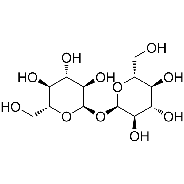
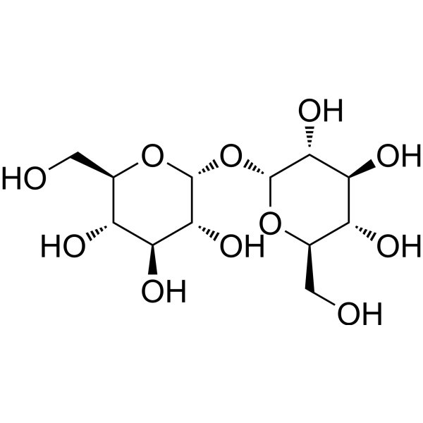
D-Trehalose is a disaccharide formed by a 1,1-glycosidic bond between two α-glucose units and is widely used as a food ingredient and pharmaceutical excipient. D-Trehalose is known to protect and stabilise proteins. For long-term storage, recombinant protein solution should be diluted further with 5% D-Trehalose, Carrier Protein.
For research use only. We do not sell to patients.
- Purity : 99.94%
- CAS No.: 99-20-7
- Formula: C12H22O11
- Molecular Weight:342.30
-
Storage:
Please store the product under the recommended conditions in the Certificate of Analysis.

To place orders, for customer services and technical support, please contact: MedChemExpress USA
Tel: 609-228-6898 E-mail: [email protected] [email protected]


-
 Biological Activity
-
Chemical Information
-
Solvent & Solubility
- Protocols
- Purity & Documentation
-
 Help & FAQs


-

HY-P70666
IL-12 Protein, Mouse (HEK293) -
HY-N1132
D-(+)-TrehaloseDisaccharide -
HY-B1779
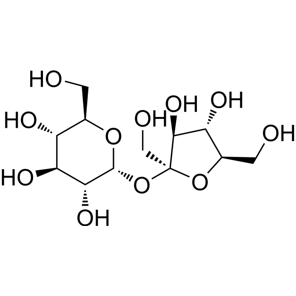
SucroseDisaccharide -

HY-N1132R
D-(+)-Trehalose (Standard)Disaccharide -

HY-13259
MG-132Proteasome Inhibitor -
HY-P73181
IL-18 Protein, Mouse (solution) -

HY-Y1891
Tween 80 -
HY-P70445
IL-4 Protein, Human -
HY-P70311
Activin A Protein, Human/Mouse/Rat (HEK293) -
HY-P71887A
CXCL14/BRAK Protein, Mouse (His) -
HY-P7044
IL-6 Protein, Human -
HY-B0389
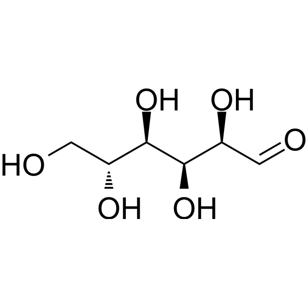
D-GlucoseMonosaccharide
Publications Citing Use of MedChemExpress (MCE) D-Trehalose, Carrier Protein
More

Biological Activity
Description
In Vitro
D-Trehalose is a typical stress metabolite that forms a unique protective film on cell surfaces under harsh environmental conditions such as high temperature, extreme cold, high osmotic pressure, and desiccation, thereby maintaining the life processes and biological characteristics of living organisms.
If a sterile solution is required, filter the prepared solution through a 0.22 μm filter before use.
1. Stabilizes cell membranes and protein structures
2. Resists desiccation
3. Pharmaceutical excipient, used as an excipient
4. Inhibits collagen degradation
Plastic tube walls strongly adsorb proteins, causing recombinant proteins to adhere to the tube walls and be difficult to separate. This results in a low actual concentration in solution, ultimately manifesting as decreased activity. When reconstituting recombinant proteins, the addition of carrier proteins can pre-block the protein binding sites on the plastic tube walls, preventing the recombinant protein from adhering to the tube walls and thus protecting protein activity. When performing serum-free culture, in vivo experiments, or protein labeling, the recombinant protein used should not contain animal or human proteins such as BSA, FBS, or HSA. Trehalose can be used as a carrier protein for long-term storage of recombinant proteins. To maintain a certain pH and salt concentration and prevent protein instability, dissolving the carrier protein in water is not recommended. Instead, a buffer with a near-neutral pH and sufficient buffering capacity, such as PBS or culture medium (RPMI1640, DMEM), is recommended.
MedChemExpress (MCE) has not independently confirmed the accuracy of these methods. They are for reference only. Further protocols information, click here.


Chemical Information
-
CAS No. 99-20-7
-
Appearance Solid
-
Molecular Weight 342.30
-
Formula C12H22O11
-
Color White to off-white
-
SMILES
OC[C@H]([C@H]([C@@H]([C@H]1O)O)O)O[C@@H]1O[C@H]2O[C@@H]([C@H]([C@@H]([C@H]2O)O)O)CO
-
Shipping
Room temperature in continental US; may vary elsewhere.
-
Storage
Please store the product under the recommended conditions in the Certificate of Analysis.


Publications (1)
-
Journal Impact Factor
-
Most Recent
-
Cell Stem Cell
Mitochondrial serine catabolism safeguards maintenance of the hematopoietic stem cell pool in homeostasis and injury. [Abstract]2024 Oct 3;31(10):1484-1500.e9. PMID: 39181130


Solvent & Solubility
In Vitro:
H2O : 100 mg/mL (292.14 mM; Need ultrasonic)
* Note: If you choose water as the stock solution, please dilute it to the working solution, then filter and sterilize it with a 0.22 μm filter before use.
* Note: If you choose water as the stock solution, please dilute it to the working solution, then filter and sterilize it with a 0.22 μm filter before use.
Concentration (start) × Volume (start) = Concentration (final) × Volume (final)
In Vivo:
For the following dissolution methods, please prepare the working solution directly:
It is recommended to prepare fresh solutions and use them promptly within a short period of time.
The percentages shown for the solvents indicate their volumetric ratio in the final prepared solution. If precipitation or phase separation occurs during preparation, heat and/or sonication can be used to aid dissolution.
Add each solvent one by one: PBS
Solubility: 100 mg/mL (292.14 mM); Clear solution; Need ultrasonic

Protocols
-
Baculovirus-insect cell protein expression and purification
Baculovirus-insect cell expression uses recombinant baculovirus to deliver a target gene into insect cells, where late or very-late viral transcription drives recombinant protein production; the method was classically demonstrated by expression of human β-interferon in baculovirus-infected insect cells. The readout is target protein accumulation, assessed by activity, fluorescence if a fluorescent reporter is used, SDS-PAGE, Western blot, or purified protein yield. The system can express soluble, secreted, membrane-associated, and multiprotein targets, but expression outcome depends on the construct, baculovirus vector, insect cell line, multiplicity of infection, infection cell density, harvest time, and target-specific stability.
-
E. coli fusion-tag soluble protein purification
The purification of soluble recombinant proteins in Escherichia coli is achieved by fusing the target protein with a solubility-enhancing affinity tag (e. g. , His-tag, GST, MBP, Fh8, CSQ-tag, or thioredoxin) to improve expression yield, prevent aggregation, and enable efficient purification via affinity chromatography. The fusion protein is expressed under inducible promoters (e. g. , IPTG-induced T7 promoter), lysed from bacterial cells, and purified using resin-based affinity chromatography (e. g. , Ni-NTA for His-tag, amylose resin for MBP, chitin resin for intein tags, or HIC for Fh8). Tags can be removed post-purification using site-specific proteases (e. g. , TEV, enterokinase) or through intracellular cleavage systems. Solubility screening using multiple fusion partners (e. g. , Expresso® system) allows optimization of expression conditions for difficult-to-express proteins.
-
Inclusion-body expression, solubilization, refolding and purification
Inclusion-body recovery uses insoluble recombinant protein aggregates from E. coli as a starting material; the workflow is cell disruption, inclusion-body isolation/washing, denaturant or mild solubilization, refolding into soluble protein, and final chromatographic purification. The readouts are soluble protein recovery, purity by SDS-PAGE/chromatography, structural recovery by methods such as circular dichroism when used, and biological activity when an assay is available.
-
Protocol For Protein Expression And Purification
Recombinant protein expression in Escherichia coli followed by purification of a His-tagged soluble protein by immobilized metal affinity chromatography (IMAC), with optional MBP fusion and TEV tag removal when the construct includes these elements. The biological readout is production of the encoded target protein, detected as an inducible band at the expected molecular mass by SDS-PAGE and quantified by total protein assay or chromatographic absorbance; the purification readout is enrichment of the target protein in elution fractions after selective binding of polyhistidine residues to immobilized Ni2+/metal-chelate resin and elution by imidazole-containing buffer. Expression is driven by an inducible bacterial expression system, commonly T7/lac-based, in which IPTG or lactose/auto-induction activates transcription and translation of the cloned gene; lower induction temperature, lower inducer concentration, induction timing, and solubility-enhancing fusion tags can influence the frac
-
Mammalian transient protein expression and purification
Mammalian transient protein expression introduces plasmid DNA into HEK293 or CHO cells for short-term recombinant protein production, allowing secreted, glycosylated, Fc-tagged, His-tagged, or membrane proteins to be produced without stable clone generation.


Purity & Documentation
-
Data Sheet (261 KB)
-
SDS (394 KB)
- English - EN (394 KB)
- Français - FR (394 KB)
- Deutsch - DE (394 KB)
- Norwegian - NO (394 KB)
- Español - ES (394 KB)
- Swedish - SV (394 KB)
- Italian - IT (394 KB)
- Korean - KR (394 KB)
- Portuguese - PT (394 KB)
-
Handling Instructions (2659 KB)


Complete Stock Solution Preparation Table
| Optional Solvent | Concentration Solvent Mass | 1 mg | 5 mg | 10 mg | 25 mg |
|---|---|---|---|---|---|
| H2O | 1 mM | 2.9214 mL | 14.6071 mL | 29.2141 mL | 73.0354 mL |
| 5 mM | 0.5843 mL | 2.9214 mL | 5.8428 mL | 14.6071 mL | |
| 10 mM | 0.2921 mL | 1.4607 mL | 2.9214 mL | 7.3035 mL | |
| 15 mM | 0.1948 mL | 0.9738 mL | 1.9476 mL | 4.8690 mL | |
| 20 mM | 0.1461 mL | 0.7304 mL | 1.4607 mL | 3.6518 mL | |
| 25 mM | 0.1169 mL | 0.5843 mL | 1.1686 mL | 2.9214 mL | |
| 30 mM | 0.0974 mL | 0.4869 mL | 0.9738 mL | 2.4345 mL | |
| 40 mM | 0.0730 mL | 0.3652 mL | 0.7304 mL | 1.8259 mL | |
| 50 mM | 0.0584 mL | 0.2921 mL | 0.5843 mL | 1.4607 mL | |
| 60 mM | 0.0487 mL | 0.2435 mL | 0.4869 mL | 1.2173 mL | |
| 80 mM | 0.0365 mL | 0.1826 mL | 0.3652 mL | 0.9129 mL | |
| 100 mM | 0.0292 mL | 0.1461 mL | 0.2921 mL | 0.7304 mL |
* Note: If you choose water as the stock solution, please dilute it to the working solution, then filter and sterilize it with a 0.22 μm filter before use.


D-Trehalose, Carrier Protein Related Classifications
HY-CP001 Related Classifications
 Powered by Bioz