



BLM-IN-1
Based on 4 publication(s) in Google Scholar
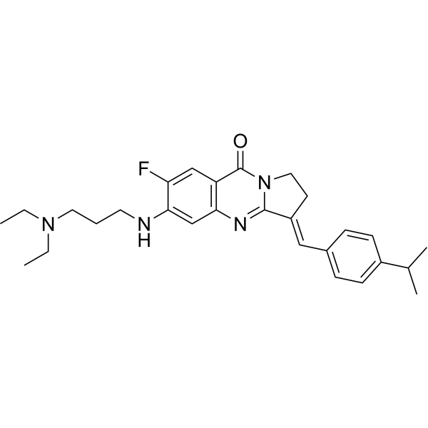
BLM-IN-1 is an effective bloom syndrome protein (BLM) inhibitor. BLM-IN-1 has a high binding affinity with a KD valueof 1.81 μM. BLM-IN-1 has good inhibitory effect for BLM with an IC50 value of 0.95 μM. BLM-IN-1 can induce cell apoptosis. BLM-IN-1can be used for the research of DNA damage and cancer.
Nur für Forschungszwecke. Wir verkaufen nicht an Patienten.
- Reinheit : 98.04%
- CAS. Nr.: 2056014-40-3
- Formel: C28H35FN4O
- Molecular Weight:462.60
-
Speicherung:Powder -20°C, 3 years , 4°C, 2 years ; In solvent -80°C, 2 years , -20°C, 1 year

To place orders, for customer services and technical support, please contact: MedChemExpress USA
Tel: 609-228-6898 E-mail: [email protected] [email protected]


-
 Biologische Aktivität
-
Chemical Information
-
Lösungsmittel & Löslichkeit
- Protokoll
- Reinheit & Dokumentation
- Verweise
-
 Help & FAQs



Publications Citing Use of MedChemExpress (MCE) BLM-IN-1
More

Biologische Aktivität
Beschreibung
IC50 & Target
IC50: 0.95 μM (BLM)[1].
In Vitro
BLM-IN-1 (compound 29) has a strong BLM binding with a KD of 1.81 μM[1].
BLM-IN-1 can inhibit BLM with an IC50 of 0.95 μM[1].
BLM-IN-1 (1.0 μM) inhibits the unwinding activity of BLM but also disrupt the BLM binding to DNA[1].
BLM-IN-1 (0-1.0 μM) regulates the homologous recombination by disrupting the recruitment of BLM to DSB sites[1].
BLM-IN-1 (1.0, 2.0 μM; 48 h) induces DNA damage response and apoptosis[1].
BLM-IN-1 (0.1, 1.0, 2.0 μM; 48 h) induces proliferation arrest and synthetic interaction[1].
MedChemExpress (MCE) has not independently confirmed the accuracy of these methods. They are for reference only.
-
Cell Line:HCT116 cells
-
Concentration:0.5, 1.0 μM
-
Incubation Time:24 h
-
Result:Obviously upregulated the level of pATM and p-ATR in damage response.
-
Cell Line:HCT116 cells
-
Concentration:1.0 μM
-
Incubation Time:24 h
-
Result:Induced the accumlation of RAD51 at DSB sites.
-
Cell Line:HCT116 cells
-
Concentration:0.1, 1.0, 2.0 μM
-
Incubation Time:48 h
-
Result:Showed a significant proliferation arrest in a dose-dependent manner in HCT116 cells.
-
Cell Line:HCT116 cells
-
Concentration:1.0, 2.0 μM
-
Incubation Time:48 h
-
Result:Increased the ratio of apoptosis cells in a concentration-dependent manner and upregulated the expression level of cleaved caspase-3, cleaved caspase-7, and cleaved PARP.


Chemical Information
-
CAS. Nr. 2056014-40-3
-
Appearance Solid
-
Molecular Weight 462.60
-
Formel C28H35FN4O
-
Color Light yellow to yellow
-
SMILES
CCN(CC)CCCNC1=CC(N=C(/C(CC2)=C/C3=CC=C(C(C)C)C=C3)N2C4=O)=C4C=C1F
-
Versand
Room temperature in continental US; may vary elsewhere.
-
Speicherung
Powder -20°C 3 years 4°C 2 years In solvent -80°C 2 years -20°C 1 year


Publications (4)
-
Journal Impact Factor
-
Most Recent
-
-
Reproduction
2026 Feb 5;171(2):xaaf018. PMID: 41643214 -
Theriogenology
2026 Jul 7:265:118070. PMID: 42424899 -
Anim Reprod Sci
BLM helicase deficiency impairs porcine oocyte maturation via induction of organelle dysfunction. [Abstract]2026 Jul:290:108196. PMID: 41990564


Lösungsmittel & Löslichkeit
In Vitro:
DMSO : 8.33 mg/mL (18.01 mM; Need ultrasonic; Hygroscopic DMSO has a significant impact on the solubility of product, please use newly opened DMSO)
Please refer to the solubility information to select the appropriate solvent. Once prepared, please aliquot and store the solution to prevent product inactivation from repeated freeze-thaw cycles.
Storage method and period of stock solution: -80°C, 2 years; -20°C, 1 year. When stored at -80°C, please use it within 2 years. When stored at -20°C, please use it within 1 year.
Please refer to the solubility information to select the appropriate solvent. Once prepared, please aliquot and store the solution to prevent product inactivation from repeated freeze-thaw cycles.
Storage method and period of stock solution: -80°C, 2 years; -20°C, 1 year. When stored at -80°C, please use it within 2 years. When stored at -20°C, please use it within 1 year.
Konzentration (Stammlösung) × Volumen (Stammlösung) = Konzentration (Ziellösung) × Volumen (Ziellösung)


Reinheit & Dokumentation
-
Data Sheet (282 KB)
-
SDS (251 KB)
- English - EN (251 KB)
- Français - FR (251 KB)
- Deutsch - DE (251 KB)
- Norwegian - NO (251 KB)
- Español - ES (251 KB)
- Swedish - SV (251 KB)
- Italian - IT (251 KB)
- Korean - KR (251 KB)
- Portuguese - PT (251 KB)
-
Handling Instructions (2659 KB)
Verweise


Complete Stock Solution Preparation Table
Please refer to the solubility information to select the appropriate solvent. Once prepared, please aliquot and store the solution to prevent product inactivation from repeated freeze-thaw cycles.
Storage method and period of stock solution: -80°C, 2 years; -20°C, 1 year. When stored at -80°C, please use it within 2 years. When stored at -20°C, please use it within 1 year.
| Optional Solvent | Concentration Solvent Mass | 1 mg | 5 mg | 10 mg | 25 mg |
|---|---|---|---|---|---|
| DMSO | 1 mM | 2.1617 mL | 10.8085 mL | 21.6169 mL | 54.0424 mL |
| 5 mM | 0.4323 mL | 2.1617 mL | 4.3234 mL | 10.8085 mL | |
| 10 mM | 0.2162 mL | 1.0808 mL | 2.1617 mL | 5.4042 mL | |
| 15 mM | 0.1441 mL | 0.7206 mL | 1.4411 mL | 3.6028 mL |


BLM-IN-1 Related Classifications
HY-111756 Related Classifications
 Powered by Bioz